
Autoimmune Hepatitis: Diagnosis and Management
Key points
- Autoimmune hepatitis: chronic immune-mediated inflammation of the liver, characterised by raised IgG, circulating autoantibodies and interface hepatitis on biopsy.
- Epidemiology: predominantly affects women (around 75%), with a bimodal age distribution peaking around puberty and again in the fifth to sixth decades.
- Type 1: the commoner form (around 80%), positive for ANA and/or anti-smooth muscle antibody, affecting adults and adolescents.
- Type 2: less common, positive for anti-LKM1 and/or anti-LC1, typically affecting children and young people, and often more aggressive.
- Presentation: highly variable - asymptomatic abnormal LFTs, chronic fatigue and jaundice, acute hepatitis, or established cirrhosis at diagnosis.
- Diagnostic triad: raised transaminases with a raised IgG, positive autoantibodies, and interface hepatitis with plasma cells on liver biopsy.
- Treatment: induction with corticosteroids (prednisolone or budesonide), then maintenance with azathioprine - check TPMT before starting.
- Associations: commonly coexists with other autoimmune disease, and may overlap with primary biliary cholangitis or primary sclerosing cholangitis.
Introduction
Autoimmune hepatitis (AIH) is a chronic, immune-mediated inflammatory disease of the liver, in which loss of tolerance to hepatocyte autoantigens drives progressive hepatocellular injury.1 It is defined by the combination of raised transaminases, elevated immunoglobulin G, circulating autoantibodies, and characteristic histology, having excluded viral, drug-induced and metabolic causes.
It is uncommon, with a UK prevalence of roughly 10-20 per 100,000, but is important because it is highly treatable: untreated, severe disease has a 10-year mortality approaching 50%, whereas appropriately treated patients often achieve a normal life expectancy. It affects women in around 75% of cases and shows a bimodal age distribution, with peaks around puberty and again in the fifth to sixth decades.
Classification and pathophysiology
Types of autoimmune hepatitis
| Feature | Type 1 | Type 2 |
|---|---|---|
| Proportion | Around 80% - the commoner form | Around 20% |
| Autoantibodies | ANA (antinuclear) and/or anti-smooth muscle antibody (ASMA); anti-soluble liver antigen (anti-SLA) in a subset | Anti-LKM1 (liver-kidney microsomal type 1) and/or anti-LC1 (liver cytosol type 1) |
| Typical age | Adults and adolescents; bimodal peaks | Children and young adults |
| Severity | Variable; often insidious | Often more aggressive, with a higher rate of presenting as acute liver failure |
| Response to treatment | Generally good, relapse common on withdrawal | Good, but relapse on withdrawal is more frequent, so treatment is usually lifelong |
A proportion of patients are seronegative, having typical clinical and histological features but no detectable autoantibodies. These patients should still be treated as autoimmune hepatitis, since they respond equally well to immunosuppression - the absence of antibodies does not exclude the diagnosis.
Pathophysiology
The prevailing model is that an environmental trigger - a virus, a drug, or molecular mimicry from a microbial antigen - initiates an immune response against hepatocyte antigens in a genetically susceptible individual. Susceptibility is strongly linked to HLA-DR3 and HLA-DR4 haplotypes; HLA-DR3 disease tends to present younger and behave more aggressively.2
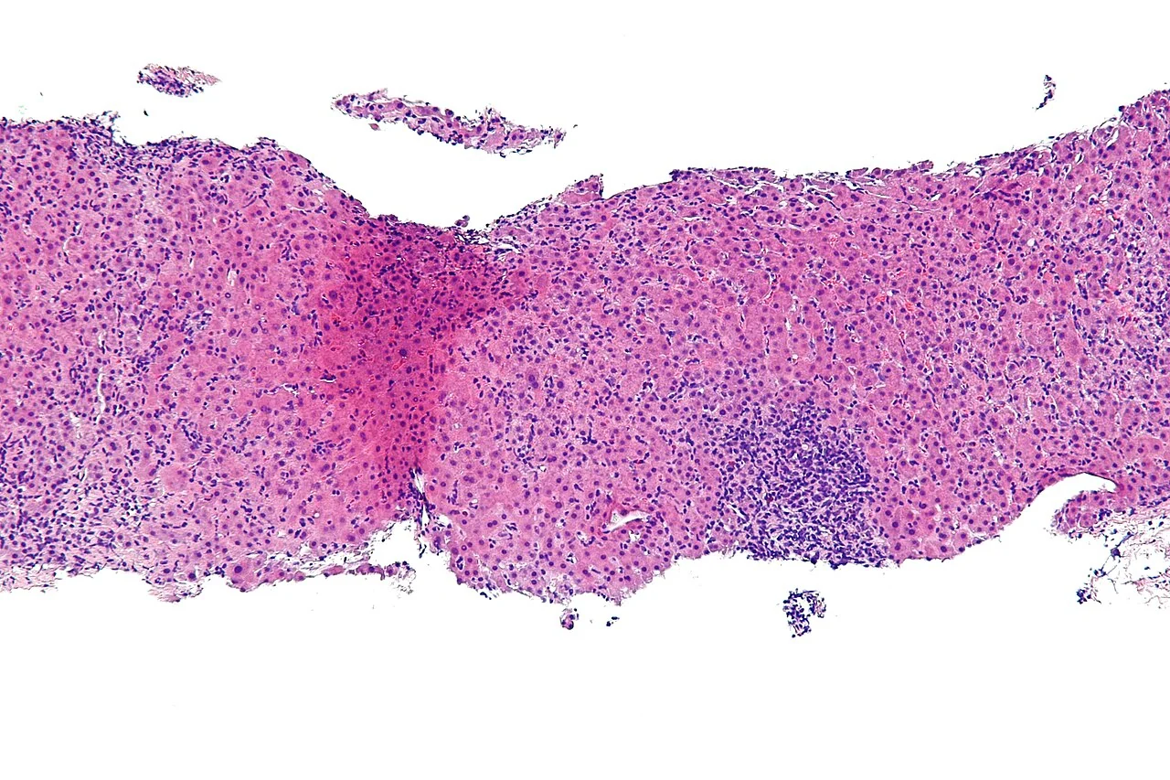
Failure of regulatory T-cell control allows CD4+ T-helper cells to drive both cytotoxic T-cell attack on hepatocytes and B-cell autoantibody production, with plasma cell infiltration and hypergammaglobulinaemia. The inflammation characteristically begins at the limiting plate, the boundary between the portal tract and the hepatic lobule, producing the hallmark lesion of interface hepatitis (historically called piecemeal necrosis). Sustained inflammation activates hepatic stellate cells, causing bridging fibrosis and eventually cirrhosis.
Clinical features
Presentation is notably heterogeneous, which is a major reason for diagnostic delay. Roughly a quarter of patients are asymptomatic at diagnosis, and up to a third already have cirrhosis.1
- Asymptomatic abnormal liver blood tests, found incidentally - around 25% of cases
- Insidious chronic presentation (the commonest): fatigue - often profound and the dominant complaint - malaise, anorexia, nausea, right upper quadrant discomfort, arthralgia, amenorrhoea and weight loss
- Acute hepatitis: jaundice, dark urine, marked transaminase elevation, sometimes indistinguishable from acute viral hepatitis
- Acute liver failure: a small but important minority present with coagulopathy and encephalopathy
- Established cirrhosis: presenting with ascites, variceal bleeding or encephalopathy in up to a third of patients at diagnosis
Examination
Findings range from an entirely normal examination to jaundice, hepatomegaly, splenomegaly and the full range of chronic liver disease stigmata - spider naevi, palmar erythema, ascites and asterixis. Cushingoid features may be present in those already on corticosteroids.
Associated autoimmune conditions
Around 40% of patients have at least one other autoimmune condition, and their presence should raise suspicion when investigating unexplained liver enzyme abnormalities:
- Autoimmune thyroid disease (Hashimoto thyroiditis, Graves disease) - the commonest association
- Coeliac disease - test all patients
- Type 1 diabetes mellitus
- Rheumatoid arthritis and other connective tissue diseases
- Inflammatory bowel disease, particularly ulcerative colitis - which should also prompt consideration of primary sclerosing cholangitis
- Vitiligo, pernicious anaemia, autoimmune haemolytic anaemia and immune thrombocytopenia
Investigations
Blood tests
- Liver enzymes: a hepatitic pattern with ALT and AST raised, often 5-20 times the upper limit of normal, and disproportionately higher than ALP
- Immunoglobulins: a raised IgG (polyclonal hypergammaglobulinaemia) is a hallmark and is present in around 85% of patients. It is also a useful marker of disease activity and treatment response
- Bilirubin: raised in icteric presentations
- Albumin and INR: markers of synthetic function; a prolonged INR indicates severe disease
- Full blood count: cytopenias may reflect hypersplenism from portal hypertension, or a coexisting autoimmune cytopenia
Autoantibodies
| Antibody | Association |
|---|---|
| ANA (antinuclear antibody) | Type 1 AIH; non-specific and found in many autoimmune conditions and in health |
| Anti-smooth muscle antibody (ASMA) | Type 1 AIH; more specific than ANA |
| Anti-LKM1 | Type 2 AIH; typically in children and young people |
| Anti-LC1 | Type 2 AIH |
| Anti-SLA/LP | Highly specific for AIH; associated with more severe disease and relapse |
| Anti-mitochondrial antibody (AMA) | Primary biliary cholangitis, not AIH - its presence should prompt reconsideration or suggest an overlap syndrome |
| pANCA | Primary sclerosing cholangitis and ulcerative colitis; may be positive in type 1 AIH |
Excluding other causes
A full non-invasive liver screen is mandatory before diagnosing autoimmune hepatitis: viral hepatitis serology (A, B, C, E, plus EBV and CMV in acute presentations), ferritin and transferrin saturation, caeruloplasmin and 24-hour urinary copper in patients under 40, alpha-1 antitrypsin level, coeliac serology and thyroid function. A careful drug history is essential, since drug-induced liver injury - particularly from nitrofurantoin, minocycline, statins, infliximab and checkpoint inhibitors - can produce an autoimmune-like hepatitis that resolves on drug withdrawal.
Imaging and biopsy
Ultrasound assesses for cirrhosis, portal hypertension and focal lesions, and excludes biliary obstruction. MRCP is performed where cholestatic features or inflammatory bowel disease raise the possibility of primary sclerosing cholangitis or an overlap syndrome.
Liver biopsy is essential to confirm the diagnosis, assess disease activity and stage fibrosis.2 Characteristic findings are:
- Interface hepatitis - lymphoplasmacytic inflammation breaching the limiting plate. The hallmark lesion
- Plasma cell infiltration in the portal tracts
- Hepatocyte rosettes and emperipolesis (one cell engulfed within another)
- Lobular hepatitis and, in severe disease, bridging or panacinar necrosis
- Variable fibrosis, up to established cirrhosis
Diagnosis is supported by the simplified International Autoimmune Hepatitis Group (IAIHG) criteria, which score autoantibody titre, IgG level, histology and the absence of viral hepatitis.
Differential diagnosis
- Viral hepatitis: especially hepatitis B, C and E - hepatitis E in particular can mimic AIH closely and must be excluded before immunosuppression
- Drug-induced liver injury: nitrofurantoin, minocycline, statins, methyldopa, anti-TNF agents and immune checkpoint inhibitors can all cause autoimmune-like hepatitis
- Primary biliary cholangitis (PBC): cholestatic enzymes, AMA positive, raised IgM rather than IgG
- Primary sclerosing cholangitis (PSC): cholestatic pattern, associated with ulcerative colitis, beading on MRCP
- Overlap syndromes: features of AIH with PBC or PSC simultaneously; treated with a combination of immunosuppression and ursodeoxycholic acid
- Wilson disease: essential to exclude in anyone under 40 - low caeruloplasmin, raised urinary copper, Kayser-Fleischer rings
- MASLD and alcohol-related liver disease
- Haemochromatosis and alpha-1 antitrypsin deficiency
- Coeliac disease, which itself causes transaminitis
Management
Treatment is immunosuppression, and the response is often dramatic. Treatment is indicated for significant transaminase elevation, raised IgG, interface hepatitis on biopsy, or any degree of fibrosis; asymptomatic patients with minimal inflammation may occasionally be monitored, but the threshold to treat is low.3
Induction of remission
- Prednisolone, typically starting at 30-60 mg daily and tapered over weeks according to biochemical response, is the standard induction agent
- Budesonide is an alternative in non-cirrhotic patients: extensive first-pass hepatic metabolism means fewer systemic steroid side effects. It must not be used in cirrhosis, where portosystemic shunting bypasses first-pass metabolism, raising systemic exposure and risking portal vein thrombosis
- Azathioprine is usually introduced early (once any jaundice is settling) as a steroid-sparing agent
Maintenance
Azathioprine monotherapy, typically 1-2 mg/kg daily, maintains remission in most patients after steroid withdrawal. Thiopurine methyltransferase (TPMT) activity must be measured before starting, since deficiency causes severe, potentially fatal myelosuppression. Monitor full blood count and liver enzymes regularly.
Treatment is generally continued for at least 2-3 years and until biochemical remission (normal transaminases and normal IgG) has been sustained for at least 24 months, with histological remission ideally confirmed before withdrawal. Even then, relapse occurs in the majority, so many patients require lifelong maintenance therapy. Second-line options for refractory or intolerant patients include mycophenolate mofetil, tacrolimus, ciclosporin and, in selected cases, rituximab or infliximab.
General measures and monitoring
- Bone protection: calcium, vitamin D and consideration of bisphosphonates, given prolonged corticosteroid exposure
- Vaccination against hepatitis A and B, influenza, pneumococcus and COVID-19 - ideally before starting immunosuppression, as live vaccines are contraindicated afterwards
- Monitoring: regular liver enzymes and IgG as markers of disease activity, and full blood count on azathioprine
- Screen for and treat associated autoimmune disease, particularly thyroid and coeliac disease
- In cirrhosis: six-monthly ultrasound surveillance for hepatocellular carcinoma, and endoscopic screening for varices
- Pregnancy: disease often improves during pregnancy and flares postpartum. Prednisolone and azathioprine are both considered acceptable in pregnancy; mycophenolate is teratogenic and must be stopped well before conception
- Liver transplantation for acute liver failure or decompensated cirrhosis, with excellent outcomes, though AIH can recur in the graft
Complications
- Cirrhosis and its decompensation - ascites, varices, hepatic encephalopathy
- Acute liver failure at presentation or during a severe flare
- Hepatocellular carcinoma, once cirrhosis is established
- Relapse on treatment withdrawal - the rule rather than the exception
- Corticosteroid toxicity: osteoporosis, diabetes, hypertension, weight gain, cataracts, skin thinning and adrenal suppression
- Azathioprine toxicity: myelosuppression (especially with TPMT deficiency), hepatotoxicity, pancreatitis, and an increased risk of skin cancer and lymphoma
- Infection from long-term immunosuppression
- Overlap syndrome development with PBC or PSC
- Recurrence of autoimmune hepatitis in a transplanted liver
Red flags
Prognosis
Autoimmune hepatitis is one of the more rewarding liver diseases to treat, because the response to immunosuppression is usually excellent. Around 80-90% of patients achieve biochemical remission, and those who do have a near-normal life expectancy.1 This contrasts starkly with untreated severe disease, where 10-year mortality approaches 50%, which is why prompt diagnosis and treatment matter so much.
Relapse after treatment withdrawal occurs in 50-80% of patients, most within the first year, so lifelong maintenance therapy is common and withdrawal should only be attempted after sustained remission. Adverse prognostic features include cirrhosis at diagnosis, presentation as acute liver failure, HLA-DR3 haplotype, anti-SLA positivity, young age at onset, and failure to normalise transaminases and IgG within 6-12 months of starting treatment.
Patients who progress to decompensated cirrhosis do well with liver transplantation, with 5-year survival around 75-90%, although disease recurs in the graft in a minority and requires ongoing immunosuppression.
References
- European Association for the Study of the Liver. EASL Clinical Practice Guidelines: autoimmune hepatitis. J Hepatol. 2015. Available here
- Gleeson D, Heneghan MA. British Society of Gastroenterology guidelines for management of autoimmune hepatitis. Gut. 2011. Available here
- Mack CL et al. Diagnosis and management of autoimmune hepatitis in adults and children: AASLD practice guidance. Hepatology. 2020. Available here
- Nephron, CC BY-SA 3.0, via Wikimedia Commons. Available here
- British Society of Gastroenterology. Guidelines on the management of abnormal liver blood tests. Gut. 2018. Available here
- BNF. Azathioprine. Available here
- British Liver Trust. Autoimmune hepatitis. Available here
This article is written for revision and education. It is not clinical guidance and must not be used to make decisions about the care of a patient. Always check current NICE guidance and local protocols.