
Vitamin D Deficiency and Osteomalacia
Key points
- Vitamin D deficiency: insufficient vitamin D to maintain normal calcium and phosphate homeostasis, causing defective mineralisation of newly formed bone matrix.
- Rickets versus osteomalacia: the same disease at different skeletal ages - rickets occurs in children before epiphyseal fusion and deforms the growth plate; osteomalacia occurs in adults after fusion and affects only remodelling bone.
- Activation pathway: skin UVB synthesis or diet, then hepatic 25-hydroxylation to calcidiol (the form measured), then renal 1-alpha-hydroxylation to calcitriol (the active form), which PTH stimulates.
- Risk factors: reduced sun exposure, covered or pigmented skin, older age, malabsorption (coeliac, Crohn's, bariatric surgery), CKD or liver disease, obesity, and enzyme-inducing anticonvulsants.
- Classic biochemistry: low 25-hydroxyvitamin D with low or low-normal calcium and phosphate, raised alkaline phosphatase, and a raised PTH (secondary hyperparathyroidism).
- Adult presentation: diffuse bone pain and tenderness, proximal myopathy with a waddling gait, and fragility fractures. Looser zones (pseudofractures) on X-ray are pathognomonic.
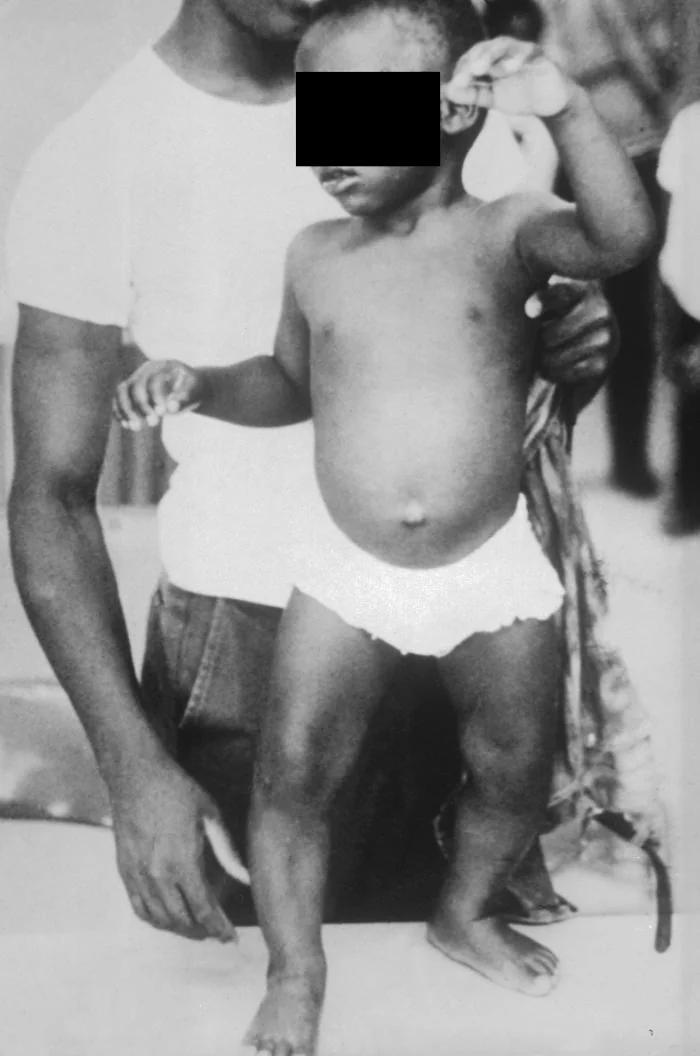
- Child presentation: bowing of the legs, widened wrists, the rachitic rosary at the costochondral junctions, craniotabes, delayed walking, delayed fontanelle closure and growth failure.
- Management: colecalciferol - a loading regimen (e.g. around 300,000 units total over 6-10 weeks) followed by maintenance, with calcium if dietary intake is poor, plus treatment of any malabsorptive cause.
- Caution: in granulomatous disease (e.g. sarcoidosis) and lymphoma, unregulated extrarenal 1-alpha-hydroxylase activity means replacement can precipitate hypercalcaemia - monitor calcium.
Introduction
Vitamin D is a fat-soluble hormone precursor whose principal role is to maintain the calcium and phosphate concentrations required to mineralise bone. When it is deficient, the body continues to lay down bone matrix (osteoid) normally, but that osteoid fails to mineralise - producing soft, weak, painful bone.1
This single pathological process produces two clinically distinct diseases, separated only by whether the growth plates are still open:
- Rickets - in children, before epiphyseal fusion. The unmineralised growth plate is soft and disorganised, so bones deform under load and growth is impaired.
- Osteomalacia - in adults, after epiphyseal fusion. There is no growth plate left to deform, so the disease presents instead with bone pain, muscle weakness and fractures.
Vitamin D deficiency is extremely common in the UK: a substantial proportion of the population has a level below the threshold at which the risk of deficiency-related bone disease rises, particularly at the end of winter, because at UK latitudes there is insufficient ambient UVB to synthesise vitamin D in the skin between roughly October and March.2 Frank rickets and osteomalacia are much rarer than biochemical deficiency, but both are still seen, and both are eminently treatable once recognised.
Physiology and pathophysiology
The activation pathway
Vitamin D requires two sequential hydroxylations to become biologically active, and each step is a point at which disease can interrupt the pathway.1
- Source. Around 80-90% comes from synthesis in the skin, where UVB converts 7-dehydrocholesterol to cholecalciferol (vitamin D3). Only a small proportion comes from the diet - oily fish, egg yolk, liver and fortified foods (and ergocalciferol, vitamin D2, from plant and fungal sources).
- Hepatic 25-hydroxylation. The liver converts cholecalciferol to 25-hydroxyvitamin D (calcidiol). This is the storage form with a long half-life, and the form measured in the laboratory - which is why 25-hydroxyvitamin D, not calcitriol, is the test used to diagnose deficiency.
- Renal 1-alpha-hydroxylation. The kidney converts calcidiol to 1,25-dihydroxyvitamin D (calcitriol), the active hormone. This step is stimulated by PTH and by hypophosphataemia, and is the rate-limiting, tightly regulated step.
- Action. Calcitriol increases intestinal absorption of calcium and phosphate (its dominant effect), promotes bone mineralisation, and increases renal calcium reabsorption.
How deficiency produces disease
Reduced calcitriol impairs intestinal calcium and phosphate absorption, so serum calcium falls. This triggers a compensatory rise in PTH (secondary hyperparathyroidism), which has three consequences that together explain the entire biochemical picture:
- Calcium is restored towards normal by mobilising it from bone - which is why calcium is often only low-normal rather than frankly low, and why a normal calcium does not exclude deficiency
- Phosphate falls further, because PTH is phosphaturic - giving the characteristic combination of low/low-normal calcium with low phosphate
- Bone turnover increases, releasing alkaline phosphatase from osteoblasts - hence the raised ALP that is such a useful clue
The end result is an inadequate calcium-phosphate product at the mineralisation front, so newly formed osteoid is deposited but not mineralised. In children this affects the growth plate as well as remodelling bone, which is why deformity is the dominant feature of rickets and pain and weakness the dominant features of osteomalacia.
Risk factors and causes
| Mechanism | Causes |
|---|---|
| Reduced skin synthesis | Insufficient sun exposure - housebound, institutionalised or hospitalised people, those who cover the skin for cultural or religious reasons, extensive sunscreen use, and UK winter months (October to March) when ambient UVB is inadequate at this latitude. Darker skin pigmentation - melanin absorbs UVB, so more exposure time is needed for the same synthesis. Older age - skin synthesises vitamin D less efficiently. |
| Reduced intake | Diets low in oily fish, eggs and fortified foods; exclusive breastfeeding without supplementation (breast milk is low in vitamin D, which is why UK guidance recommends supplementing breastfed infants); restrictive or vegan diets without fortified alternatives |
| Malabsorption (vitamin D is fat-soluble) | Coeliac disease, Crohn's disease, pancreatic exocrine insufficiency, cholestatic liver disease, short bowel syndrome, and bariatric surgery - especially malabsorptive procedures such as Roux-en-Y gastric bypass |
| Impaired hydroxylation | Chronic kidney disease - loss of renal 1-alpha-hydroxylase activity, so calcitriol cannot be produced even when 25-hydroxyvitamin D is adequate (this is renal osteodystrophy, and it requires an active analogue such as alfacalcidol rather than plain vitamin D). Severe liver disease - impaired 25-hydroxylation. |
| Increased catabolism | Enzyme-inducing drugs - phenytoin, carbamazepine, phenobarbital, rifampicin - accelerate vitamin D breakdown via CYP450 induction; also long-term corticosteroids and some antiretrovirals |
| Sequestration | Obesity - vitamin D is fat-soluble and is sequestered in adipose tissue, lowering the circulating level for a given intake, so higher replacement doses are often needed |
| Increased demand | Pregnancy and lactation, and periods of rapid growth in infancy and adolescence |
| Inherited (rare) | X-linked hypophosphataemic rickets (PHEX mutation causing renal phosphate wasting through raised FGF23 - the commonest inherited rickets, and note it is resistant to plain vitamin D), vitamin D-dependent rickets type 1 (1-alpha-hydroxylase deficiency) and type 2 (receptor resistance) |
Clinical features
Most vitamin D deficiency is asymptomatic and detected biochemically. Symptomatic disease presents differently in children and adults.
Osteomalacia (adults)
- Diffuse, poorly localised bone pain and tenderness - classically in the pelvis, ribs, spine and proximal long bones. The bones may be tender to direct pressure, which is an unusual and useful finding.
- Proximal myopathy - symmetrical weakness of the hip girdle producing a characteristic waddling gait, difficulty rising from a chair and difficulty climbing stairs. Vitamin D is required for normal muscle function, and this weakness improves with replacement.
- Fragility fractures - and an increased tendency to fall, from the combination of muscle weakness and poor proprioception
- Symptoms of hypocalcaemia if calcium falls significantly - perioral and peripheral paraesthesiae, cramps, and in severe cases tetany with Chvostek's and Trousseau's signs, seizures or laryngospasm
- Fatigue and low mood, which are common and non-specific
Rickets (children)
- Bowing of the legs (genu varum) once weight-bearing begins, or knock-knees (genu valgum) in older children
- Widening of the wrists and ankles - palpable expansion of the soft, unmineralised metaphyses
- Rachitic rosary - visible or palpable beading at the costochondral junctions, from metaphyseal expansion of the anterior ribs
- Craniotabes - abnormally soft skull bones in infancy, and delayed closure of the anterior fontanelle; also frontal bossing
- Harrison's sulcus - a horizontal groove along the lower border of the chest where the softened ribs are drawn in by the diaphragm
- Delayed motor milestones (particularly delayed walking), delayed dentition, and growth failure with short stature
- Hypocalcaemic seizures - which may be the presenting feature in infancy, sometimes before any skeletal signs are apparent
- Increased fracture risk and, in severe untreated disease, pelvic deformity
Investigations
Biochemistry
Serum 25-hydroxyvitamin D is the diagnostic test. Calcitriol (1,25-dihydroxyvitamin D) is not used to diagnose deficiency, because secondary hyperparathyroidism drives 1-alpha-hydroxylation and can keep it normal or even raised despite profound deficiency.2
| Level | Interpretation | Action |
|---|---|---|
| Below 25 nmol/L | Deficient - at risk of rickets or osteomalacia | Treat with a loading regimen |
| 25 to 50 nmol/L | Insufficient - may be inadequate in some people, particularly with other risk factors or established bone disease | Consider treatment or maintenance supplementation, guided by risk factors and symptoms |
| Above 50 nmol/L | Sufficient for musculoskeletal health in most people | Advise maintenance supplementation where risk factors persist |
| Test | Vitamin D deficiency | Why |
|---|---|---|
| 25-hydroxyvitamin D | Low | The primary abnormality |
| Calcium | Low or low-normal | Reduced intestinal absorption, partly compensated by secondary hyperparathyroidism mobilising bone calcium |
| Phosphate | Low | Reduced intestinal absorption plus the phosphaturic effect of the raised PTH |
| Alkaline phosphatase (ALP) | Raised | Increased osteoblastic activity and bone turnover - a key discriminator from osteoporosis, in which ALP is normal |
| PTH | Raised (secondary hyperparathyroidism) | An appropriate compensatory response to the falling calcium |
Further tests are directed at the underlying cause: U&Es and eGFR for renal impairment, LFTs for liver disease, coeliac serology (anti-tissue transglutaminase antibodies) and full blood count with ferritin, B12 and folate where malabsorption is suspected, and a magnesium level, since concurrent hypomagnesaemia impairs both PTH secretion and its action.
Imaging
Imaging is not required to diagnose deficiency, but is used where there is bone pain, deformity or suspected fracture.
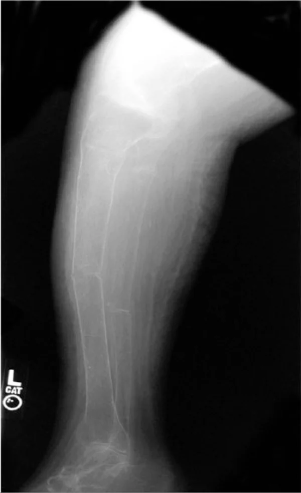
- Looser zones (pseudofractures) - narrow transverse translucent bands running perpendicular to the cortex, representing unmineralised osteoid at sites of stress. Classically seen in the pubic rami, medial femoral neck, ribs, scapula and lateral scapular border. They are essentially pathognomonic of osteomalacia.
- Generalised osteopenia with coarse, indistinct trabeculae and blurred cortical margins ('washed out' bones)
- In rickets - cupping, splaying and fraying of the metaphyses (classically at the distal radius and ulna at the wrist, and around the knee), a widened growth plate, and delayed bone age
- Vertebral changes - biconcave 'cod-fish' vertebrae from soft endplates
- DEXA scan - often shows low bone density, but this reflects reduced mineral rather than osteoporosis and should be repeated after vitamin D replacement before diagnosing osteoporosis or starting a bisphosphonate
- Bone biopsy with tetracycline labelling is the definitive test showing a widened, unmineralised osteoid seam, but is essentially never needed in practice
Management
Vitamin D replacement
Colecalciferol (vitamin D3) is the preferred preparation, being more effective than ergocalciferol at raising and sustaining 25-hydroxyvitamin D. Treatment of established deficiency has two phases.3,4
| Phase | Typical regimen | Notes |
|---|---|---|
| Loading (treatment) phase | A total of approximately 300,000 units of colecalciferol given over 6 to 10 weeks - for example 50,000 units weekly for 6 weeks, or 40,000 units weekly for 7 weeks, or 4,000 units daily for 10 weeks | Used for symptomatic deficiency or a level below 25 nmol/L. Rapid loading is what relieves bone pain and myopathy. |
| Maintenance phase | 800 to 2,000 units daily (occasionally up to 4,000 units), started around a month after loading and continued long term where risk factors persist | Higher maintenance doses are often needed in obesity, malabsorption and with enzyme-inducing drugs |
| Calcium | Add a calcium supplement if dietary intake is inadequate (roughly under 700 mg daily) | Not needed routinely if the diet is adequate - and correcting vitamin D alone can unmask a calcium requirement as bone begins to remineralise |
| Children and infants | Weight- and age-dependent regimens; UK guidance recommends a routine daily supplement for all breastfed infants, and for formula-fed infants taking under 500 mL a day | Paediatric dosing should follow the BNF for Children and specialist guidance |
Special situations
- Chronic kidney disease - plain colecalciferol cannot be 1-alpha-hydroxylated, so an active analogue (alfacalcidol or calcitriol) is required, alongside phosphate binders and management of renal bone disease
- Severe liver disease - impaired 25-hydroxylation may also warrant an active analogue
- Malabsorption - much higher oral doses, or occasionally parenteral vitamin D, plus treatment of the underlying cause (a gluten-free diet in coeliac disease, pancreatic enzyme replacement in exocrine insufficiency)
- After bariatric surgery - lifelong supplementation with monitoring of vitamin D, calcium, iron and B12
- Hypocalcaemic tetany or seizures - treat the hypocalcaemia acutely first with intravenous calcium gluconate under cardiac monitoring, then correct vitamin D; also check and correct magnesium
- X-linked hypophosphataemic rickets - phosphate supplements with an active vitamin D analogue, or burosumab; plain vitamin D is ineffective
Monitoring and lifestyle advice
- Recheck calcium around a month after loading, particularly in anyone at risk of hypercalcaemia, and recheck 25-hydroxyvitamin D after 3 to 6 months - not sooner, as it takes that long to plateau
- Expect ALP to rise transiently as bone remineralises briskly before falling to normal; this is a sign of response, not deterioration
- Advise on sunlight - short, regular periods of exposure to arms and legs in late spring and summer, without burning
- Advise on diet - oily fish, egg yolks, red meat, liver and fortified cereals and spreads
- Do not start a bisphosphonate before vitamin D is replete, as it risks precipitating hypocalcaemia
Complications
- Fragility fractures - including insufficiency fractures through Looser zones, and hip fracture in older adults
- Falls - from proximal myopathy and impaired neuromuscular function, compounding fracture risk
- Permanent skeletal deformity in children if rickets is untreated - bowed legs, short stature and, historically, pelvic deformity complicating later childbirth
- Growth failure and delayed motor development in children
- Hypocalcaemic seizures, tetany and laryngospasm - particularly in infants, and occasionally the presenting feature
- Dilated cardiomyopathy - a rare but recognised and potentially fatal complication of severe infantile hypocalcaemic rickets
- Secondary hyperparathyroidism, which if prolonged and severe can become tertiary (autonomous)
- Chronic pain and functional impairment in adults, often mislabelled for years as fibromyalgia or non-specific musculoskeletal pain
- Dental abnormalities - enamel hypoplasia and delayed dentition
Red flags
Prognosis
The prognosis of nutritional vitamin D deficiency is excellent, and the response to treatment is one of the more satisfying in medicine. Bone pain and proximal myopathy typically begin to improve within weeks of adequate loading, and biochemistry normalises over weeks to a few months - with ALP often rising transiently first as bone remineralises before settling.
In children, established bone deformity can remodel substantially with treatment, particularly if rickets is corrected while growth continues, and most children treated early achieve normal growth and a normal skeleton. Deformity that persists into adulthood, or where treatment was very delayed, may require orthopaedic correction.
The main determinants of a poorer outcome are delayed diagnosis - adults are often treated for non-specific chronic pain for years before osteomalacia is considered - and failure to identify an underlying cause. Deficiency will simply recur if untreated coeliac disease, ongoing malabsorption, chronic kidney disease or an enzyme-inducing drug is left unaddressed, which is why the priority after replacement is establishing why the patient became deficient and arranging appropriate long-term maintenance.
References
- Holick MF. Vitamin D deficiency. New England Journal of Medicine. 2007. Available here
- NICE Clinical Knowledge Summaries. Vitamin D deficiency in adults. Available here
- Royal Osteoporosis Society. Vitamin D and bone health: a practical clinical guideline for patient management. Available here
- BNF. Colecalciferol, ergocalciferol and alfacalcidol - indications and dosing. Available here
- NICE NG56 / PH56. Vitamin D: supplement use in specific population groups. Available here
- CDC Public Health Image Library (PHIL ID 2435), public domain, via Wikimedia Commons. Available here
- Leslie Gamache and Mark R. Burge, CC BY 3.0, via Wikimedia Commons. Available here
This article is written for revision and education. It is not clinical guidance and must not be used to make decisions about the care of a patient. Always check current NICE guidance and local protocols.