
Carpal Tunnel Syndrome
Key points
- Definition: compression of the median nerve beneath the flexor retinaculum - the commonest entrapment neuropathy, affecting around 3 to 5% of adults.
- Who: women about three times more often than men, peaking between 40 and 60, and very commonly in pregnancy.
- Symptoms: numbness and tingling in the thumb, index, middle and radial half of the ring finger, characteristically waking the patient at night and relieved by shaking the hand.
- Sensation over the thenar eminence is spared: the palmar cutaneous branch leaves the median nerve proximal to the tunnel and passes over the retinaculum - the finding that distinguishes it from a proximal median lesion.
- Motor supply: LOAF - lateral two Lumbricals, Opponens pollicis, Abductor pollicis brevis and Flexor pollicis brevis. Test thumb abduction.
- Diagnosis: clinical. Nerve conduction studies confirm and grade severity where the diagnosis is uncertain or surgery is planned.
- First-line treatment: a neutral wrist splint worn at night for at least 4 weeks, with treatment of any underlying cause.
- Surgery: carpal tunnel decompression for severe or persistent symptoms, sensory loss or thenar wasting - and as an emergency in acute compression after a distal radius fracture.
Introduction
Carpal tunnel syndrome is compression of the median nerve as it passes through the carpal tunnel at the wrist. It is the commonest entrapment neuropathy by a wide margin, with a prevalence of around 3 to 5% in adults, and carpal tunnel decompression is one of the most frequently performed operations in the NHS.2
It is worth learning properly for two reasons beyond its frequency. It is an unusually clean example of applied anatomy - almost every clinical feature can be derived from where the nerve runs and what it supplies. And it is the classic setting for a common examination error: attributing hand numbness to the carpal tunnel when the pattern actually indicates a cervical radiculopathy or an ulnar neuropathy.
Anatomy
The carpal tunnel is a fibro-osseous canal. Its floor and walls are formed by the concave arch of the carpal bones - the scaphoid tubercle and trapezium on the radial side, the pisiform and hook of hamate on the ulnar side - and its roof by the flexor retinaculum (transverse carpal ligament), a thick, inelastic band spanning between them.
Ten structures pass through it: the median nerve, and nine flexor tendons - the four tendons of flexor digitorum superficialis, the four of flexor digitorum profundus, and flexor pollicis longus. The tunnel is a fixed volume with non-compliant walls, so anything that increases its contents or reduces its size raises the pressure inside it directly.
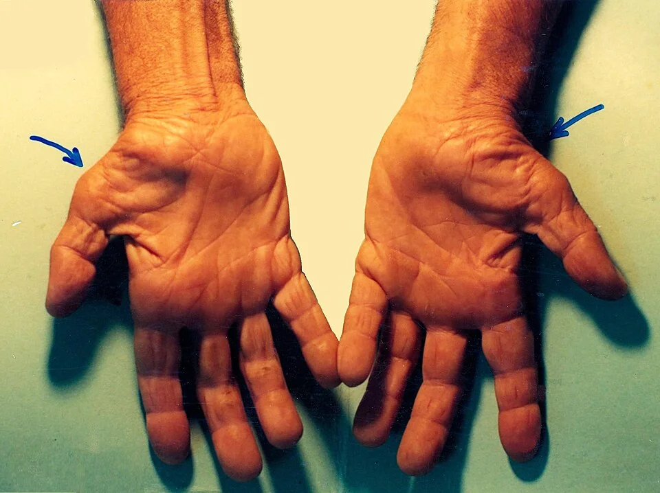
In the hand, the median nerve supplies sensation to the palmar surface of the thumb, index and middle fingers and the radial half of the ring finger, together with the dorsal skin over their distal phalanges. Its motor supply is remembered as LOAF: the lateral two Lumbricals, Opponens pollicis, Abductor pollicis brevis and Flexor pollicis brevis. Abductor pollicis brevis is the muscle that matters clinically, because it is the most reliably median-innervated and is what you test and what visibly wastes.
Why the symptoms occur when they do
Raised pressure in the tunnel first impairs venous outflow in the epineurium, then arterial inflow, producing nerve ischaemia. This causes reversible conduction block and paraesthesiae; sustained compression leads to segmental demyelination and eventually to axonal loss, at which point wasting and permanent sensory loss develop. This explains the nocturnal predominance - people sleep with the wrist flexed, which raises tunnel pressure several-fold, and fluid redistributes centrally when lying flat - and why shaking the hand, which restores perfusion, relieves the symptoms within a minute or two.
Causes and risk factors
Most cases are idiopathic, but a secondary cause should be actively considered, particularly if the patient is young, male, or the presentation is bilateral or atypical.
| Category | Examples |
|---|---|
| Idiopathic | The majority. Associated with obesity, a squarer wrist ratio and increasing age. |
| Occupational and mechanical | Repetitive forceful wrist flexion and extension, sustained grip, and use of vibrating tools. The evidence for keyboard use alone as a cause is weak. |
| Endocrine and metabolic | Pregnancy (symptoms in up to half of pregnant women, usually in the third trimester); hypothyroidism; acromegaly; diabetes mellitus; obesity; the menopause |
| Inflammatory | Rheumatoid arthritis with flexor tenosynovitis; gout; other inflammatory arthropathies |
| Infiltrative | Amyloidosis, including dialysis-related beta-2 microglobulin amyloid; mucopolysaccharidoses; sarcoidosis |
| Traumatic and structural | Distal radius fracture (which can cause acute compression); lunate dislocation; malunion; ganglion; anomalous muscles; a persistent median artery |
| Fluid retention | Heart failure, renal failure and dialysis, and the combined oral contraceptive |
| Other | Hereditary neuropathy with liability to pressure palsies; any space-occupying lesion within the tunnel |
Clinical features
- Numbness, tingling and burning in the median distribution - thumb, index, middle and the radial half of the ring finger. Patients frequently describe the whole hand as numb, so ask specifically about the little finger.
- Nocturnal symptoms that wake the patient, typically in the early hours - the most characteristic feature. Relief by hanging the hand out of bed, shaking or flicking it is the flick sign, and is reasonably specific.
- Provocation by sustained wrist posture - driving, holding a phone or a newspaper, cycling, or using a hairdryer
- Pain radiating proximally into the forearm and occasionally to the elbow or shoulder - a well-recognised trap that leads to the neck or shoulder being blamed
- Clumsiness and weakness - dropping objects, difficulty with buttons, zips and picking up coins
- Bilateral in around half of patients, though usually worse in the dominant hand
Examination
- Inspect both hands together for thenar wasting - flattening of the eminence at the base of the thumb. Compare the two sides directly, since bilateral wasting is easy to miss.
- Sensation in the median distribution, and confirm the thenar eminence is spared
- Motor - test thumb abduction by asking the patient to point the thumb vertically away from the palm against resistance, palpating abductor pollicis brevis. Weakness here is the most useful motor sign.
- Phalen's test - maximal wrist flexion held for 60 seconds reproduces the symptoms
- Tinel's test - percussion over the flexor retinaculum produces tingling in the median distribution. Reasonably specific but insensitive.
- Durkan's carpal compression test - firm thumb pressure over the tunnel for 30 seconds; generally the most accurate of the three provocation tests
- Examine the neck and perform a full upper limb neurological examination, and look for the features of hypothyroidism, acromegaly and rheumatoid arthritis
Differential diagnosis
| Condition | Distinguishing features |
|---|---|
| Cervical radiculopathy (C6 or C7) | Neck pain, symptoms extending above the wrist into the forearm and arm, reduced biceps or triceps reflex, worse on neck movement, positive Spurling's test. Can coexist with carpal tunnel syndrome - the "double crush" phenomenon. |
| Proximal median nerve compression (pronator teres syndrome) | Thenar eminence sensation is also lost, because the palmar cutaneous branch is affected; forearm pain on resisted pronation; night symptoms are less prominent |
| Ulnar neuropathy | Little finger and ulnar half of the ring finger affected; wasting of the interossei and hypothenar eminence; positive Froment's sign |
| Peripheral polyneuropathy | Symmetrical glove-and-stocking distribution, feet affected too, absent ankle jerks - typically diabetic |
| Thoracic outlet syndrome | Whole-limb symptoms, often ulnar-predominant, positional and related to arm elevation, sometimes with vascular features |
| Thumb base (first carpometacarpal) osteoarthritis | Pain rather than numbness, localised to the base of the thumb, with a positive grind test and squaring of the thumb base |
| De Quervain's tenosynovitis | Radial wrist pain over the first extensor compartment, positive Finkelstein's test, no sensory symptoms |
| Motor neurone disease | Painless progressive wasting with fasciculations and no sensory symptoms - a rare but important cause of thenar wasting |
Investigations
Carpal tunnel syndrome is a clinical diagnosis, and a typical presentation in a middle-aged woman does not require investigation before a trial of splinting.1
- Nerve conduction studies - confirm the diagnosis, grade severity, and exclude a more proximal lesion or a generalised neuropathy. Indicated where the diagnosis is uncertain, where features are atypical, before surgery in many units, and where there is a medicolegal or occupational dimension. Note that around 10% of patients with genuine carpal tunnel syndrome have normal studies, so a negative result does not exclude it in the face of a convincing history.
- Blood tests - TFTs, HbA1c or fasting glucose, and consider a pregnancy test. Test more widely (including for paraproteins) if amyloidosis or an inflammatory arthropathy is suspected.
- Ultrasound - increasingly used, measuring the cross-sectional area of the median nerve at the tunnel inlet; it also identifies a ganglion, tenosynovitis or an anomalous structure
- Radiographs or MRI - only where trauma, a mass or a bony abnormality is suspected
Management
Treat the cause and modify the load
Correcting hypothyroidism, optimising diabetes control, supporting weight loss and modifying provoking activities all help, and in pregnancy the symptoms usually resolve spontaneously within weeks of delivery - so conservative management is nearly always the right answer there.
Conservative treatment
| Treatment | Evidence and practical notes |
|---|---|
| Neutral wrist splint worn at night | First-line for mild to moderate symptoms. Worn for at least 4 weeks, ideally 8, before judging response. It works by preventing the wrist flexion that raises tunnel pressure during sleep - so it must hold the wrist in neutral, not in extension.5 |
| Corticosteroid injection into the carpal tunnel | Produces significant relief in the majority at 1 to 3 months, with benefit declining thereafter. Useful where splinting fails, where surgery must be deferred (for example in pregnancy), and as a pointer that decompression is likely to help.4 |
| Short course of oral corticosteroid | Modest, short-lived benefit; occasionally used where injection is not available |
| Activity and ergonomic modification | Reasonable advice, though the evidence for workplace interventions alone is limited |
| Not effective | NSAIDs, diuretics, pyridoxine (vitamin B6), ultrasound therapy and yoga have little or no evidence of benefit, and diuretics and pyridoxine should not be used |
Surgery
Carpal tunnel decompression divides the flexor retinaculum, immediately relieving the pressure. It is usually performed as a day case under local anaesthetic through a short palmar incision; endoscopic release gives a faster return to work but a slightly higher rate of nerve injury, and long-term outcomes are equivalent.3,6
- Indications - severe symptoms; failure of conservative treatment after an adequate trial; persistent sensory loss; thenar wasting or weakness; and severe changes on nerve conduction studies
- Do not wait for wasting to develop before referring - by that point recovery is often incomplete
- Postoperatively - the wound is protected for around 2 weeks, with early finger movement encouraged; return to light work within 2 weeks and heavier work by 6 to 12 weeks
Complications
- Permanent sensory loss and thenar wasting from prolonged untreated compression, with lasting loss of fine pinch and dexterity
- Scar tenderness and pillar pain - discomfort in the thenar and hypothenar eminences either side of the incision, common in the first few months and usually self-limiting
- Incomplete release, which is the commonest reason for persistent symptoms after surgery
- Recurrence, in around 3 to 5%
- Nerve injury - to the palmar cutaneous branch, producing a painful neuroma at the scar, or to the recurrent motor branch, causing thenar weakness
- Bowstringing of the flexor tendons and loss of grip strength, usually temporary
- Wound infection, and rarely complex regional pain syndrome
Persistent symptoms after decompression should prompt reconsideration of the diagnosis before assuming a technical failure - cervical radiculopathy, a polyneuropathy or a proximal median lesion may have been the real problem, or may coexist.
Red flags
Prognosis
Mild carpal tunnel syndrome frequently improves spontaneously, particularly where a reversible cause such as pregnancy, hypothyroidism or weight gain is corrected. Around a third of untreated mild cases resolve without intervention, which is part of the justification for a trial of splinting before referral.
Surgical decompression is highly effective, relieving symptoms in around 75 to 90% of patients, with the nocturnal paraesthesiae often disappearing within a few nights of the operation. Recovery of sensation is usually good, though it may take months. Recovery of thenar bulk and strength is far less reliable once wasting is established - which is the clinical justification for referring on the basis of weakness rather than waiting for the patient to complain about it.
The predictors of a poorer outcome are long duration of symptoms, severe axonal loss on nerve conduction studies, diabetes, and coexisting cervical or peripheral neuropathy. The practical implication is the same one that runs through compressive neuropathies generally: the deficit present at the time of decompression largely determines the deficit afterwards, so the timing of referral matters more than the choice of technique.
References
- NICE Clinical Knowledge Summaries. Carpal tunnel syndrome. Available here
- Padua L, Coraci D, Erra C et al. Carpal tunnel syndrome: clinical features, diagnosis, and management. The Lancet Neurology. 2016. Available here
- British Society for Surgery of the Hand. Evidence for surgical treatment: carpal tunnel syndrome. Available here
- Marshall SC, Tardif G, Ashworth NL. Local corticosteroid injection for carpal tunnel syndrome. Cochrane Database of Systematic Reviews. 2007. Available here
- Page MJ, Massy-Westropp N, O'Connor D, Pitt V. Splinting for carpal tunnel syndrome. Cochrane Database of Systematic Reviews. 2012. Available here
- Graham B, Peljovich AE, Afra R et al. The American Academy of Orthopaedic Surgeons evidence-based clinical practice guideline on management of carpal tunnel syndrome. Journal of Bone and Joint Surgery. 2016. Available here
This article is written for revision and education. It is not clinical guidance and must not be used to make decisions about the care of a patient. Always check current NICE guidance and local protocols.