
Childhood Hip and Leg Disorders
Key points
- Age is the discriminator: developmental dysplasia in infancy, transient synovitis and Perthes' from 4 to 10, and slipped upper femoral epiphysis from 10 to 16.
- Exclude septic arthritis first: a febrile child who will not weight bear needs urgent assessment, aspiration and washout - not observation.
- Knee pain means examine the hip: a quarter of children with a slipped upper femoral epiphysis present complaining only of knee pain, with an apparently normal knee.
- DDH screening: Ortolani and Barlow tests at the newborn and 6 to 8 week checks, with selective ultrasound for breech presentation, a family history or abnormal examination.
- Imaging in infancy: ultrasound, not radiography, under about 4.5 months - the femoral head has not yet ossified.
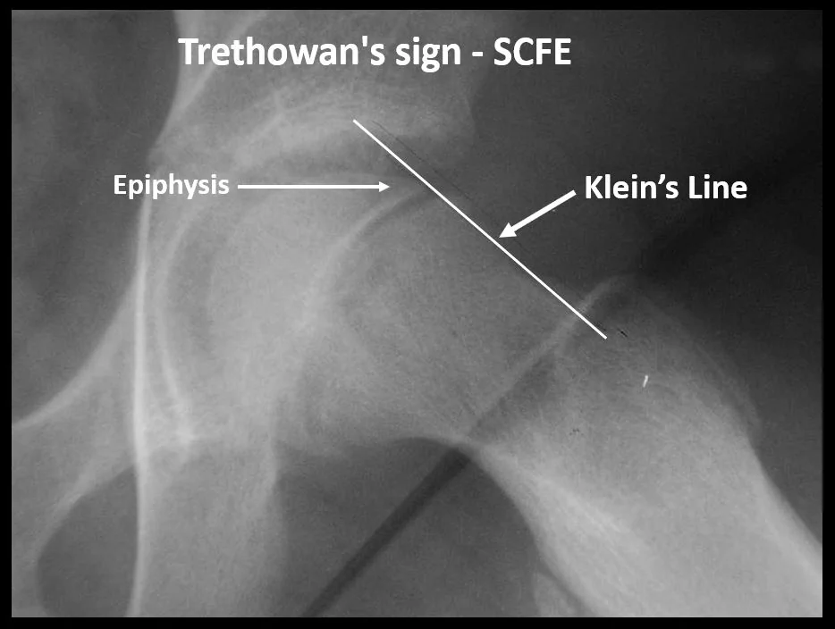
- SUFE needs a frog-leg lateral: the slip may be invisible on the AP film. Look for Klein's line failing to intersect the epiphysis - Trethowan's sign.
- SUFE management: stop weight bearing immediately, refer urgently, and fix in situ with a single cannulated screw. Do not attempt to reduce a chronic slip.
- Any limp under 3, or a limp with systemic symptoms: warrants investigation - consider infection, malignancy and non-accidental injury.
Introduction
A limping child is one of the most common paediatric musculoskeletal presentations and one of the highest-stakes, because the differential ranges from a self-limiting viral synovitis to septic arthritis, leukaemia and non-accidental injury. Around 1.5 to 3% of children present with a limp at some point.1
The most efficient way through it is by age, because the four conditions that dominate the topic occupy almost non-overlapping age bands. Layered on top of that are two questions asked of every child regardless of age: is the child febrile and systemically unwell, and can the child weight bear? Those two questions identify almost everyone who needs urgent action.
The third principle is anatomical: hip pathology refers to the knee via the obturator nerve. A child brought with knee pain and a normal knee has a hip problem until the hip has been examined and, if necessary, imaged. Missing a slipped upper femoral epiphysis for this reason remains one of the classic avoidable errors in orthopaedics.
The limping child: a framework
| Age | Common causes | Must not miss |
|---|---|---|
| 0 to 3 years | Developmental dysplasia of the hip; toddler's fracture of the tibia; soft tissue injury | Septic arthritis and osteomyelitis; non-accidental injury; malignancy; neuromuscular disease presenting as delayed or abnormal walking |
| 4 to 10 years | Transient synovitis (by far the commonest); Perthes' disease; trauma | Septic arthritis; osteomyelitis; juvenile idiopathic arthritis; leukaemia; bone tumour |
| 10 to 16 years | Slipped upper femoral epiphysis; Osgood-Schlatter disease; osteochondritis dissecans; overuse and sports injury | SUFE presenting as knee pain; osteosarcoma and Ewing sarcoma; septic arthritis; avulsion fracture |
| Any age | Trauma; reactive arthritis; viral myositis; footwear or foreign body | Septic arthritis; malignancy; child protection concerns |
Developmental dysplasia of the hip
DDH describes a spectrum from a shallow, dysplastic acetabulum through subluxation to frank dislocation. It affects 1 to 3 per 1000 live births in its more severe forms, is around six times more common in girls, involves the left hip more often (reflecting the usual intrauterine position), and is bilateral in about a fifth.
Risk factors
- Breech presentation - the strongest risk factor, particularly breech at or after 36 weeks or at delivery, regardless of eventual mode of delivery
- A first-degree family history of DDH
- Female sex and being firstborn
- Oligohydramnios, multiple pregnancy and high birth weight - all reducing intrauterine space
- Other packaging deformities - congenital torticollis, talipes, plagiocephaly
- Swaddling with the legs extended and adducted, which is a modifiable risk factor worth advising against
Detection
The UK Newborn and Infant Physical Examination (NIPE) programme screens all babies at birth and again at 6 to 8 weeks, using two manoeuvres performed on a relaxed infant:3
- Ortolani's test - the hip is flexed and gently abducted while lifting the greater trochanter forwards; a palpable clunk indicates a dislocated hip relocating into the acetabulum
- Barlow's test - the flexed hip is adducted with gentle posterior pressure; a clunk indicates a located but dislocatable hip
Both tests become unreliable after about 3 months as soft tissues tighten. Later presentations are detected by asymmetric skin creases in the thigh or groin, limited abduction in flexion (often the most reliable sign after infancy), apparent limb shortening with the knees at unequal heights when the hips and knees are flexed (Galeazzi sign), and, once walking, a painless limp, toe walking or a waddling Trendelenburg gait.
Management
- Under 6 months - a Pavlik harness, holding the hips flexed and abducted so the femoral head is directed into the acetabulum, worn for several weeks with regular ultrasound monitoring. Success rates are high when started early.4
- 6 to 18 months, or failed harness - closed reduction under anaesthetic with adductor tenotomy and a hip spica cast, sometimes with arthrogram
- Older children - open reduction, often with femoral shortening or a pelvic (Salter or Pemberton) osteotomy
- The major complication of treatment is avascular necrosis of the femoral head, which is why the harness must not force excessive abduction, and why the position is checked on ultrasound
Transient synovitis and septic arthritis
Transient synovitis (irritable hip) is the commonest cause of hip pain in children aged 3 to 10. It is a self-limiting sterile inflammation of the synovium, typically occurring 1 to 2 weeks after a viral upper respiratory tract infection. The child limps or refuses to walk, holds the hip flexed and externally rotated, and has restricted internal rotation - but is systemically well, with at most a low-grade temperature.
It is a diagnosis of exclusion, and the condition that must be excluded is septic arthritis, which shares the presentation and destroys a joint within days. The Kocher criteria were developed precisely to separate them.2
| Criterion | Number present | Probability of septic arthritis |
|---|---|---|
| Non-weight-bearing on the affected side | 0 of 4 | Under 1% |
| Temperature above 38.5 °C | 1 of 4 | About 3% |
| ESR above 40 mm/hour | 2 of 4 | About 40% |
| White cell count above 12 x10^9/L | 3 of 4 | About 93% |
| (A CRP above 20 mg/L is often used as a fifth predictor) | 4 of 4 | About 99% |
Once septic arthritis is genuinely excluded, transient synovitis is managed with rest, simple analgesia and NSAIDs, and it settles within one to two weeks. Arrange follow-up: a proportion of children initially labelled irritable hip turn out to have early Perthes' disease, and persistence beyond a couple of weeks warrants repeat radiographs.
Perthes' disease
Legg-Calve-Perthes disease is idiopathic avascular necrosis of the proximal femoral epiphysis, followed by revascularisation and remodelling over a period of two to four years. It typically affects children aged 4 to 8 (range 3 to 12), is four to five times commoner in boys, and is bilateral in 10 to 15% - though bilateral simultaneous involvement should prompt consideration of a skeletal dysplasia or hypothyroidism instead.
Associations include low birth weight, short stature and delayed bone age, passive smoking, socioeconomic deprivation and hyperactivity. The underlying cause remains unclear, and is probably a combination of a precarious epiphyseal blood supply and repeated minor insults.5
Presentation and stages
- Insidious onset over weeks to months of a limp, which may be painless, with hip, groin, thigh or knee pain
- Restricted abduction and internal rotation on examination, with muscle spasm, and eventually thigh wasting and limb shortening
- Waldenstrom's stages describe the natural history: initial necrosis, then fragmentation, then reossification, then remodelling - the whole process taking 2 to 4 years
Radiographs (AP and frog-leg lateral) may be normal early on. The earliest changes are widening of the joint space and increased density of the epiphysis, followed by a subchondral fracture line (the crescent sign), then fragmentation, and later flattening and broadening of the head (coxa plana and coxa magna). MRI or bone scan detects the disease before radiographic change appears, and is used where the diagnosis is suspected but the film is normal.
Management
- The goal is containment - keeping the softened femoral head within the acetabulum so that it remodels into a spherical shape
- Younger children (under about 6) with limited involvement are managed conservatively: analgesia, activity restriction, physiotherapy to preserve range of movement, and regular review. Most do well.
- Older children and those with extensive head involvement or loss of containment may need bracing in abduction or surgical containment with a femoral varus or pelvic osteotomy
- Maintaining hip range of movement is the single most important part of conservative treatment, and losing abduction is the trigger for escalation
Prognosis depends chiefly on age at onset - children under 6 do substantially better - together with the extent of epiphyseal involvement (classified by the Herring lateral pillar system) and the range of movement maintained. A deformed, non-spherical head predisposes to femoroacetabular impingement and to osteoarthritis in early adulthood.
Slipped upper femoral epiphysis
SUFE (also called SCFE) is displacement of the femoral head through the growth plate. The epiphysis stays in the acetabulum while the metaphysis and neck displace anteriorly and rotate externally - so relative to the neck, the head slips posteriorly and inferiorly. It is essentially a Salter-Harris type I fracture through a physis weakened during the adolescent growth spurt.
Who gets it
- Age 10 to 16 - typically boys aged 12 to 15 and girls aged 10 to 14, corresponding to the growth spurt
- Boys more than girls, roughly 2 to 1
- Obesity - the single strongest modifiable risk factor, present in the majority
- Bilateral in 20 to 40%, often sequentially, with the second slip usually within 18 months of the first
- Endocrinopathy - hypothyroidism, growth hormone deficiency or its treatment, hypogonadism, panhypopituitarism and renal osteodystrophy. Screen for these if the child is outside the usual age range, is short or underweight, or if the slip is bilateral at presentation.
Presentation and examination
- Usually insidious over weeks, with a limp and vague groin, thigh or knee discomfort; sometimes acute-on-chronic after minor trauma
- Around a quarter present with knee pain alone and a completely normal knee - the reason the hip must always be examined
- Loss of internal rotation is the earliest and most consistent sign
- Obligatory external rotation on passive hip flexion (Drehmann's sign) - as the hip is flexed the leg swings involuntarily into external rotation and abduction. This is close to pathognomonic and easy to elicit.
- In severe or unstable slips the leg lies shortened and externally rotated, and the child cannot weight bear
Imaging
Request AP and frog-leg lateral views of both hips. The lateral is essential because a mild slip, which displaces posteriorly, may be entirely invisible on the AP film - and imaging both hips allows comparison and detects an asymptomatic contralateral slip.
- Trethowan's sign - Klein's line, drawn along the superior border of the femoral neck on the AP view, normally intersects part of the epiphysis. In SUFE it passes above it without intersecting.
- Widening and irregularity of the physis, and loss of epiphyseal height on the AP view
- The classic description of the appearance is "ice cream slipping off the cone"
- MRI detects a pre-slip or an early slip where radiographs are equivocal
Management
- Stop weight bearing immediately - a stable slip can become unstable, and that transition dramatically worsens the prognosis. Admit the child and use a wheelchair or bed rest; do not send them home to await an outpatient appointment.7
- Urgent orthopaedic referral
- Surgical fixation in situ with a single cannulated screw across the physis, which prevents further slip and promotes physeal closure
- Do not attempt to reduce a chronic slip - forcible reduction disrupts the remaining blood supply and precipitates avascular necrosis. Severe slips may later need a corrective osteotomy once healed.
- Consider prophylactic fixation of the contralateral hip in younger children, in those with an endocrinopathy, and where follow-up is likely to be unreliable
Complications include avascular necrosis, chondrolysis (acute loss of articular cartilage with a stiff painful hip), femoroacetabular impingement from the residual deformity, limb length discrepancy, and osteoarthritis in early adult life.
Other conditions worth knowing
| Condition | Features | Management |
|---|---|---|
| Osgood-Schlatter disease | Traction apophysitis of the tibial tuberosity in active adolescents aged 10 to 15. A tender, prominent tibial tuberosity, worse with running and jumping, relieved by rest. | Self-limiting over months to a couple of years. Activity modification, analgesia, quadriceps stretching. Imaging is not needed. The prominence may be permanent. |
| Sinding-Larsen-Johansson disease | The same process at the inferior pole of the patella | As for Osgood-Schlatter |
| Osteochondritis dissecans | A fragment of subchondral bone and cartilage separating, usually from the lateral aspect of the medial femoral condyle. Vague knee pain, effusion, and locking if the fragment detaches. | Rest and activity restriction in skeletally immature children; surgical fixation or removal for unstable or loose fragments |
| Talipes equinovarus (clubfoot) | Congenital fixed equinus, varus and forefoot adduction. May be positional (correctable) or structural. | Ponseti method - serial casting from the first weeks of life, usually with percutaneous Achilles tenotomy, then a foot abduction brace. Highly effective if started early. |
| Genu varum and valgum | Physiological varum (bow legs) until about 2 years, physiological valgum (knock knees) peaking at 3 to 4, with adult alignment by about 7 | Reassurance if symmetrical, painless and following the expected pattern. Investigate if asymmetrical, painful, progressive, or with short stature - consider Blount's disease, rickets and skeletal dysplasia. |
| In-toeing | Metatarsus adductus in infancy, internal tibial torsion in toddlers, and increased femoral anteversion in early school age (the child sits in a W position) | Almost all resolve spontaneously. Reassure; refer only if severe, asymmetrical, or causing functional problems. |
| Growing pains | Bilateral, poorly localised leg pain in the evening or at night in a 3 to 12 year old, relieved by massage, with a completely normal examination and no limp | A diagnosis of exclusion. Reassurance. Unilateral pain, a limp, daytime symptoms or any abnormal sign means it is not growing pains. |
Assessment and investigation
- History - age, onset and duration, trauma, fever and systemic symptoms, recent viral illness, ability to weight bear, night pain, weight loss, developmental milestones and birth history including breech presentation
- Observe the gait before touching the child - antalgic, Trendelenburg, toe walking, or a circumducting gait
- Examine the whole child - both hips, knees, ankles and feet, the spine, and the abdomen; look for rash, bruising, lymphadenopathy and organomegaly; measure and plot height and weight
- Bloods - FBC with film, CRP, ESR and blood cultures if infection or malignancy is a possibility. A film is important: leukaemia can present with bone pain and limp before the count is obviously abnormal.
- Radiographs - AP pelvis and frog-leg lateral of both hips for any adolescent hip presentation, and for suspected Perthes'
- Ultrasound - for a hip effusion, and for DDH in infants
- MRI - for early Perthes', osteomyelitis, a suspected tumour, or an equivocal slip
- Joint aspiration - urgently where septic arthritis is suspected, before antibiotics
Comparing the three hip conditions
| Transient synovitis | Perthes' disease | Slipped upper femoral epiphysis | |
|---|---|---|---|
| Typical age | 3 to 10 | 4 to 8 | 10 to 16 |
| Sex | Boys somewhat more | Boys 4 to 5 times more | Boys about twice as often |
| Body habitus | No association | Often small for age | Usually obese |
| Onset | Acute, over days, after a viral illness | Insidious, over weeks to months | Insidious over weeks, or acute after minor trauma |
| Systemic features | Well, afebrile or low-grade temperature | Well | Well |
| Key examination finding | Restricted internal rotation, mild | Restricted abduction and internal rotation | Obligatory external rotation on hip flexion; loss of internal rotation |
| Radiograph | Normal | Initially normal, then sclerosis, crescent sign, fragmentation | Klein's line fails to intersect the epiphysis; best seen on frog-leg lateral |
| Management | Rest, NSAIDs, follow-up | Containment - conservative if young, surgical if older or extensive | Non-weight bearing and urgent fixation in situ |
Prognosis
Transient synovitis resolves completely within one to two weeks in the great majority, with a small recurrence rate and no long-term sequelae - provided the diagnosis was correct.
DDH treated early with a Pavlik harness has excellent outcomes, with normal hip development in most; late diagnosis requires more extensive surgery and carries a substantially higher risk of residual dysplasia, avascular necrosis and early osteoarthritis. This is the entire rationale for newborn screening.
Perthes' disease in a child under 6 with limited head involvement usually results in a well-remodelled hip; older children and those with extensive involvement frequently develop a deformed head, with symptomatic osteoarthritis in the fourth or fifth decade. SUFE treated promptly while stable does well, with over 90% of hips remaining satisfactory; an unstable slip carries up to a 50% risk of avascular necrosis, and both groups have an increased lifetime risk of impingement and arthritis. In all of these, the determinant of outcome is how early the diagnosis was made - which is a reasonable summary of the whole topic.
References
- NICE Clinical Knowledge Summaries. Acute childhood limp. Available here
- Kocher MS, Zurakowski D, Kasser JR. Differentiating between septic arthritis and transient synovitis of the hip in children: an evidence-based clinical prediction algorithm. Journal of Bone and Joint Surgery. 1999. Available here
- Public Health England. Newborn and infant physical examination (NIPE) screening programme handbook. Available here
- NICE Clinical Knowledge Summaries. Developmental dysplasia of the hip. Available here
- Perry DC, Hall AJ. The epidemiology and etiology of Perthes disease. Orthopedic Clinics of North America. 2011. Available here
- Loder RT, Richards BS, Shapiro PS et al. Acute slipped capital femoral epiphysis: the importance of physeal stability. Journal of Bone and Joint Surgery. 1993. Available here
- British Orthopaedic Association Standards for Trauma and Orthopaedics (BOAST). Management of children with suspected slipped upper femoral epiphysis. Available here
This article is written for revision and education. It is not clinical guidance and must not be used to make decisions about the care of a patient. Always check current NICE guidance and local protocols.