
Acromegaly
Key points
- Acromegaly: the clinical syndrome caused by excess growth hormone (GH) in adults, after the epiphyses have fused. The same process before fusion causes gigantism.
- Cause: a GH-secreting pituitary somatotroph adenoma in over 95% of cases. Ectopic GHRH or GH secretion from a neuroendocrine tumour is rare.
- Mechanism: GH stimulates hepatic and tissue production of IGF-1, which drives most of the tissue overgrowth and metabolic disturbance seen in the syndrome.
- Presentation: insidious over years - acral enlargement (hands, feet, rings, shoes), coarsening facial features, macroglossia, prognathism, deep voice and excessive sweating. Often diagnosed late.
- Diagnosis: raised IGF-1 as the screening test, confirmed with an oral glucose tolerance test showing failure of GH to suppress below 1 microgram/L, then MRI pituitary to find the adenoma.
- Management: trans-sphenoidal surgery is first line. Medical therapy (somatostatin analogues, pegvisomant, dopamine agonists) and radiotherapy are used when surgery fails or is not possible.
- Complications: hypertension, cardiomyopathy, type 2 diabetes, obstructive sleep apnoea, colonic polyps with increased colorectal cancer risk, and visual field loss from chiasm compression.
- Prognosis: untreated acromegaly roughly doubles mortality, mostly from cardiovascular disease. Normalising IGF-1 restores life expectancy close to that of the general population.
Introduction
Acromegaly is the clinical syndrome that results from chronic excess growth hormone (GH) secretion in adults, after the epiphyseal growth plates have fused.1 Because bones can no longer grow in length once the epiphyses are closed, the excess GH instead causes soft tissue and bone thickening, producing the characteristic acral and facial overgrowth.
When the same process occurs before epiphyseal fusion, in children and adolescents, the result is gigantism - excessive linear growth as well as the soft tissue changes, producing an unusually tall adult.
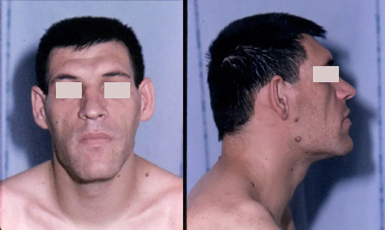
Acromegaly is rare, with an incidence of around 3-4 per million per year and a prevalence of around 60 per million.2 It affects men and women equally and is usually diagnosed in the fourth or fifth decade. Because the changes develop extremely slowly, over 5-10 years on average, diagnosis is often delayed - old photographs comparing the face over time are a classic and useful clue.
Aetiology and pathophysiology
Over 95% of cases are caused by a GH-secreting pituitary adenoma arising from somatotroph cells of the anterior pituitary.1 Most are macroadenomas (1 cm or larger) by the time of diagnosis, reflecting the slow, insidious onset of symptoms.
| Cause | Detail |
|---|---|
| Pituitary somatotroph adenoma | Over 95% of cases. Usually sporadic; rarely part of MEN1, McCune-Albright syndrome or Carney complex, or caused by an activating GNAS mutation |
| Ectopic GHRH secretion | Very rare - bronchial or pancreatic neuroendocrine tumours secreting growth hormone-releasing hormone, which drives pituitary somatotroph hyperplasia and GH excess |
| Ectopic GH secretion | Extremely rare - GH produced directly by a neuroendocrine tumour outside the pituitary |
GH is secreted in a pulsatile fashion under hypothalamic control (stimulated by GHRH, inhibited by somatostatin) and acts on the liver and peripheral tissues to stimulate production of insulin-like growth factor 1 (IGF-1).1 Unlike GH, which fluctuates through the day and is difficult to interpret from a single sample, IGF-1 is stable throughout the day, giving an integrated measure of GH activity over the preceding 24 hours - this is why IGF-1, not GH, is used as the screening test.
Most of the clinical syndrome - soft tissue and bone overgrowth, organomegaly, insulin resistance and the increased cardiovascular and neoplastic risk - is mediated by IGF-1 rather than by GH itself.
Clinical features
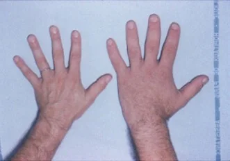
Features develop so gradually that patients and their families often do not notice the change, and the diagnosis is frequently made incidentally, by a dentist, optician or when old photographs are compared.
| System | Features |
|---|---|
| Acral (hands and feet) | Enlargement of the hands and feet - increasing ring, glove and shoe size; broad, spade-like hands; thickened, doughy skin |
| Face | Coarsening of facial features, frontal bossing, prognathism (protrusion of the lower jaw), interdental separation as the jaw grows, macroglossia, a broadened nose and thickened lips |
| Voice and airway | Deepening of the voice from laryngeal and vocal cord thickening; obstructive sleep apnoea from macroglossia and soft tissue overgrowth of the upper airway |
| Skin | Increased sweating (hyperhidrosis) and oily skin (seborrhoea), skin tags, thickened greasy skin |
| Musculoskeletal | Carpal tunnel syndrome from soft tissue swelling around the median nerve, proximal myopathy, arthralgia and secondary osteoarthritis from abnormal joint loading, back pain |
| Cardiovascular | Hypertension, acromegalic cardiomyopathy (biventricular hypertrophy progressing to dysfunction), arrhythmias |
| Metabolic | Insulin resistance and type 2 diabetes mellitus - GH is directly diabetogenic |
| Reproductive | Menstrual irregularity, reduced libido, erectile dysfunction - from co-existing hyperprolactinaemia (the adenoma may co-secrete prolactin) or from compression of normal gonadotroph function |
| Visual | Bitemporal hemianopia from superior compression of the optic chiasm by a macroadenoma |
| Organomegaly | Thyroid enlargement (goitre), hepatomegaly, splenomegaly, cardiomegaly |
Complications
Acromegaly is a multisystem disease, and screening for its complications is part of routine work-up as well as ongoing follow-up.
- Cardiovascular disease - hypertension, left ventricular hypertrophy and acromegalic cardiomyopathy, arrhythmias and heart failure. This is the leading cause of premature death in untreated acromegaly.
- Type 2 diabetes mellitus and insulin resistance - occurs in around a third of patients, from the direct diabetogenic action of GH
- Obstructive sleep apnoea - very common, from macroglossia and pharyngeal soft tissue overgrowth; screen with a sleep study
- Colonic polyps and increased colorectal cancer risk - IGF-1 promotes colonic mucosal proliferation; colonoscopy is recommended at diagnosis and periodically thereafter
- Visual field defects - bitemporal hemianopia from optic chiasm compression by a macroadenoma; formal perimetry is required if the tumour abuts the chiasm
- Hypopituitarism - compression of the surrounding normal pituitary by a macroadenoma can cause secondary hypothyroidism, hypoadrenalism and hypogonadism
- Osteoarthritis - from abnormal joint loading and cartilage overgrowth; often persists even after biochemical cure
- Carpal tunnel syndrome - from soft tissue swelling compressing the median nerve at the wrist
Investigations
Biochemical screening
Serum IGF-1 is the initial screening test.1,2 Because IGF-1 is stable throughout the day (unlike GH, which is secreted in pulses and is often undetectable between bursts), a single sample gives a reliable integrated measure of GH activity. A normal age- and sex-matched IGF-1 makes acromegaly very unlikely.
Confirmatory test
If IGF-1 is raised, the diagnosis is confirmed with an oral glucose tolerance test (OGTT) measuring GH at baseline and every 30 minutes after a 75 g glucose load.1 In healthy individuals, glucose suppresses GH secretion. Failure of GH to suppress below 1 microgram/L (1 μg/L) confirms autonomous GH secretion and the diagnosis of acromegaly.
Localising the source
Once GH excess is biochemically confirmed, arrange MRI of the pituitary to identify and characterise the adenoma (size, cavernous sinus invasion, proximity to the optic chiasm).1 If MRI shows no pituitary lesion, consider an ectopic source - measure GHRH and image the chest and abdomen for a neuroendocrine tumour.
Assessing the pituitary and complications
- Full anterior pituitary hormone profile (prolactin, TSH/free T4, cortisol/short Synacthen test, LH/FSH with sex hormones) - to detect co-secretion (prolactin is co-secreted in a significant minority) and hypopituitarism from mass effect
- Formal visual field testing (perimetry) if the adenoma abuts or compresses the optic chiasm
- HbA1c and fasting glucose - screen for diabetes and impaired glucose tolerance
- Blood pressure and echocardiogram - screen for hypertension and cardiomyopathy
- Sleep study if symptoms suggest obstructive sleep apnoea
- Colonoscopy - screen for colonic polyps, given the increased risk of colorectal neoplasia
Management
The aims of treatment are to normalise GH and IGF-1, remove or control the tumour mass, preserve normal pituitary function, and reverse or prevent complications.
Surgery
Trans-sphenoidal surgery is first-line treatment for almost all patients, including most macroadenomas.1 It offers the chance of a rapid biochemical cure, is curative in the majority of microadenomas and a good proportion of non-invasive macroadenomas, and immediately relieves any mass effect on the optic chiasm. Cure rates are lower where the tumour invades the cavernous sinus.
Medical therapy
Used when surgery does not achieve biochemical control, while awaiting the effect of radiotherapy, or as primary treatment where surgery is unsuitable.
| Drug class | Example | Notes |
|---|---|---|
| Somatostatin analogues | Octreotide, lanreotide (long-acting depot injections) | First-line medical therapy. Bind somatostatin receptors on the tumour to suppress GH secretion; also shrink tumour volume in many patients. Side effects include gallstones and gastrointestinal upset.4 |
| GH receptor antagonist | Pegvisomant | Blocks the peripheral GH receptor, so GH itself remains high but IGF-1 falls - GH levels cannot be used to monitor treatment; use IGF-1 instead. Does not shrink the tumour, so used when somatostatin analogues fail to control IGF-1. Requires monitoring of liver function.4 |
| Dopamine agonists | Cabergoline | Least effective option; reserved for mild disease or as an add-on, particularly where there is co-secretion of prolactin |
Radiotherapy
Stereotactic or conventional radiotherapy is reserved for residual or recurrent disease after surgery that has not responded adequately to medical therapy, or where surgery is not possible.1 It controls tumour growth reliably, but the fall in GH and IGF-1 is slow, over years, and it carries a substantial long-term risk of hypopituitarism, so lifelong pituitary function monitoring is required afterwards.
Monitoring
After treatment, IGF-1 (and OGTT-suppressed GH where relevant) is rechecked periodically to confirm and maintain biochemical control, alongside surveillance MRI, pituitary function testing, and ongoing screening for cardiovascular, metabolic and colonic complications.
Red flags
Prognosis
Untreated acromegaly is associated with roughly double the mortality of the general population, driven principally by cardiovascular disease, cerebrovascular disease, respiratory complications and malignancy.2 Because the condition develops so slowly, many patients have already accumulated years of cardiovascular and metabolic damage by the time of diagnosis.
Normalising IGF-1 and controlling GH restores life expectancy close to that of the general population.1,2 Soft tissue swelling, sweating and headache tend to improve quickly after successful treatment, but established bone and cartilage overgrowth (facial appearance, jaw prognathism, joint disease) is largely irreversible, so patients should be counselled that treatment halts progression and improves systemic health rather than fully reversing the physical changes. Lifelong follow-up is required, both to detect recurrence and to manage the cardiovascular, metabolic and neoplastic complications.
References
- Katznelson L, Laws ER Jr, Melmed S et al. Acromegaly: an Endocrine Society clinical practice guideline. Journal of Clinical Endocrinology & Metabolism. 2014. Available here
- Melmed S. Acromegaly. New England Journal of Medicine. Available here
- Society for Endocrinology. Clinical guidance - acromegaly. Available here
- BNF. Octreotide, lanreotide, pegvisomant and cabergoline - indications and monitoring. Available here
- NHS England. Clinical commissioning policy: pegvisomant for acromegaly. Available here
- Chanson P, Salenave S, CC BY 2.0, via Wikimedia Commons. Available here
This article is written for revision and education. It is not clinical guidance and must not be used to make decisions about the care of a patient. Always check current NICE guidance and local protocols.