
Visual Field Defects
Key points
- The central principle: the pattern of field loss localises the lesion along the visual pathway with unusual precision, because the anatomy is orderly and well understood.
- Monocular loss: a defect confined to one eye localises the lesion anterior to the chiasm - retina or optic nerve.
- Bitemporal hemianopia: loss of both temporal fields from compression of the decussating nasal fibres at the chiasm - most often a pituitary adenoma.
- Homonymous defects: the same side of the field lost in both eyes localises the lesion behind the chiasm, on the side opposite to the field loss.
- The congruity rule: the further back the lesion, the more congruous (identical between the eyes) the defect. Optic tract lesions are incongruous; occipital lesions are exactly matching.
- PITS: Parietal lesions give Inferior quadrantanopia; Temporal lesions give Superior quadrantanopia - the Meyer's loop 'pie in the sky'.
- Macular sparing: an occipital lesion may spare the central few degrees, because the macular representation at the occipital pole receives a dual blood supply from the middle cerebral artery.
- Vertical versus horizontal: neurological defects respect the vertical midline; retinal and glaucomatous defects respect the horizontal midline. This single rule separates the two groups.
Introduction
Few areas of clinical medicine allow a lesion to be localised as precisely from the bedside as the visual pathway does. The reason is that the pathway is long, anatomically orderly, and passes through regions with distinct vascular supplies and distinct patterns of disease. A field chart is therefore not simply a record of how much vision has been lost, but a map pointing at a particular few centimetres of brain.
The visual field is the whole area seen by one eye when it is fixating straight ahead. In a normal eye it extends about 60° superiorly, 75° inferiorly, 60° nasally and 100° temporally - the asymmetry reflecting the brow, cheek and nose. Fields are conventionally drawn as the patient sees them, so a left homonymous hemianopia means the patient cannot see to their left, which corresponds to a lesion in the right occipital lobe.
Everything that follows rests on one anatomical fact: light from the temporal half of the visual field falls on the nasal retina, and vice versa, because the eye's optics invert and reverse the image. Fibres from the nasal retina decussate at the chiasm; fibres from the temporal retina do not. Getting this the right way round is the whole of chiasmal localisation.
Anatomy of the visual pathway
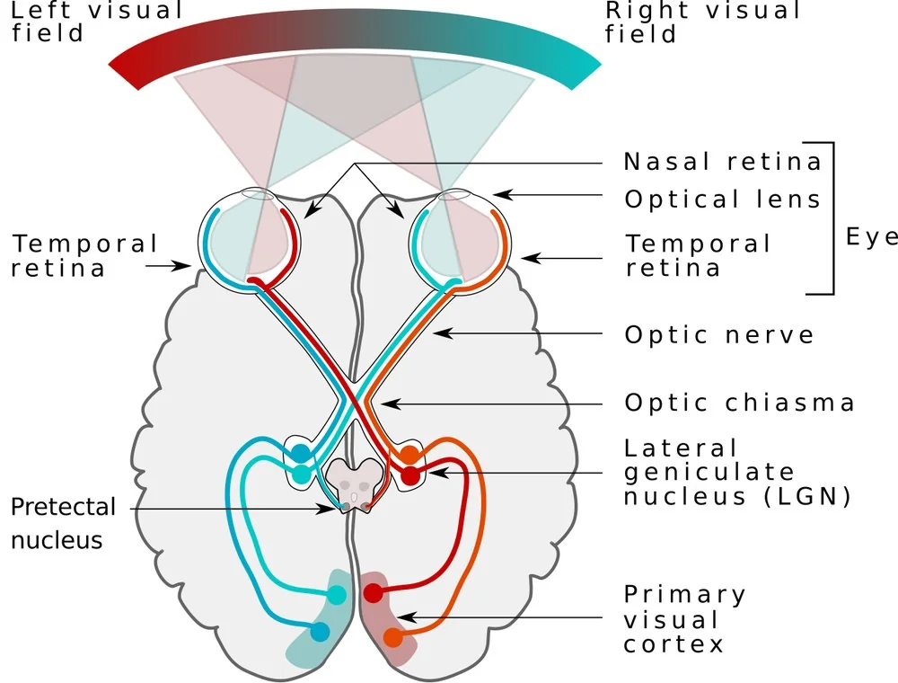
- Retina - ganglion cell axons converge on the optic disc. Fibres from the temporal retina arch above and below the macula as the arcuate bundles; fibres from the nasal retina run radially; fibres from the macula form the papillomacular bundle entering the disc temporally.
- Optic nerve - carries all the fibres from one eye. Any lesion here produces a defect confined to that eye and, if significant, a relative afferent pupillary defect.
- Optic chiasm - fibres from the nasal half of each retina decussate; temporal fibres remain uncrossed. The chiasm sits above the pituitary fossa and below the floor of the third ventricle, with the internal carotid arteries laterally.
- Optic tract - now carries fibres representing the contralateral half of the visual field from both eyes. From here on, every lesion produces a homonymous defect.
- Lateral geniculate nucleus - the thalamic relay. Fibres also branch before the LGN to the pretectal nucleus for the pupillary light reflex and to the superior colliculus.
- Optic radiations - the inferior fibres, representing the superior visual field, sweep forward around the temporal horn as Meyer's loop before turning back; the superior fibres, representing the inferior visual field, run more directly through the parietal lobe
- Primary visual cortex (V1) - in the occipital lobe around the calcarine sulcus. The superior field projects below the sulcus and the inferior field above it. The macula is represented at the occipital pole and occupies a disproportionately large area.
Prechiasmal defects
Any lesion of the retina or optic nerve produces a defect confined to one eye, and if the optic nerve is significantly affected there will be a relative afferent pupillary defect.
| Pattern | Description | Typical causes |
|---|---|---|
| Central scotoma | A blind area at fixation, with reduced acuity and colour vision | Optic neuritis, toxic and nutritional optic neuropathy, Leber hereditary optic neuropathy, macular disease |
| Centrocaecal scotoma | Central scotoma extending to the blind spot | Toxic and nutritional optic neuropathy - ethambutol, methanol, B12 deficiency, tobacco and alcohol |
| Altitudinal defect | Loss of the upper or lower half, respecting the horizontal midline | Anterior ischaemic optic neuropathy (classically inferior altitudinal), branch retinal artery occlusion |
| Arcuate (Bjerrum) scotoma | An arc sweeping from the blind spot around fixation, respecting the horizontal midline | Glaucoma, and any optic neuropathy affecting an arcuate bundle |
| Nasal step | A step-shaped defect at the nasal horizontal midline | Glaucoma - one of the earliest detectable defects |
| Enlarged blind spot | Expansion of the physiological blind spot | Papilloedema, optic disc drusen, myelinated nerve fibres, peripapillary atrophy |
| Generalised constriction | Concentric narrowing with preserved central acuity | Retinitis pigmentosa, advanced glaucoma, panretinal photocoagulation, vigabatrin toxicity |
| Total monocular blindness | Complete loss in one eye | Complete optic nerve lesion - trauma, transection, severe ischaemia, central retinal artery occlusion |
Chiasmal defects
At the chiasm, fibres from the nasal retina of each eye cross. These fibres carry the temporal half of each visual field. A lesion compressing the chiasm from below or above therefore knocks out both temporal fields, producing a bitemporal hemianopia.
- Pituitary adenoma - the classic cause, compressing the chiasm from below. Because it pushes upward, it first affects the inferonasal fibres, which carry the superior temporal field, so the defect characteristically begins as a bitemporal superior quadrantanopia and progresses downward.
- Craniopharyngioma - compresses from above, so the defect typically starts in the inferior temporal fields and progresses upward
- Meningioma of the tuberculum sellae or planum sphenoidale
- Internal carotid artery aneurysm - can compress the chiasm laterally, damaging the uncrossed temporal fibres and producing a rare binasal defect
- Pituitary apoplexy - sudden haemorrhage into an adenoma, causing acute headache, visual loss, ophthalmoplegia and hypopituitarism. This is a neurosurgical and endocrine emergency requiring urgent hydrocortisone.
- Other - Rathke cleft cyst, germinoma, glioma, sarcoidosis, and trauma
Retrochiasmal defects
Behind the chiasm, each side of the brain carries the contralateral half of the visual field from both eyes. Every lesion here therefore produces a homonymous defect on the side opposite the lesion, with normal visual acuity in each eye and no relative afferent pupillary defect.
| Site | Defect | Congruity | Associated features |
|---|---|---|---|
| Optic tract | Contralateral homonymous hemianopia | Incongruous | May have a contralateral relative afferent pupillary defect, and later a band or bow-tie optic atrophy |
| Lateral geniculate nucleus | Contralateral homonymous hemianopia, sometimes a wedge-shaped sectoranopia | Variable | Rare; usually vascular, from the anterior or lateral choroidal artery |
| Temporal lobe (Meyer's loop) | Contralateral superior homonymous quadrantanopia - 'pie in the sky' | Incongruous | Temporal lobe epilepsy, formed hallucinations, memory and language disturbance |
| Parietal lobe | Contralateral inferior homonymous quadrantanopia - 'pie on the floor' | More congruous | Visual inattention or neglect (right parietal), Gerstmann syndrome (left parietal), asymmetrical optokinetic nystagmus |
| Occipital cortex | Contralateral homonymous hemianopia, often with macular sparing | Exactly congruous | Posterior cerebral artery infarct; may be an isolated finding with no other neurological signs |
| Occipital pole | Congruous homonymous central scotomas | Congruous | The macular representation is at the pole; small infarcts here produce a purely central defect |
| Bilateral occipital | Cortical blindness | - | Normal pupils and normal fundi. Anton syndrome is cortical blindness with denial of blindness and confabulation. |
Macular sparing
An occipital infarct in the territory of the posterior cerebral artery frequently spares the central 5-10° of the field. The usual explanation is that the occipital pole, where the macula is represented, receives a dual blood supply from terminal branches of the middle cerebral artery as well as the posterior cerebral artery, so it survives a PCA occlusion. The clinical consequence is important: a patient can have a dense homonymous hemianopia yet read 6/6 and appear superficially normal, which is why the field must be tested rather than assumed from the acuity.
Assessment
Confrontation testing
- Sit about one metre from the patient at the same eye level
- Test each eye separately - the patient covers their left eye, you close your right, and each fixates the other's open eye
- Bring a target - a moving finger, or ideally a red-topped pin, which is more sensitive - in from the periphery in each of the four quadrants, and ask them to say when they first see it. Compare with your own field.
- Test across the vertical and horizontal midlines deliberately, since the midline the defect respects is the key information
- Map the blind spot with a red pin, comparing it with your own, to detect enlargement
- Test for visual inattention by presenting both hands simultaneously - extinction on one side indicates parietal neglect even when each field is individually intact
- Count fingers in each quadrant as a rapid screen where a formal comparison is not possible
- In children or uncooperative patients, use a bright toy and watch for a saccade towards it
Formal perimetry
- Automated static perimetry (Humphrey) - the standard for glaucoma and neurological field assessment, testing predetermined points at varying intensity. A 24-2 or 30-2 programme covers the central field; a 10-2 programme is used for advanced glaucoma and macular disease.
- Goldmann kinetic perimetry - a moving target of known size and brightness mapped manually. Better for very peripheral defects, for patients unable to perform automated testing, and for large neurological defects.
- Esterman binocular field test - the DVLA standard for assessing fitness to drive
- Amsler grid - a bedside test of the central 10°, used for macular disease and metamorphopsia
- Optical coherence tomography - structural correlate; retinal nerve fibre layer and ganglion cell loss usually precede detectable field loss4,5
Investigations and management
- Formal perimetry in both eyes to define the defect precisely - the confrontation test screens, but the chart localises
- Full ophthalmic examination - acuity, colour vision, pupils for an RAPD, and dilated fundoscopy for glaucomatous cupping, disc swelling, pallor and retinal disease
- MRI brain and orbits with contrast - for any defect that localises at or behind the chiasm, or any unexplained optic neuropathy. MRI of the pituitary specifically for a bitemporal defect.
- Urgent CT head where an acute stroke is suspected, following the local stroke pathway
- Pituitary function tests for chiasmal compression
- Vascular risk assessment - blood pressure, glucose, lipids, ECG and carotid imaging where the defect is homonymous and of vascular onset
- Bloods - ESR and CRP in anyone over 50 with an altitudinal defect, to exclude giant cell arteritis; B12 and folate for a centrocaecal defect
- Referral - urgent neurosurgical referral for chiasmal compression, stroke pathway for acute homonymous defects, and ophthalmology for glaucomatous and retinal causes
Practical consequences for the patient
- Driving - a homonymous hemianopia or quadrantanopia normally means the DVLA visual field standard is not met, and the patient must notify the DVLA and stop driving. Bitemporal loss and advanced glaucomatous field loss have the same implication. The standard is assessed by an Esterman binocular field test.3
- Reading difficulty - a right homonymous hemianopia disrupts reading in English because the reader cannot see the next word; a left hemianopia disrupts finding the start of the next line
- Collisions and falls - patients bump into door frames and people on the affected side, and are at higher risk of falls
- Rehabilitation - visual scanning training, prism spectacles and occupational therapy assessment can improve function significantly, and referral is worthwhile even where the defect is permanent
- Certification of visual impairment where the criteria are met, and referral to low vision services
- Recovery - homonymous defects after stroke improve in around a third of patients, mostly within the first three months, after which further recovery is unlikely
Putting it together
A practical sequence for any field chart put in front of you: first, is the defect in one eye or both? Second, if both, is it homonymous or heteronymous? Third, does it respect the vertical or the horizontal midline? Fourth, if homonymous, is it congruous, and is it superior or inferior? Those four questions take under a minute and will localise almost any lesion that appears in an exam or a clinic.
The wider clinical point is that visual field loss is systematically under-detected, because patients compensate. Binocular overlap conceals monocular defects entirely, scanning eye movements fill in hemianopic fields, and central acuity - the number that gets recorded - can remain 6/6 throughout. Pituitary tumours are frequently diagnosed only when a routine optician's field test picks up what years of normal acuity concealed.1,2
That makes confrontation testing worth doing properly rather than as a token gesture, particularly in anyone with headache, unexplained visual complaints, endocrine symptoms or a suspected stroke. It costs a minute, requires no equipment, and is one of the few bedside tests that will point directly at a specific structure inside the skull.
References
- Kedar S, Zhang X, Lynn MJ et al. Congruency in homonymous hemianopia. American Journal of Ophthalmology. 2007. Available here
- Zhang X, Kedar S, Lynn MJ et al. Homonymous hemianopias: clinical-anatomic correlations in 904 cases. Neurology. 2006. Available here
- DVLA. Assessing fitness to drive: a guide for medical professionals. Available here
- NICE NG81. Glaucoma: diagnosis and management. 2017, updated 2022. Available here
- Royal College of Ophthalmologists. Clinical guidance and resources. Available here
This article is written for revision and education. It is not clinical guidance and must not be used to make decisions about the care of a patient. Always check current NICE guidance and local protocols.