
Diabetic Ketoacidosis: The Triad, the Fluids and the Potassium
Key points
- The diagnostic triad: ketonaemia 3.0 mmol/L or above, glucose above 11 mmol/L or known diabetes, and bicarbonate below 15 mmol/L or venous pH below 7.3. All three are needed.
- Euglycaemic DKA: the glucose can be normal - on SGLT2 inhibitors, in pregnancy, with starvation or alcohol, or if insulin was taken before arrival. Check ketones in any unwell diabetic patient.
- Fluids first: 0.9% sodium chloride, starting with 1 litre over 1 hour, or 500 ml over 10 to 15 minutes if the systolic blood pressure is below 90 mmHg.
- Fixed rate insulin: 0.1 units/kg/hour of soluble insulin by intravenous infusion. Continue the patient's usual long-acting insulin; stop their short-acting.
- Potassium: none if above 5.5 mmol/L, 40 mmol/L if 3.5 to 5.5, and senior help if below 3.5. Insulin drives potassium into cells and the level falls fast.
- Add glucose, do not stop insulin: once the glucose falls below 14 mmol/L, run 10% glucose alongside the saline. Insulin must continue until the ketones have cleared.
- Resolution: ketones below 0.6 mmol/L and venous pH above 7.3. Only then convert to subcutaneous insulin, and only when the patient is eating and drinking.
- The complications that kill: hypokalaemia in adults and cerebral oedema in children and young adults. Both are largely consequences of treatment.
Introduction
Diabetic ketoacidosis is a life-threatening emergency caused by absolute insulin deficiency, in which uncontrolled lipolysis and hepatic ketogenesis produce a severe metabolic acidosis. It is the defining acute complication of type 1 diabetes, though it also occurs in type 2 diabetes - particularly under severe physiological stress, in ketosis-prone type 2 diabetes, and with SGLT2 inhibitors.
Around 25% of people with new type 1 diabetes present in DKA, and it remains a common presentation in those already diagnosed. UK mortality in adults is now below 1%, but that figure conceals the fact that most deaths are from complications of treatment rather than from the acidosis itself - hypokalaemia in adults and cerebral oedema in children.
Pathophysiology
Two hormonal changes drive everything: absolute insulin deficiency and a rise in counter-regulatory hormones - glucagon, cortisol, catecholamines and growth hormone. Together they produce two parallel processes.
- Hyperglycaemia - unrestrained hepatic gluconeogenesis and glycogenolysis with failure of peripheral glucose uptake. Once glucose exceeds the renal threshold, an osmotic diuresis develops, causing profound loss of water and of sodium, potassium, magnesium and phosphate. A typical adult is 5 to 7 litres depleted.
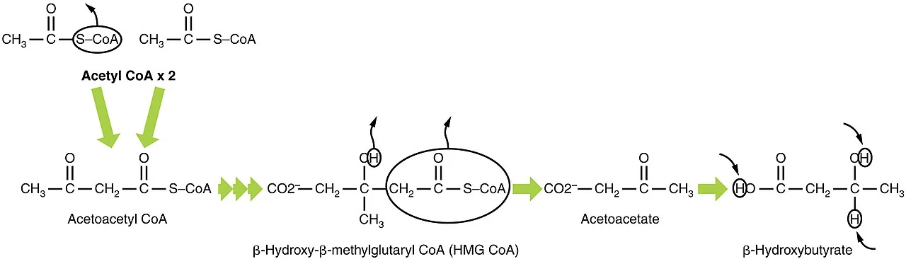
- Ketogenesis - without insulin, hormone-sensitive lipase releases free fatty acids from adipose tissue. The liver oxidises these to acetoacetate and beta-hydroxybutyrate, both strong organic acids, and to acetone. Accumulation overwhelms buffering and produces a raised anion gap metabolic acidosis.
Diagnosis and severity
Note the second criterion carefully: a normal glucose does not exclude DKA in someone with known diabetes. This is the basis of euglycaemic DKA.
| Domain | Marker of severity |
|---|---|
| Ketones | Blood ketones above 6 mmol/L |
| Acid-base | Bicarbonate below 5 mmol/L, or venous pH below 7.1 |
| Anion gap | Above 16 |
| Potassium | Below 3.5 mmol/L on admission |
| Consciousness | GCS below 12, or an abnormal AVPU score |
| Oxygenation | Saturations below 92% on air |
| Circulation | Systolic BP below 90 mmHg, pulse above 100 or below 60 |
Precipitants
A precipitant should be sought in every patient, and identifying it is part of the treatment.
- Infection - the commonest, particularly urinary, respiratory and skin. Remember that patients may be normothermic or hypothermic despite sepsis.
- Insulin omission or inadequate dosing - deliberate or inadvertent, including pump failure, and in young women sometimes deliberate omission for weight control
- New-onset type 1 diabetes - around a quarter of presentations
- Myocardial infarction - which may be silent, so obtain an ECG in every patient
- Drugs - corticosteroids, SGLT2 inhibitors, thiazides, antipsychotics, cocaine and alcohol
- Pregnancy - DKA develops at lower glucose levels and progresses faster, and threatens the fetus
- Pancreatitis, trauma, surgery and any severe intercurrent illness
Clinical features
Symptoms
- Polyuria, polydipsia and weight loss over the preceding days
- Nausea and vomiting - which worsens the dehydration
- Abdominal pain - common, sometimes severe enough to mimic an acute abdomen. It usually resolves with treatment of the DKA, but do not assume this if pain persists after correction, since pancreatitis and surgical pathology also precipitate DKA.
- Weakness, lethargy and confusion
- Reduced conscious level in severe cases
Signs
- Dehydration - dry mucous membranes, reduced skin turgor, sunken eyes, prolonged capillary refill
- Tachycardia and hypotension
- Kussmaul respiration - deep, sighing hyperventilation, the respiratory compensation for the acidosis
- Ketotic breath - a sweet smell often described as pear drops or nail varnish remover. A substantial minority of people genetically cannot smell it.
- Reduced conscious level - correlating with osmolality more than with pH
- Signs of the precipitant - a chest or urinary source, cellulitis, a foot ulcer
- Normothermia or hypothermia - fever is often absent even with significant infection
Investigations
- Capillary blood glucose and blood ketones - immediately, at the bedside
- Venous blood gas - for pH, bicarbonate and potassium. A venous sample is sufficient and avoids repeated arterial punctures; an arterial gas is not required unless there is respiratory compromise.
- U&Es - and note that the sodium is often falsely low because hyperglycaemia draws water into the extracellular space. Corrected sodium rises by about 2.4 mmol/L for every 5.5 mmol/L of glucose above normal, and a rising corrected sodium during treatment is expected.
- Full blood count - a leucocytosis is usual in DKA itself and does not by itself indicate infection
- CRP, blood cultures, urinalysis and urine culture - to identify a precipitant
- ECG - for silent myocardial infarction and for the changes of hyper- or hypokalaemia
- Chest X-ray
- Amylase or lipase - amylase is frequently raised in DKA without pancreatitis, so interpret cautiously and use lipase where available
- Pregnancy test in women of childbearing age
- Lactate - often modestly raised from hypoperfusion
- Weight - needed to calculate the insulin rate; use an estimate if it cannot be measured
Management
UK practice follows the Joint British Diabetes Societies guideline. Treatment runs on three parallel tracks - fluid, insulin and potassium - alongside treating the precipitant.1
Immediate assessment
- ABCDE assessment with large-bore intravenous access and continuous monitoring
- Confirm the diagnosis with bedside glucose and ketones and a venous gas
- Weigh the patient or estimate the weight
- Involve the diabetes specialist team early, and critical care if there are markers of severity
- Consider a nasogastric tube if there is vomiting with a reduced conscious level, and a urinary catheter if no urine has been passed after an hour or if the patient is incontinent
Fluid replacement
| Bag | Volume and duration |
|---|---|
| 1 | 1 litre over 1 hour (no potassium) |
| 2 | 1 litre with potassium over 2 hours |
| 3 | 1 litre with potassium over 2 hours |
| 4 | 1 litre with potassium over 4 hours |
| 5 | 1 litre with potassium over 4 hours |
| 6 | 1 litre with potassium over 6 hours |
- If the systolic BP is below 90 mmHg - give 500 ml of 0.9% sodium chloride over 10 to 15 minutes and repeat if it remains below 90. Seek senior help if more than 1 litre is needed, and consider another cause of shock such as sepsis or a cardiac event.
- Give fluid more cautiously in young people aged 18 to 25 (cerebral oedema risk), the elderly, pregnancy, heart failure and renal failure
- Reassess continuously - the aim is to correct the deficit over 24 to 48 hours, not as fast as possible
Insulin
- Fixed rate intravenous insulin infusion (FRIII) at 0.1 units/kg/hour, made up as 50 units of human soluble insulin in 50 ml of 0.9% sodium chloride
- No bolus dose is given in adults
- Continue the patient's usual long-acting (basal) insulin at the normal dose and time. This prevents rebound ketosis when the infusion stops and is one of the most frequently omitted steps.
- Stop short-acting subcutaneous insulin while the infusion runs
- When capillary glucose falls below 14 mmol/L, start 10% glucose at 125 ml/hour alongside the saline, and continue the insulin infusion. The insulin is treating the ketosis, not the glucose - stopping it early is a classic and dangerous error.
Potassium
Monitoring and targets
| Parameter | Target rate of change |
|---|---|
| Blood ketones | Fall by at least 0.5 mmol/L per hour |
| Venous bicarbonate | Rise by at least 3.0 mmol/L per hour |
| Capillary glucose | Fall by at least 3.0 mmol/L per hour |
| Potassium | Maintained between 4.0 and 5.5 mmol/L |
- Hourly capillary glucose and blood ketones
- Venous gas at 60 minutes, 2 hours, and then 2-hourly
- Hourly fluid balance, and continuous cardiac monitoring in severe cases
- If targets are not being met, first check the insulin infusion is actually running and connected - a disconnected or blocked line is the commonest explanation
Resolution and conversion
- DKA has resolved when ketones are below 0.6 mmol/L and venous pH is above 7.3
- Bicarbonate should not be used alone as the endpoint, because large volumes of 0.9% sodium chloride cause a hyperchloraemic metabolic acidosis that keeps the bicarbonate low after the ketosis has cleared. This is self-limiting and does not need treating.
- Convert to subcutaneous insulin only when the patient is eating and drinking
- Give the subcutaneous rapid-acting insulin with a meal and continue the intravenous infusion for 30 to 60 minutes afterwards - stopping the infusion before the subcutaneous insulin has taken effect causes rebound ketosis
- If the patient is not eating, continue a variable rate intravenous insulin infusion with substrate rather than stopping insulin
Things not to do
- Do not give sodium bicarbonate routinely - it does not improve outcome, worsens intracellular acidosis and hypokalaemia, and may increase the risk of cerebral oedema. It is reserved for extreme acidosis on senior advice.
- Do not give an insulin bolus in adults
- Do not stop the insulin when the glucose normalises - add glucose instead
- Do not omit the patient's usual long-acting insulin
- Do not use urine ketones to monitor progress
- Do not forget venous thromboembolism prophylaxis - DKA is strongly prothrombotic
Complications
- Hypokalaemia - the commonest treatment-related cause of death in adults, causing arrhythmia and respiratory muscle weakness
- Hyperkalaemia - present at the outset and itself arrhythmogenic
- Cerebral oedema - occurring in around 0.5 to 1% of paediatric DKA with a mortality approaching 25%, and rare but recognised in young adults. Suspect it with headache, irritability, deteriorating consciousness, bradycardia with hypertension, or new focal neurology, typically 4 to 12 hours into treatment.
- Hypoglycaemia - from continuing insulin without adding glucose
- Hyperchloraemic metabolic acidosis - from large-volume saline; benign and self-limiting
- Pulmonary oedema and ARDS - from over-rapid fluid replacement
- Venous thromboembolism - substantially increased risk
- Aspiration pneumonia - in the vomiting patient with a reduced conscious level
- Acute kidney injury
- Hypophosphataemia and hypomagnesaemia - usually not clinically significant, and routine phosphate replacement is not recommended
- Rhinocerebral mucormycosis - rare, but classically associated with DKA. Suspect it with facial pain, black nasal or palatal eschar and orbital signs; it is rapidly fatal without urgent ENT involvement, liposomal amphotericin and surgical debridement.
Red flags
Prevention and prognosis
Mortality from DKA in UK adults is now below 1%, though it rises substantially with age, comorbidity and in those presenting with severe acidosis or hypokalaemia. In children, the dominant risk is cerebral oedema, which accounts for most deaths.
The most important intervention is prevention, and every admission should be treated as a failure worth analysing. Before discharge:
- Identify and address the precipitant
- Review and reinforce sick day rules - never stop insulin, check glucose and ketones frequently, maintain fluid and carbohydrate intake, and know when to seek help
- Ensure the patient has a blood ketone meter and strips, and knows how to use them
- Review insulin regimen, technique and injection sites, and pump function if relevant
- Arrange specialist diabetes team follow-up before discharge
- Ask about psychological factors - depression, diabetes distress, disordered eating and deliberate insulin omission are common contributors to recurrent DKA and will not be volunteered unless asked about directly
- Consider social factors - the cost of prescriptions, food insecurity, homelessness and caring responsibilities all contribute
Recurrent DKA identifies a group at very high risk: repeated episodes are associated with a substantially increased mortality over the following years, and the driver is far more often psychosocial than educational. Responding to a third admission with another leaflet about sick day rules misses the point - what is usually needed is psychological support, practical help and continuity with a named team.
References
- Joint British Diabetes Societies for Inpatient Care. The management of diabetic ketoacidosis in adults. Available here
- British Society for Paediatric Endocrinology and Diabetes (BSPED). Interim guideline for the management of children and young people under 18 with diabetic ketoacidosis. Available here
- NICE NG17. Type 1 diabetes in adults: diagnosis and management. 2015, updated 2022. Available here
- NICE NG18. Diabetes (type 1 and type 2) in children and young people. 2015, updated 2023. Available here
- MHRA Drug Safety Update. SGLT2 inhibitors: monitor ketones in blood during treatment interruption for surgical procedures or acute serious medical illness. Available here
- NICE Clinical Knowledge Summaries. Diabetes - type 1: diabetic ketoacidosis. Available here
- BNF. Soluble insulin and potassium chloride - intravenous use. Available here
- OpenStax College, Anatomy and Physiology, CC BY 3.0, via Wikimedia Commons. Available here
This article is written for revision and education. It is not clinical guidance and must not be used to make decisions about the care of a patient. Always check current NICE guidance and local protocols.