
Intestinal Ischaemia: Acute and Chronic Mesenteric Ischaemia and Ischaemic Colitis
Key points
- Three syndromes: acute mesenteric ischaemia (a surgical emergency), chronic mesenteric ischaemia (mesenteric angina), and ischaemic colitis (the commonest and mildest).
- Acute mesenteric ischaemia: sudden, severe, generalised abdominal pain that is classically out of proportion to the clinical findings, with a soft abdomen early on.
- Commonest cause: embolus to the superior mesenteric artery, typically from atrial fibrillation; other causes are thrombosis, non-occlusive low flow and venous thrombosis.
- Investigation: urgent CT angiography is the investigation of choice; do not delay it for other tests. A raised lactate is a late and unreliable sign.
- Ischaemic colitis: transient hypoperfusion of a watershed area, classically the splenic flexure, causing crampy left-sided pain then bloody diarrhoea.
- Watershed areas: the splenic flexure (Griffiths point) and rectosigmoid junction (Sudeck point) lie at the boundaries between arterial territories.
- Chronic mesenteric ischaemia: postprandial abdominal pain, food fear and weight loss in a patient with widespread atherosclerosis.
- Prognosis: acute mesenteric ischaemia has a mortality of 50-80%, driven almost entirely by delayed diagnosis; ischaemic colitis usually resolves conservatively.
Introduction
Intestinal ischaemia results from inadequate blood supply to the bowel. It encompasses three clinically distinct syndromes that share a common mechanism but differ enormously in presentation, urgency and prognosis, and separating them is the key to managing them correctly.1
| Syndrome | Onset | Territory | Severity |
|---|---|---|---|
| Acute mesenteric ischaemia | Sudden, over hours | Small bowel, usually superior mesenteric artery | Surgical emergency; mortality 50-80% |
| Chronic mesenteric ischaemia | Gradual, over months | Two or more mesenteric vessels | Chronic, disabling; risk of acute-on-chronic infarction |
| Ischaemic colitis | Over hours to days | Colon, especially watershed areas | Commonest and usually mildest; often self-limiting |
Relevant anatomy
The gut is supplied by three unpaired branches of the aorta: the coeliac trunk (foregut - to the second part of the duodenum), the superior mesenteric artery (SMA) (midgut - to two thirds of the way along the transverse colon), and the inferior mesenteric artery (IMA) (hindgut - distal transverse colon to the upper rectum).
The boundaries between these territories are watershed areas with the least collateral supply, and are therefore most vulnerable to hypoperfusion: the splenic flexure (Griffiths point), at the SMA-IMA junction, and the rectosigmoid junction (Sudeck point), between the IMA and the internal iliac supply. This anatomy explains why ischaemic colitis so characteristically affects the splenic flexure.
Acute mesenteric ischaemia
Causes
| Mechanism | Proportion | Typical patient |
|---|---|---|
| Arterial embolism | Around 50% | Sudden onset in a patient with atrial fibrillation, recent myocardial infarction, valvular disease or endocarditis. Classically lodges just distal to the SMA origin |
| Arterial thrombosis | Around 25% | Pre-existing atherosclerosis, often with preceding symptoms of chronic mesenteric ischaemia; more insidious onset |
| Non-occlusive mesenteric ischaemia | Around 20% | Critically ill patients with a low-flow state: shock, sepsis, heart failure, dialysis, or vasopressor use. No occluding lesion is found |
| Mesenteric venous thrombosis | Around 5% | Younger patients with a prothrombotic state: thrombophilia, malignancy, oral contraceptives, portal hypertension, intra-abdominal sepsis. More gradual onset |
Clinical features
The hallmark is severe, acute, poorly localised abdominal pain that is strikingly out of proportion to the physical findings. Early on the abdomen is characteristically soft and non-tender despite the patient being in agony - a discrepancy that is the single most important clue and the reason the diagnosis is so often missed.2
- Sudden, severe, diffuse central abdominal pain, often colicky at first then constant
- Nausea and vomiting, and often forceful diarrhoea early in the course
- The classic triad of abdominal pain, atrial fibrillation and no abdominal signs
- As infarction progresses (typically after 6-12 hours), pain may transiently improve as the bowel becomes necrotic and the nerves die - a dangerous false reassurance
- Late features: peritonism, guarding, rigidity, abdominal distension, per rectum bleeding, shock, sepsis and multi-organ failure. By this stage transmural infarction has occurred and mortality is very high
Ischaemic colitis
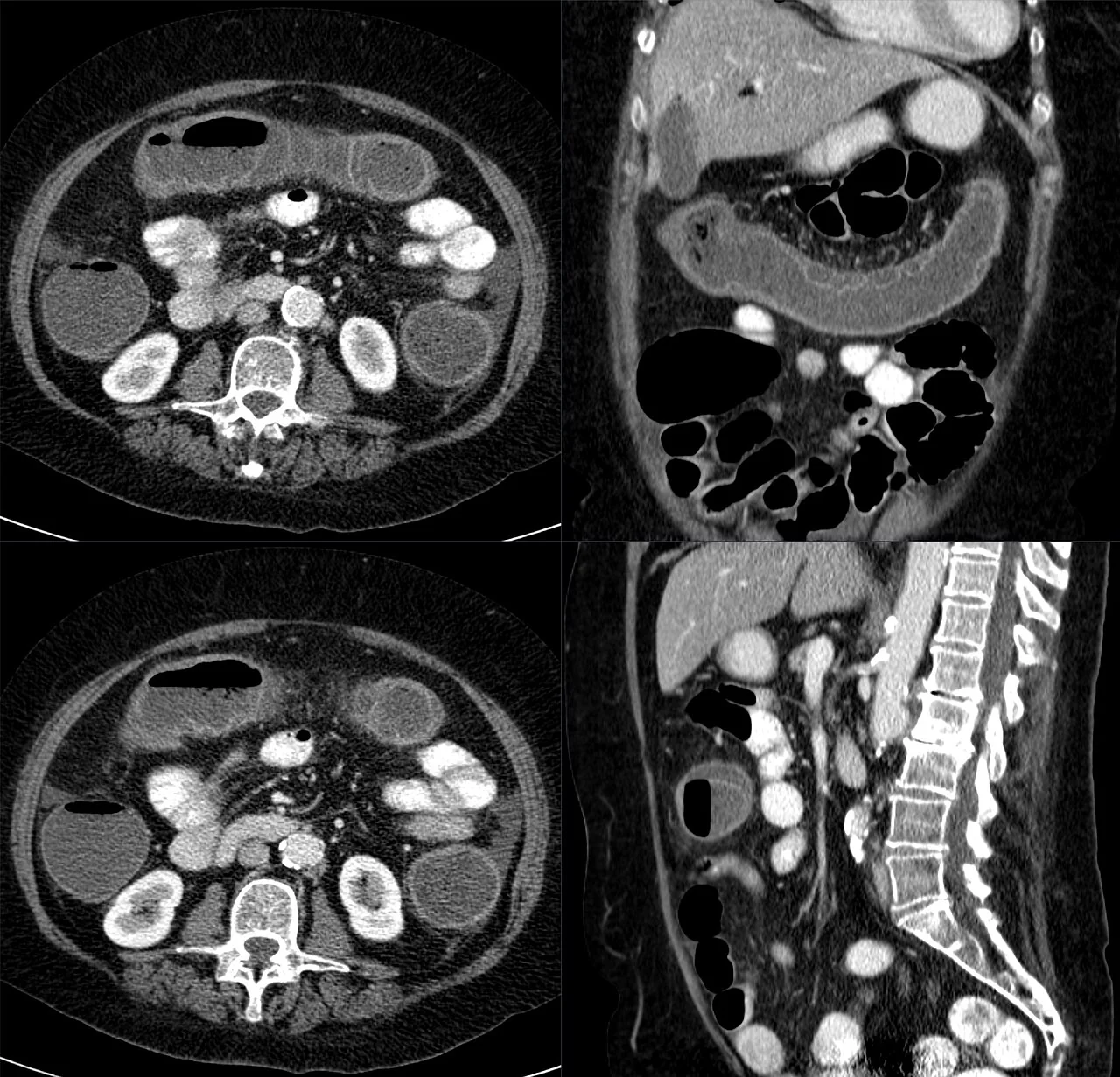
Ischaemic colitis is the commonest form of intestinal ischaemia and results from transient hypoperfusion of the colon, usually without a demonstrable arterial occlusion. It typically affects older patients with vascular risk factors, and in most cases the ischaemia is mucosal and reversible rather than transmural.3
Precipitants
- Hypotension from any cause: sepsis, heart failure, haemorrhage, dehydration, cardiac surgery or dialysis
- Aortic or cardiac surgery, particularly aortic aneurysm repair with sacrifice of the inferior mesenteric artery
- Drugs: vasopressors, digoxin, diuretics, NSAIDs, combined oral contraceptives, and cocaine
- Thrombophilia and vasculitis in younger patients
- Strenuous exertion, classically long-distance running
- Constipation or an obstructing colonic lesion causing raised intraluminal pressure
Clinical features
The classic sequence is sudden crampy left-sided abdominal pain, often left iliac fossa, followed within 24 hours by loose stool mixed with bright red blood. Unlike acute mesenteric ischaemia, the pain is usually mild to moderate, there is localised tenderness over the affected segment, and the patient is not systemically collapsed. Bleeding is rarely heavy enough to need transfusion.
Chronic mesenteric ischaemia
Also known as intestinal or mesenteric angina, this results from atherosclerotic stenosis of the mesenteric vessels. Because of the rich collateral circulation, symptoms usually require significant stenosis of at least two of the three main mesenteric arteries.1
The classic triad is:
- Postprandial abdominal pain, beginning 15-60 minutes after eating and lasting 1-2 hours, as the metabolic demand of digestion outstrips the available blood supply
- Food fear (sitophobia) - the patient learns to avoid eating, or eats only small amounts, to prevent the pain
- Weight loss, which is often marked and can prompt an initial suspicion of malignancy
Patients almost always have widespread atherosclerosis elsewhere - coronary, cerebrovascular or peripheral arterial disease - and are frequently current smokers. An abdominal bruit may be audible. The main risks are progressive malnutrition and acute-on-chronic thrombosis causing infarction.
Investigations
Imaging
CT angiography of the mesenteric vessels is the investigation of choice for both acute and chronic mesenteric ischaemia, and should be arranged urgently on clinical suspicion.2 It demonstrates the occluding embolus or thrombus, and signs of bowel compromise such as wall thickening, reduced mucosal enhancement, pneumatosis intestinalis (gas within the bowel wall), portal venous gas and free fluid. In ischaemic colitis, CT typically shows segmental colonic wall thickening with fat stranding in a watershed distribution.
Plain abdominal X-ray is usually normal early and is of limited value; the classic "thumbprinting" of mucosal oedema and any evidence of pneumatosis are late findings. Catheter mesenteric angiography remains the gold standard for chronic disease and allows simultaneous intervention.
Blood tests
- Venous or arterial lactate: may be raised, but a normal lactate does not exclude the diagnosis and elevation is a late marker of established infarction. Never use it to rule out mesenteric ischaemia
- Full blood count: leucocytosis is common but non-specific
- Arterial blood gas: metabolic acidosis with a raised anion gap, again a late finding
- Amylase, creatine kinase and LDH: may be raised, non-specifically
- Urea and electrolytes, CRP, clotting, group and save, and an ECG to identify atrial fibrillation
- Thrombophilia screen in younger patients or suspected venous thrombosis
Endoscopy
Colonoscopy or flexible sigmoidoscopy is used in ischaemic colitis once the patient is stable, showing pale, oedematous mucosa with petechial haemorrhage and sometimes a single longitudinal ulcer, in a clearly demarcated segmental distribution with an abrupt transition to normal mucosa. Biopsies help exclude infection and inflammatory bowel disease. Endoscopy is avoided if there is any suspicion of transmural infarction or peritonism because of the perforation risk.
Differential diagnosis
- Perforated viscus, including perforated peptic ulcer
- Ruptured or leaking abdominal aortic aneurysm - always consider in an older patient with abdominal pain and shock
- Acute pancreatitis - severe epigastric pain radiating to the back with raised lipase
- Bowel obstruction and strangulated hernia
- Inflammatory bowel disease or infective colitis - the main differentials for ischaemic colitis, distinguished by segmental watershed distribution and rapid resolution
- Diverticulitis, which can look similar on CT
- Myocardial infarction, particularly inferior MI presenting with epigastric pain
- Diabetic ketoacidosis and other metabolic causes of abdominal pain with acidosis
Management
Acute mesenteric ischaemia
This is a surgical emergency requiring simultaneous resuscitation and definitive vascular management:2
- ABCDE assessment with aggressive IV fluid resuscitation and correction of acidosis and electrolytes
- Urgent senior surgical and vascular review, with early critical care involvement
- Broad-spectrum IV antibiotics, given the risk of bacterial translocation from ischaemic bowel
- Anticoagulation with intravenous heparin unless contraindicated
- Nil by mouth, nasogastric decompression, analgesia and catheterisation with strict fluid balance
- Revascularisation: embolectomy, surgical bypass, or endovascular thrombectomy, thrombolysis or stenting, depending on the cause and local expertise
- Laparotomy to assess bowel viability and resect frankly necrotic bowel. A planned second-look laparotomy at 24-48 hours is frequently performed, since viability is difficult to judge at the first operation
Mesenteric venous thrombosis is managed differently: anticoagulation alone is often sufficient, with surgery reserved for infarction. In non-occlusive ischaemia, the priority is correcting the underlying low-flow state - treating sepsis or cardiac failure and withdrawing vasopressors where possible - as there is no occlusion to remove.
Ischaemic colitis
Most cases (around 80%) settle with conservative management: bowel rest, IV fluids, correction of the precipitating hypoperfusion, review and withdrawal of causative drugs, and antibiotics if there are systemic features. Symptoms typically resolve within a few days. Surgery is required for peritonitis, perforation, massive bleeding, fulminant colitis, or failure to improve, and involves resection of the affected segment.
Chronic mesenteric ischaemia
Management combines aggressive cardiovascular risk factor modification - smoking cessation, statin, antiplatelet therapy, blood pressure and diabetes control - with revascularisation, usually by endovascular angioplasty and stenting, or surgical bypass in selected patients. Nutritional support is important given the degree of weight loss, and revascularisation both relieves symptoms and reduces the risk of acute infarction.
Complications
- Bowel infarction and perforation with faecal peritonitis
- Sepsis, septic shock and multi-organ failure from bacterial translocation
- Short bowel syndrome following extensive small bowel resection, with lifelong dependence on parenteral nutrition in severe cases
- Colonic stricture - a common late sequela of ischaemic colitis, causing obstructive symptoms weeks to months later
- Chronic ischaemic colitis with persistent segmental inflammation
- Malnutrition and cachexia in chronic mesenteric ischaemia
- Reperfusion injury following revascularisation, with metabolic derangement and cardiovascular instability
- Recurrent thromboembolism if the underlying source, such as atrial fibrillation, is not addressed
Red flags
Prognosis
Acute mesenteric ischaemia carries a mortality of 50-80%, one of the highest of any abdominal emergency, and this figure is driven overwhelmingly by delay in diagnosis.2 Outcomes improve dramatically if revascularisation is achieved within around 6 hours, before transmural infarction develops, which is why the threshold for CT angiography must be very low. Survivors of extensive resection may face lifelong short bowel syndrome.
Ischaemic colitis has a far better prognosis, with around 80% resolving on conservative management alone and no long-term sequelae, although a minority develop a late colonic stricture or chronic segmental colitis. Right-sided ischaemic colitis, which reflects SMA territory involvement, carries a worse prognosis than the classic left-sided form and more often requires surgery.
Chronic mesenteric ischaemia responds well to revascularisation, with symptom relief and weight gain in the majority, but untreated it progresses to malnutrition and carries a substantial risk of acute-on-chronic thrombosis and infarction.
References
- Clair DG, Beach JM. Mesenteric Ischemia. N Engl J Med. 2016. Available here
- Bala M et al. Acute mesenteric ischemia: updated guidelines of the World Society of Emergency Surgery. World J Emerg Surg. 2022. Available here
- Brandt LJ et al. ACG clinical guideline: epidemiology, risk factors, patterns of presentation, diagnosis, and management of colon ischemia. Am J Gastroenterol. 2015. Available here
- Hellerhoff, CC BY-SA 4.0, via Wikimedia Commons. Available here
- British Society of Gastroenterology. Guidelines on the management of acute lower gastrointestinal bleeding. Gut. 2019. Available here
- NICE Clinical Knowledge Summaries (CKS). Atrial fibrillation. 2024. Available here
- Gnanapandithan K, Feuerstadt P. Review Article: Mesenteric Ischemia. Curr Gastroenterol Rep. 2020. Available here
This article is written for revision and education. It is not clinical guidance and must not be used to make decisions about the care of a patient. Always check current NICE guidance and local protocols.