
Mallory-Weiss Tear: Diagnosis and Management
Key points
- Mallory-Weiss tear: a longitudinal mucosal laceration at or near the gastro-oesophageal junction, caused by a sudden rise in intra-abdominal pressure.
- Classic history: haematemesis that follows an episode of forceful retching or vomiting - the first vomit contains no blood, later ones do.
- Precipitants: alcohol binges, hyperemesis gravidarum, gastroenteritis, bulimia nervosa, severe coughing and, rarely, CPR.
- Depth: the tear involves mucosa and submucosa only; it does not perforate the full thickness of the oesophageal wall.
- Diagnosis: upper GI endoscopy, which confirms the tear and excludes other causes of upper GI bleeding.
- Management: usually conservative - around 90% stop bleeding spontaneously; endoscopic haemostasis for active bleeding.
- Key differential: Boerhaave syndrome, a full-thickness oesophageal rupture, which is a surgical emergency with a very different prognosis.
- Prognosis: excellent, with rapid healing and a low rebleeding rate; recurrence is uncommon.
Introduction
A Mallory-Weiss tear is a longitudinal laceration of the mucosa and submucosa at or just below the gastro-oesophageal junction, classically produced by a sudden, forceful rise in intra-abdominal and intraluminal pressure during vomiting or retching.1
It accounts for around 5-10% of cases of upper gastrointestinal bleeding, making it one of the more common causes after peptic ulcer disease.2 It is important to recognise because, unlike most other causes of upper GI bleeding, it is usually self-limiting and carries an excellent prognosis.
Pathophysiology
During vomiting, a sudden and marked increase in intra-abdominal pressure is transmitted to the stomach and distal oesophagus. If the gastro-oesophageal junction does not relax in a coordinated way, the resulting pressure gradient across the junction shears the mucosa, producing a longitudinal tear.1
The tear typically extends through the mucosa and submucosa, exposing the underlying submucosal arteries and veins, which is the source of the bleeding. Crucially, it does not extend through the muscular layer, which is what distinguishes it from Boerhaave syndrome (a full-thickness rupture). The presence of a hiatus hernia increases the pressure gradient across the junction and is a recognised predisposing factor.
Risk factors and precipitants
Anything causing repeated, forceful vomiting or retching can precipitate a tear:2
- Alcohol excess, particularly binge drinking - the classic association
- Hyperemesis gravidarum and severe pregnancy-related vomiting
- Infectious gastroenteritis with protracted vomiting
- Bulimia nervosa and self-induced vomiting
- Severe or paroxysmal coughing
- Chemotherapy-induced or postoperative vomiting
- Cardiopulmonary resuscitation and blunt abdominal trauma (rare)
Predisposing factors that increase the likelihood or severity of bleeding include hiatus hernia, older age, chronic liver disease with portal hypertension, coagulopathy, and anticoagulant or antiplatelet therapy.
Clinical features
The classic history is of haematemesis following an episode of forceful vomiting or retching, where the initial vomit contains no blood and subsequent vomits do. This sequence is highly characteristic and is the single most useful diagnostic clue in the history.
Other features include:
- Haematemesis, which may be fresh red blood, streaks of blood, or coffee-ground vomit
- Melaena, if enough blood has passed distally
- Epigastric or retrosternal pain, though this is usually mild
- Light-headedness, syncope or signs of hypovolaemia if bleeding is significant
- Features of the precipitating cause: alcohol intoxication, pregnancy, or gastroenteritis
Most patients are haemodynamically stable at presentation. Significant haemodynamic compromise is uncommon and should prompt consideration of an alternative or additional diagnosis.
Differential diagnosis
The key differentials are the other causes of upper GI bleeding, and in particular Boerhaave syndrome:
| Feature | Mallory-Weiss tear | Boerhaave syndrome |
|---|---|---|
| Depth of injury | Mucosa and submucosa only | Full-thickness oesophageal rupture |
| Dominant symptom | Haematemesis after retching | Severe chest or epigastric pain after vomiting |
| Systemic upset | Usually well, haemodynamically stable | Rapidly unwell: shock, sepsis, subcutaneous emphysema |
| Investigation | Upper GI endoscopy | CT with oral contrast or contrast swallow |
| Management | Usually conservative | Surgical emergency - resuscitation, antibiotics, urgent repair |
| Mortality | Very low | High, rising sharply with delayed diagnosis |
Other differentials include peptic ulcer disease, oesophageal varices (particularly relevant in patients with alcohol excess, who may have both), oesophagitis, gastritis and malignancy.3
Investigations
Initial investigations are those for any upper GI bleed: full blood count, urea and electrolytes, liver function tests, clotting screen, and group and save or crossmatch. A Glasgow-Blatchford score is calculated to guide the urgency of endoscopy and whether the patient can be managed as an outpatient; many patients with a Mallory-Weiss tear score low.3
Endoscopy
Upper GI endoscopy is the diagnostic investigation and confirms the diagnosis by direct visualisation. The tear appears as a single (occasionally multiple) longitudinal mucosal laceration at the gastro-oesophageal junction, often best seen on retroflexion of the endoscope in the gastric cardia.
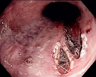
Endoscopy also allows exclusion of coexisting pathology, which matters particularly in patients with alcohol-related liver disease, in whom varices may be present alongside the tear. If Boerhaave syndrome is suspected clinically, CT with oral contrast is performed instead of proceeding directly to endoscopy.
Management
Management follows the general principles of upper GI bleeding, but the great majority of tears require no specific intervention.
Supportive management
Around 90% of Mallory-Weiss tears stop bleeding spontaneously and heal within 48-72 hours.1 Treatment therefore consists of IV access, fluid resuscitation and transfusion if required, correction of coagulopathy, and antiemetics to break the cycle of ongoing vomiting. Acid suppression with a PPI is commonly given to aid mucosal healing, although the evidence for this is limited.
Endoscopic therapy
Endoscopic haemostasis is indicated where there is active bleeding or a visible vessel at endoscopy. Options include adrenaline injection, mechanical clips, band ligation and thermal coagulation, often used in combination. Clipping and band ligation are generally preferred over thermal methods, given the relatively thin wall at this site.
Further measures
Angiographic embolisation of the left gastric artery is an option for the rare case of ongoing bleeding despite endoscopic therapy, with surgical oversewing of the tear a last resort. Addressing the underlying precipitant is an important part of care: alcohol advice and support, treatment of hyperemesis, or eating disorder services for patients with bulimia nervosa.
Complications
- Significant haemorrhage requiring transfusion, more likely in patients with portal hypertension or coagulopathy
- Rebleeding, which occurs in a small minority, typically within the first 24 hours
- Hypovolaemic shock and its sequelae in severe cases
- Aspiration pneumonia from vomiting, particularly in intoxicated or obtunded patients
- Progression to full-thickness perforation is very rare, but any patient with severe pain, sepsis or surgical emphysema should be reassessed for Boerhaave syndrome
Red flags
Prognosis
The prognosis is excellent. The great majority of tears stop bleeding without intervention and heal completely within a few days, leaving no long-term sequelae, and recurrence is uncommon provided the precipitating cause is addressed.2 Mortality is low and, where it occurs, usually reflects comorbidity or coexisting pathology such as portal hypertension rather than the tear itself. Outcomes are worse in patients who are anticoagulated, have chronic liver disease, or present with haemodynamic compromise.
References
- Rawla P, Devasahayam J. Mallory-Weiss Syndrome. StatPearls. 2023. Available here
- Kim HS. Endoscopic management of Mallory-Weiss tearing. Clin Endosc. 2015. Available here
- NICE CG141. Acute upper gastrointestinal bleeding in over 16s: management. 2012 (updated 2016). Available here
- British Society of Gastroenterology. Management of acute upper gastrointestinal bleeding. Gut. 2019. Available here
- Samir, CC BY-SA 3.0, via Wikimedia Commons. Available here
- NHS. Vomiting blood (haematemesis). 2023. Available here
- Turner AR, Turner SD. Boerhaave Syndrome. StatPearls. 2023. Available here
This article is written for revision and education. It is not clinical guidance and must not be used to make decisions about the care of a patient. Always check current NICE guidance and local protocols.