
Ascites and Spontaneous Bacterial Peritonitis
Key points
- Ascites: pathological accumulation of fluid within the peritoneal cavity; cirrhosis with portal hypertension accounts for around 80% of cases.
- SAAG: serum-ascites albumin gradient = serum albumin minus ascitic albumin. 11 g/L or above indicates portal hypertension; below 11 g/L points elsewhere.
- Mechanism in cirrhosis: portal hypertension causes splanchnic vasodilatation, reducing effective circulating volume and activating the renin-angiotensin-aldosterone system, causing sodium and water retention.
- Medical management: dietary sodium restriction plus spironolactone first-line (targeting hyperaldosteronism), with furosemide added if needed.
- Large-volume paracentesis: for tense or refractory ascites, with IV albumin cover (around 8 g per litre drained) to prevent post-paracentesis circulatory dysfunction.
- SBP: infection of ascitic fluid without a surgical source, defined by an ascitic neutrophil count above 250 cells/mm³, regardless of culture result.
- SBP treatment: IV antibiotics (e.g. piperacillin-tazobactam or cefotaxime) plus IV albumin, which reduces hepatorenal syndrome and mortality.
- Key rule: perform a diagnostic ascitic tap in every patient with ascites who is admitted or deteriorates - SBP is frequently silent.
Introduction
Ascites is the pathological accumulation of fluid within the peritoneal cavity. It is the commonest complication of cirrhosis and marks the transition to decompensated disease, carrying a median survival of roughly 2 years and a 50% two-year mortality once it becomes refractory.1
Around 80% of cases are due to cirrhosis with portal hypertension, with malignancy, heart failure and, less commonly, infection or pancreatic disease accounting for most of the remainder. Its development should always prompt two questions: what is causing it, and is it infected?
Causes and the SAAG
The most useful diagnostic tool for classifying ascites is the serum-ascites albumin gradient (SAAG), calculated as serum albumin minus ascitic fluid albumin, from samples taken on the same day. It has largely replaced the older transudate/exudate distinction because it more reliably identifies portal hypertension.2
| SAAG 11 g/L or above (portal hypertension present) | SAAG below 11 g/L (portal hypertension absent) |
|---|---|
| Cirrhosis - by far the commonest cause | Peritoneal malignancy and peritoneal carcinomatosis |
| Alcoholic hepatitis | Tuberculous peritonitis |
| Right heart failure and constrictive pericarditis | Pancreatitis (pancreatic ascites) |
| Budd-Chiari syndrome (hepatic vein thrombosis) | Nephrotic syndrome |
| Portal vein thrombosis | Serositis in connective tissue disease |
| Massive hepatic metastases | Bowel obstruction or infarction |
| Fulminant hepatic failure | Biliary or chylous ascites |
Pathophysiology in cirrhosis
Ascites in cirrhosis develops through a sequence that explains why the treatment works the way it does:1
- Portal hypertension raises sinusoidal hydrostatic pressure, increasing lymph production beyond the capacity of the thoracic duct to return it, so fluid weeps into the peritoneal cavity
- Splanchnic vasodilatation, driven largely by nitric oxide released in response to portal hypertension, pools blood in the splanchnic bed
- This reduces the effective circulating volume, despite total body fluid being increased - the kidney perceives underfilling
- Activation of the renin-angiotensin-aldosterone system, sympathetic nervous system and antidiuretic hormone follows, causing avid sodium and water retention
- Hypoalbuminaemia from impaired hepatic synthesis lowers plasma oncotic pressure, compounding fluid shift into the peritoneum
- Retained sodium and water accumulate as further ascites, perpetuating the cycle
This secondary hyperaldosteronism is why spironolactone, an aldosterone antagonist, is the first-line diuretic in cirrhotic ascites rather than a loop diuretic - it targets the primary mechanism. It also explains why dietary sodium restriction is effective, and why ADH-driven water retention produces the dilutional hyponatraemia that marks advanced disease.
Clinical features
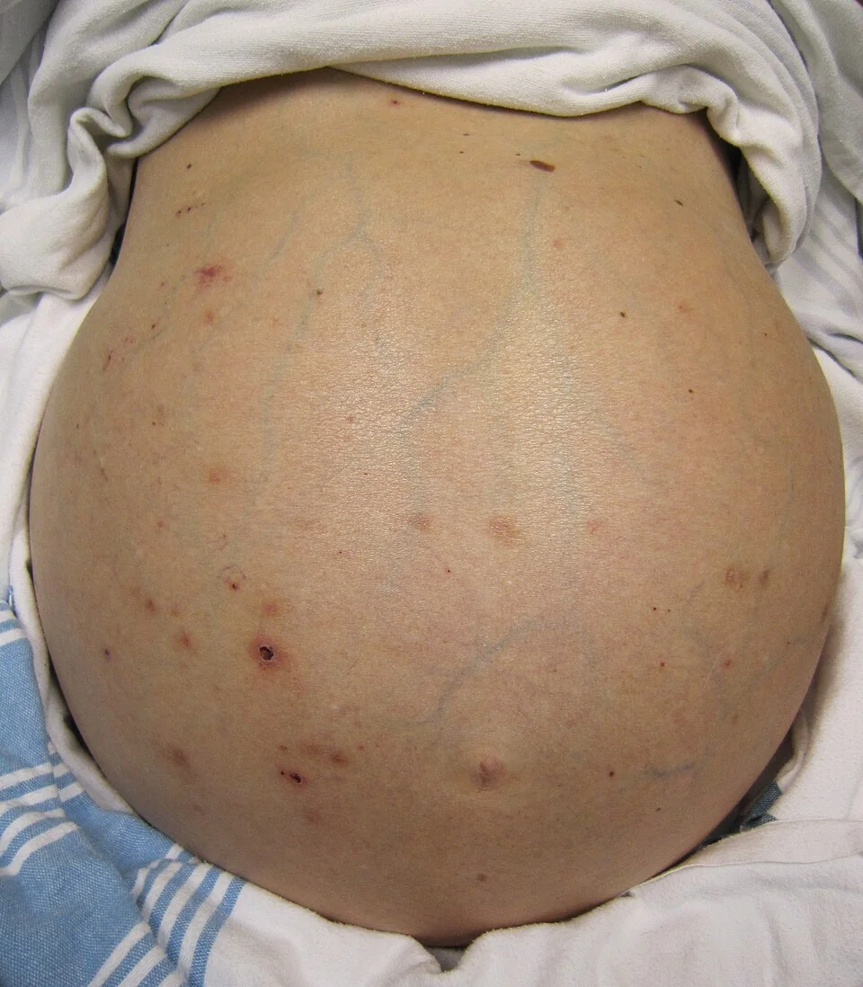
Patients typically report progressive abdominal distension, weight gain, early satiety, and sometimes breathlessness from diaphragmatic splinting or a coexisting hepatic hydrothorax.
Examination
- Shifting dullness - the most useful bedside sign, detectable with roughly 1.5 litres or more of fluid
- Fluid thrill in tense ascites
- Flank fullness and an everted umbilicus
- Stigmata of chronic liver disease and portal hypertension - spider naevi, palmar erythema, jaundice, caput medusae, splenomegaly
- Signs pointing to a non-hepatic cause: raised JVP and peripheral oedema in heart failure, cachexia and lymphadenopathy in malignancy, a Sister Mary Joseph nodule in intra-abdominal malignancy
Spontaneous bacterial peritonitis
SBP is notoriously silent or subtle - the classic teaching that it presents with fever and abdominal pain is misleading, since a substantial minority have neither. Suspect it in any patient with ascites who deteriorates in any way:
- Fever, abdominal pain or tenderness (present in only about two thirds)
- New or worsening hepatic encephalopathy - often the only clue
- Unexplained deterioration in renal function
- Ascites that has become diuretic-resistant
- Hypotension, tachycardia or frank sepsis
- Diarrhoea, vomiting or ileus
- No symptoms at all - which is precisely why a diagnostic tap is mandatory rather than discretionary
Investigations
Diagnostic ascitic tap (paracentesis)
This is the single most important investigation and should be performed in every patient presenting with new ascites, and in every patient with known ascites who is admitted to hospital or deteriorates.3 It is a safe procedure even with a raised INR - coagulopathy is not a contraindication, and routine correction with blood products before the tap is not required.
Send ascitic fluid for:
| Test | Interpretation |
|---|---|
| Neutrophil (polymorph) count | Above 250 cells/mm³ diagnoses SBP, regardless of culture result. This is the key number |
| Albumin | Used with a same-day serum albumin to calculate the SAAG |
| Culture (inoculated into blood culture bottles at the bedside) | Improves yield substantially. Often negative even in genuine SBP (culture-negative neutrocytic ascites) - treat on the neutrophil count |
| Total protein | Ascitic protein below 15 g/L identifies patients at high risk of SBP who warrant prophylaxis |
| Cytology | For malignant cells if peritoneal malignancy is suspected; requires a large volume and repeated samples |
| Glucose, LDH, amylase | A low glucose with high LDH and multiple organisms suggests secondary bacterial peritonitis from a perforated viscus. High amylase suggests pancreatic ascites |
| Acid-fast bacilli and adenosine deaminase | For suspected tuberculous peritonitis |
Other investigations
- Bloods: full blood count, urea and electrolytes (renal function and sodium), liver function, albumin, INR, CRP, glucose and blood cultures
- Abdominal ultrasound: confirms ascites, assesses the liver for cirrhosis and focal lesions, and Doppler examines portal and hepatic vein patency to exclude thrombosis and Budd-Chiari syndrome
- CT abdomen and pelvis: where malignancy or secondary peritonitis is suspected
- Echocardiogram: if right heart failure or constrictive pericarditis is a possibility
- Urinary sodium and 24-hour urinary sodium excretion, to assess adherence to sodium restriction and guide diuretic dosing
Management of ascites
General measures
- Treat the underlying cause - alcohol abstinence, antiviral therapy, treatment of heart failure or malignancy
- Dietary sodium restriction to around 80-90 mmol (roughly 5-6 g salt) per day. This is the foundation of treatment and its absence is a common reason for apparent diuretic failure
- Fluid restriction is not routinely needed unless there is significant hyponatraemia (sodium below about 125 mmol/L)
- Daily weights - aim for weight loss of no more than 0.5 kg/day without peripheral oedema, or 1 kg/day with oedema, to avoid precipitating renal impairment
- Avoid NSAIDs (impair renal prostaglandins and worsen sodium retention and renal failure) and ACE inhibitors/ARBs (worsen hypotension and renal perfusion)
Diuretics
Spironolactone is first-line, typically starting at 100 mg daily and titrated up to 400 mg, targeting the secondary hyperaldosteronism that drives sodium retention. Furosemide (starting at 40 mg, up to 160 mg) is added if the response is inadequate, conventionally maintaining a 100:40 ratio to preserve potassium balance.1
Monitor urea, electrolytes and creatinine closely. Diuretics should be stopped if there is significant hyponatraemia, acute kidney injury, hyperkalaemia (with spironolactone), or hepatic encephalopathy. Gynaecomastia is a common and dose-limiting side effect of spironolactone; amiloride is a less effective alternative.
Large-volume paracentesis
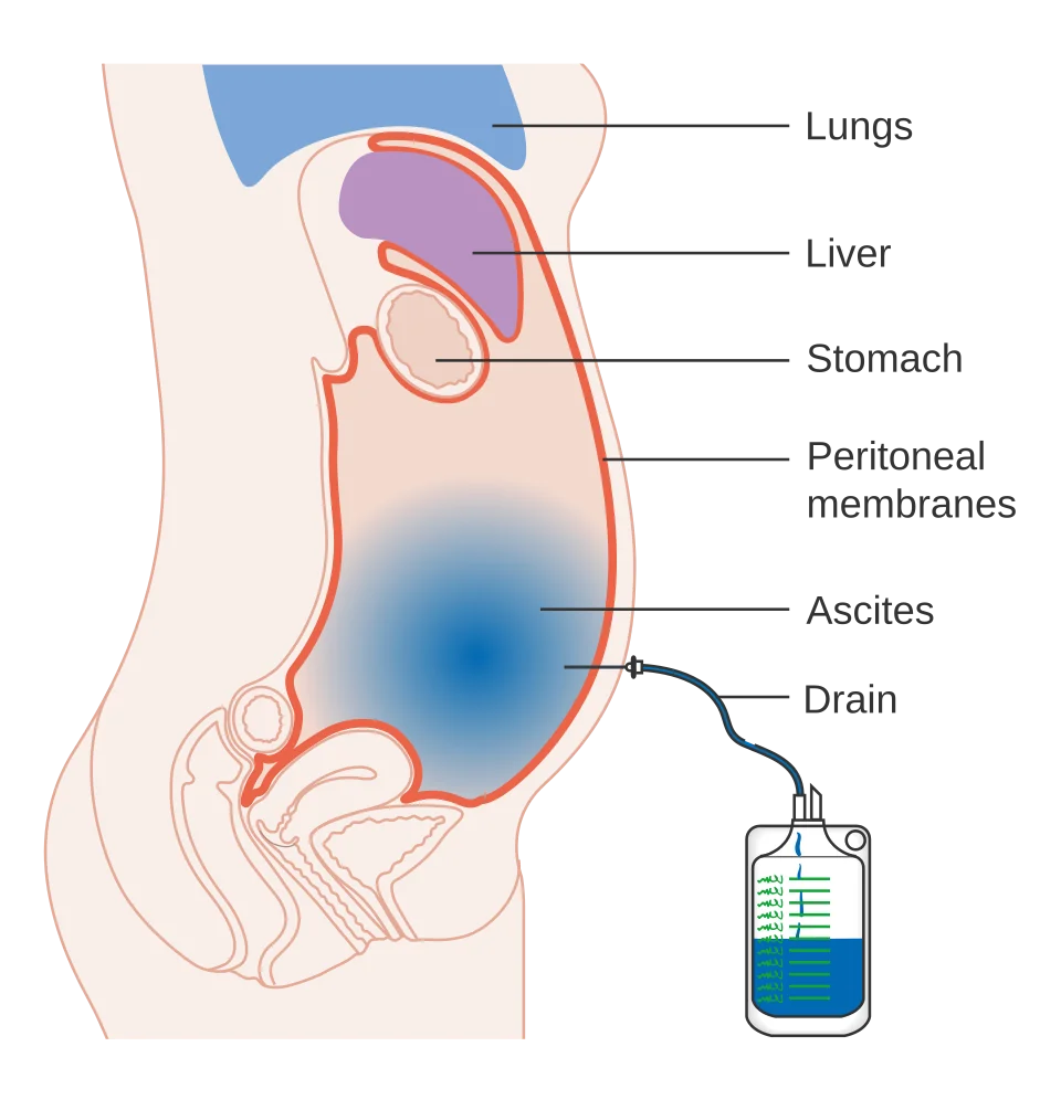
Therapeutic paracentesis is used for tense ascites causing discomfort or breathlessness, and for refractory ascites not controlled by maximal diuretics. Intravenous albumin cover of approximately 8 g per litre of ascites removed should be given whenever more than 5 litres is drained, to prevent post-paracentesis circulatory dysfunction - a fall in effective circulating volume that precipitates renal failure, hyponatraemia and increased mortality.
Refractory ascites
Defined as ascites that cannot be mobilised despite maximal diuretics and sodium restriction, or that recurs rapidly after paracentesis. Options are repeated large-volume paracentesis with albumin, transjugular intrahepatic portosystemic shunt (TIPS) - effective but risks precipitating hepatic encephalopathy and worsening liver function - and liver transplantation, which is the definitive treatment and should be considered in all such patients. A permanent tunnelled drain may be used for palliation, particularly in malignant ascites.
Management of spontaneous bacterial peritonitis
SBP is infection of ascitic fluid without an identifiable intra-abdominal surgical source, arising from bacterial translocation across the gut wall. The commonest organisms are Escherichia coli, Klebsiella and streptococci - typically a monomicrobial growth.3
Treatment
- Start empirical IV antibiotics immediately once the neutrophil count exceeds 250 cells/mm³ - do not wait for culture results. Typical regimens are piperacillin-tazobactam or a third-generation cephalosporin such as cefotaxime, guided by local policy and rationalised on sensitivities
- Give IV albumin - 1.5 g/kg at diagnosis and 1 g/kg on day 3. This is not merely supportive: albumin substantially reduces the incidence of hepatorenal syndrome and improves mortality, and is one of the highest-yield interventions in hepatology
- Stop diuretics and non-selective beta-blockers during the acute episode, as both worsen haemodynamics and renal perfusion
- Avoid nephrotoxins, particularly NSAIDs and aminoglycosides
- Monitor renal function closely and repeat the ascitic tap at 48 hours if the response is uncertain - a fall of less than 25% in neutrophil count suggests treatment failure or secondary peritonitis
Prophylaxis
Secondary prophylaxis with an oral quinolone (ciprofloxacin) or co-trimoxazole is given indefinitely to all patients who have had an episode of SBP, since recurrence approaches 70% within a year without it. Primary prophylaxis is offered to patients with ascitic protein below 15 g/L with advanced liver disease, and antibiotic prophylaxis is given to all patients with cirrhosis and upper GI bleeding. Any patient who has had SBP should be assessed for liver transplantation.
Complications
- Spontaneous bacterial peritonitis and its recurrence
- Hepatorenal syndrome - functional renal failure, frequently precipitated by SBP, large-volume paracentesis without albumin, or over-diuresis
- Dilutional hyponatraemia, a marker of advanced disease and poor prognosis
- Post-paracentesis circulatory dysfunction
- Hepatic hydrothorax - ascitic fluid tracking into the pleural space, usually right-sided; it can itself become infected (spontaneous bacterial empyema)
- Umbilical hernia with risk of incarceration or rupture, from raised intra-abdominal pressure
- Abdominal compartment syndrome and respiratory compromise in tense ascites
- Malnutrition and sarcopenia, often masked by the weight of the ascites
- Cellulitis of the abdominal wall and leakage from paracentesis sites
Red flags
Prognosis
The development of ascites marks the transition to decompensated cirrhosis and is a watershed prognostic event: median survival falls to roughly 2 years, and around 15% of patients die within a year of its first appearance.1 Refractory ascites carries a still poorer outlook, with 50% mortality at 6-12 months.
Spontaneous bacterial peritonitis carries an in-hospital mortality of around 20-30%, and even among survivors, one-year mortality approaches 50-70% without transplantation. Recurrence is the rule without prophylaxis. These figures explain why an episode of SBP, refractory ascites, or hepatorenal syndrome should each trigger prompt assessment for liver transplantation, which remains the only intervention capable of substantially altering the trajectory.
As always in cirrhosis, treating the underlying cause can change the picture entirely - sustained alcohol abstinence or cure of hepatitis C can allow ascites to resolve and the patient to recompensate.
References
- European Association for the Study of the Liver. EASL Clinical Practice Guidelines for decompensated cirrhosis. J Hepatol. 2018. Available here
- Runyon BA. Management of adult patients with ascites due to cirrhosis: AASLD practice guideline. Hepatology. 2013. Available here
- Aithal GP et al. British Society of Gastroenterology guidelines on the management of ascites in cirrhosis. Gut. 2021. Available here
- NICE NG50. Cirrhosis in over 16s: assessment and management. 2016. Available here
- NICE Clinical Knowledge Summaries (CKS). Cirrhosis. 2023. Available here
- BNF. Spironolactone. Available here
- James Heilman, MD, CC BY-SA 3.0, via Wikimedia Commons. Available here
- Cancer Research UK, CC BY-SA 4.0, via Wikimedia Commons. Available here
- British Liver Trust. Ascites. Available here
This article is written for revision and education. It is not clinical guidance and must not be used to make decisions about the care of a patient. Always check current NICE guidance and local protocols.