
Compartment Syndrome
Key points
- Definition: raised pressure within a closed fascial compartment exceeds capillary perfusion pressure, compromising blood flow to the muscle and nerve contained within it.
- Diagnosis: clinical, made on examination - it is not a radiological diagnosis, and imaging should never delay treatment.
- Earliest signs: pain out of proportion to the injury, and pain on passive stretch of the muscles in the affected compartment. Escalating analgesia requirement is a red flag.
- Classic association: tibial shaft fracture, but also supracondylar fracture of the humerus in children, forearm fractures, crush injury and tight casts.
- Exam trap: distal pulses are usually preserved until very late. A normal pulse does not exclude compartment syndrome.
- Pressure thresholds: absolute compartment pressure above 30 mmHg, or a delta pressure (diastolic BP minus compartment pressure) below 30 mmHg, is diagnostic.
- Management: remove all circumferential dressings, keep the limb at heart level, give analgesia, and proceed to urgent fasciotomy of all affected compartments.
- Complications: Volkmann's ischaemic contracture, rhabdomyolysis and AKI, permanent nerve damage, amputation, and death from the systemic effects of muscle necrosis.
Introduction
Compartment syndrome occurs when pressure within a closed fascial compartment rises high enough to compromise the perfusion of the muscle and nerve contained within it. It is one of the true orthopaedic emergencies: left unrecognised, it causes irreversible muscle necrosis and nerve damage within hours, not days.2
It is a favourite exam topic because the diagnosis is made almost entirely at the bedside, and because the classic teaching point - that distal pulses are usually normal - directly contradicts the intuition that a well-perfused hand or foot means the limb is safe. Recognising the early clinical picture, rather than waiting for the late one, is the entire skill being tested.
A companion article on fracture principles introduces compartment syndrome briefly as a complication to watch for after any high-energy limb injury. This article is the full reference: pathophysiology, causes, the clinical features that matter, how and when to measure compartment pressure, and the emergency management that follows.
Pathophysiology
Skeletal muscle is organised into compartments bounded by relatively inelastic fascia. This fascia is functionally important - it allows muscles to generate force efficiently - but it also means the compartment has a fixed volume. Any process that increases the volume of its contents, or reduces the volume of the compartment itself, raises the pressure inside it.
As compartment pressure rises, it first impedes venous outflow, since veins are thin-walled and collapse at lower pressures than arteries. Blood continues to enter the compartment through the arterial supply but cannot leave as effectively, so venous congestion increases, capillary hydrostatic pressure rises, and fluid is forced out of the vasculature into the tissue. This further increases compartment volume and therefore pressure - a self-perpetuating vicious cycle of rising pressure, venous congestion and swelling.
Perfusion of the tissue within the compartment depends on the difference between arterial inflow pressure and tissue pressure. As compartment pressure rises towards diastolic pressure, this perfusion gradient falls, and capillary flow to muscle and nerve becomes inadequate long before the compartment pressure comes anywhere near systemic arterial pressure. This is the key mechanistic point behind the exam teaching that distal pulses remain palpable in all but the most advanced, neglected cases: the pressure needed to stop capillary flow to muscle is far lower than the pressure needed to occlude a major artery.
Muscle is exquisitely sensitive to ischaemia. Irreversible changes can begin within four to six hours of onset, and nerve conduction fails within a similar window. This narrow therapeutic window is why compartment syndrome is treated as a time-critical surgical emergency rather than something to observe and reassess in the morning.
Causes
Anything that increases the contents of a compartment or restricts its ability to expand can precipitate compartment syndrome. Fractures are the commonest trigger, but the diagnosis should be considered after any significant limb trauma or vascular insult.
Increased compartment contents
- Fractures, especially the tibial shaft (the classic association, seen in up to a fifth of tibial shaft fractures6), supracondylar fracture of the humerus in children, and forearm (both-bone) fractures
- Crush injury, with direct muscle damage and swelling
- Reperfusion injury after vascular repair, embolectomy, or release of a prolonged tourniquet or prolonged limb ischaemia
- Bleeding into a compartment, from a bleeding disorder, anticoagulation, or an arterial injury
- IV fluid or drug infiltration/extravasation into the soft tissues
- Intensive eccentric exercise, which causes an acute rise in muscle volume and can precipitate acute compartment syndrome even without trauma
- Snake bite and other envenomation, and severe soft tissue infection
Reduced compartment volume or external constriction
- Tight circumferential casts, dressings or splints - the commonest iatrogenic cause, and one that must always be considered in a patient who develops pain after cast application
- Circumferential burns, where the burnt skin itself becomes inelastic and constricting (eschar)
- Prolonged limb compression, for example lying on a limb for an extended period while unconscious or intoxicated
Clinical features
The traditional teaching describes six features, the 6 Ps: pain, paraesthesia, pallor, poikilothermia, pulselessness and paralysis. This list is useful as a memory aid but is misleading if the features are treated as equally weighted or equally timed, because the Ps that make the mnemonic memorable are largely late and unreliable signs.
Pain out of proportion to the injury, and pain on passive stretch of the muscles within the affected compartment, are the earliest and most reliable clinical signs. Pain that seems disproportionate to the radiographic severity of a fracture, or that continues to worsen despite adequate analgesia and immobilisation, should always raise the suspicion of compartment syndrome. Passive stretch of the muscle bellies within the compartment - for example passive extension of the fingers in a forearm compartment syndrome, or passive plantarflexion/dorsiflexion of the toes and ankle in the leg - reproduces and exacerbates the pain, because stretching ischaemic muscle within a rigid, high-pressure space provokes a disproportionate pain response.
Paraesthesia in the distribution of nerves running through the compartment is an early-to-intermediate sign, reflecting nerve ischaemia before irreversible damage occurs, and should be taken seriously rather than dismissed as positional. Pallor, poikilothermia (a cold limb) and pulselessness are late signs, appearing only once compartment pressure has risen high enough to compromise larger vessels - by which point substantial and often irreversible muscle and nerve damage has already occurred. Paralysis, or weakness of the muscles within the compartment, is a very late and ominous sign indicating advanced ischaemia.
| Feature | Timing | What it reflects |
|---|---|---|
| Pain out of proportion to injury | Earliest | Rising tissue pressure and evolving ischaemia |
| Pain on passive stretch of the compartment | Earliest | The single most sensitive bedside sign |
| Escalating analgesia requirement | Early | Pain not controlled by a dose that would normally suffice |
| Paraesthesia | Early-intermediate | Nerve ischaemia within the compartment |
| Tense, swollen, shiny compartment | Intermediate | Raised tissue volume and pressure on examination |
| Pallor and poikilothermia (cold limb) | Late | Compromise of larger vessels - substantial damage has occurred |
| Pulselessness | Very late | Pressure has risen to compromise arterial inflow - a pre-terminal sign for the limb |
| Paralysis | Very late | Advanced muscle and nerve ischaemia |
Analgesia requirement as a clinical clue
A patient whose opioid requirement is escalating disproportionately to what the injury would normally demand - needing repeated top-up doses, or doses far above what a similar fracture would usually require, to achieve any relief - should prompt urgent reassessment of the limb. This escalating requirement is a direct consequence of the disproportionate pain that defines early compartment syndrome, and in a patient who cannot otherwise communicate clearly (for example a sedated or confused patient), an unexplained rise in analgesia or opioid consumption may be the only available clue.
Examination
Examination should be systematic and, where compartment syndrome is a possibility, repeated at short, defined intervals rather than performed once and filed away.
- Inspection: the compartment is tense, swollen and often shiny or tightly stretched; overlying skin may show blistering in advanced cases
- Palpation: the compartment feels firm or 'wood-like' compared with the contralateral limb, and is markedly tender
- Pain on passive stretch: reproduces or worsens the pain when the muscles of the affected compartment are passively stretched - for example passive dorsiflexion of the toes and ankle for the deep posterior and anterior compartments of the leg, or passive extension of the fingers and wrist for the volar compartment of the forearm
- Sensation: test light touch and two-point discrimination in the distribution of nerves running through the compartment, comparing with the other side
- Motor function: active movement of the muscles within the compartment, noting that weakness is a late finding and its absence does not reassure
- Pulses and capillary refill: check and document, but interpret with caution - do not let a normal pulse override a convincing clinical picture
- Serial neurovascular observations: repeated assessment at short intervals (commonly every 30-60 minutes in a patient at high risk) to detect a deteriorating trend, since a single normal assessment does not exclude evolving compartment syndrome
Any patient with a high-risk injury - particularly a tibial shaft fracture, a supracondylar humeral fracture in a child, or a limb after prolonged ischaemia and revascularisation - should have these findings documented explicitly and reviewed at each observation3,4, not simply recorded as 'neurovascularly intact' without detail.
Investigations
Compartment syndrome is, first and last, a clinical diagnosis. Investigations play a supporting role: confirming the diagnosis when examination is equivocal or unreliable, and assessing the systemic consequences of established muscle damage. None should be allowed to delay definitive treatment when the clinical picture is convincing.
Intracompartmental pressure measurement
A needle manometer (or an equivalent handheld pressure monitor) is inserted into the compartment in question to measure tissue pressure directly. This is not required when the clinical diagnosis is clear, but it is valuable when the patient cannot be reliably examined - for example when unconscious, sedated or intubated on an intensive care unit, receiving epidural or regional analgesia that masks pain, or in a young child whose examination is difficult to interpret.
- Absolute compartment pressure above 30 mmHg is generally accepted as diagnostic5
- Delta pressure (diastolic blood pressure minus compartment pressure) below 30 mmHg is a more physiologically meaningful threshold, since it accounts for the patient's own perfusion pressure, and is diagnostic when below this level6
- Measurements should be taken within 5 cm of the fracture site, and ideally in more than one compartment, since pressure can vary across the limb
- A single normal reading does not exclude evolving compartment syndrome, and pressures should be repeated if clinical suspicion persists
Blood tests
- Creatine kinase (CK) - markedly elevated with significant muscle damage, and useful for tracking the extent of injury and response to treatment
- U&Es - to detect evolving hyperkalaemia and acute kidney injury from myoglobin release and rhabdomyolysis
- Clotting screen - relevant if bleeding into the compartment is a possible cause, or before urgent surgery
- Urinalysis - myoglobinuria classically produces urine that is positive for blood on dipstick without red cells on microscopy
Management
Compartment syndrome is a surgical emergency. Once suspected, management proceeds in parallel rather than in sequence: immediate conservative steps are taken at the bedside while theatre is arranged, without waiting for confirmatory tests.
Immediate steps
- Remove all circumferential dressings, bandages and casts down to skin - this alone can be sufficient to relieve pressure in early or borderline cases and must never be deferred
- Keep the limb at the level of the heart - not elevated above it. Elevating the limb reduces arterial inflow pressure without proportionately reducing venous pressure, further narrowing the perfusion gradient across the compartment and worsening ischaemia.
- Give adequate analgesia, typically titrated intravenous opioids
- Correct hypotension and optimise arterial perfusion pressure
- Alert the senior orthopaedic team immediately and arrange emergency theatre - this diagnosis does not wait for the next scheduled trauma list
Fasciotomy
Definitive treatment is urgent surgical fasciotomy: open decompression of all affected compartments through long skin and fascial incisions, without waiting for imaging or for pressures to be formally confirmed once the clinical picture is convincing. Delay of even a few hours materially worsens outcomes, and fasciotomy performed within about six hours of onset gives the best chance of full recovery.2
In the leg, all four compartments (anterior, lateral, superficial posterior and deep posterior) are at risk from a single injury and are conventionally decompressed together, typically through two incisions - one lateral, releasing the anterior and lateral compartments, and one medial, releasing the superficial and deep posterior compartments. The principle in any limb is the same: every compartment at risk is opened, the fascia is divided along its full length, and the muscle is inspected for viability.
Wounds are usually left open at the initial operation because the swollen muscle continues to expand for some days; primary closure at this stage would simply recreate a closed, constricted space. A planned second look is performed at 24-48 hours to reassess muscle viability and debride any necrotic tissue, and the wound is subsequently closed directly, closed gradually as swelling resolves, or covered with a split-skin graft once definitive closure is not possible without tension.
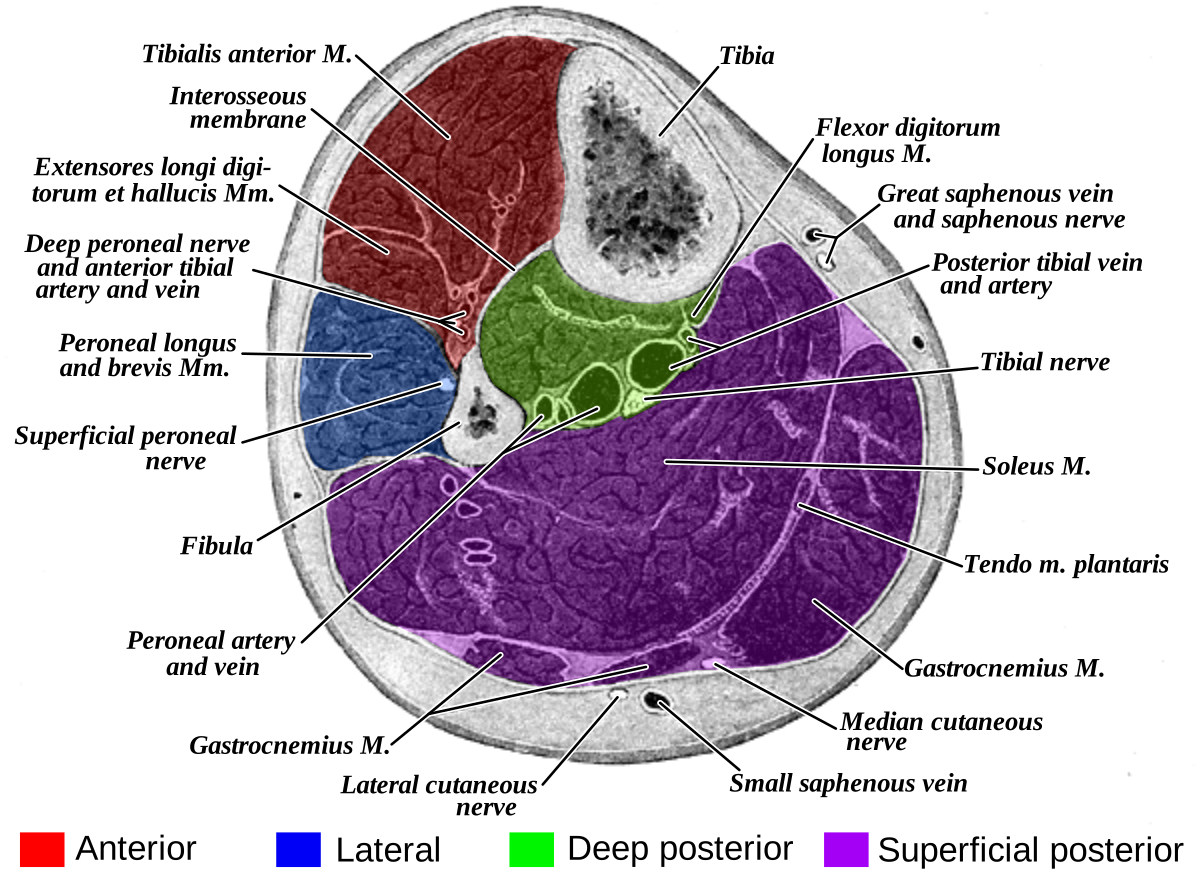
| Compartment | Key muscles | Nerve | Passive stretch test |
|---|---|---|---|
| Anterior | Tibialis anterior, extensor hallucis longus, extensor digitorum longus | Deep peroneal nerve | Passive plantarflexion of the toes/ankle |
| Lateral | Peroneus longus and brevis | Superficial peroneal nerve | Passive inversion of the ankle |
| Superficial posterior | Gastrocnemius, soleus | Sural nerve (sensory) | Passive dorsiflexion of the ankle |
| Deep posterior | Flexor digitorum longus, flexor hallucis longus, tibialis posterior | Tibial nerve | Passive dorsiflexion of the toes/ankle |
Complications
Outcome is determined almost entirely by how quickly the diagnosis is made and acted upon. The complications below are largely preventable with prompt recognition and fasciotomy, and largely inevitable without it.
- Volkmann's ischaemic contracture - fixed flexion contracture of the fingers, wrist and sometimes elbow following unrecognised forearm compartment syndrome, caused by replacement of infarcted muscle with inelastic fibrous tissue. It is a life-changing, largely irreversible deformity, which is precisely why early recognition and fasciotomy of the forearm are so heavily emphasised.
- Rhabdomyolysis and myoglobinuria - release of myoglobin and intracellular contents from necrotic muscle, causing pigment-induced acute kidney injury; managed with aggressive intravenous fluid resuscitation and monitoring of renal function and potassium8
- Hyperkalaemia - from cell lysis, with risk of cardiac arrhythmia, particularly on reperfusion of a previously ischaemic compartment
- Permanent nerve damage - sensory loss and motor weakness in the distribution of nerves that ran through the affected compartment, from prolonged ischaemia
- Chronic pain and contracture even after fasciotomy, if decompression was delayed
- Infection, including of open fasciotomy wounds and, in severe cases, of necrotic muscle
- Amputation - required in severe or significantly delayed cases where muscle necrosis is extensive and the limb is not salvageable or viable tissue would not sustain function
- Death - from the systemic effects of severe rhabdomyolysis, hyperkalaemia and reperfusion injury, particularly after crush injury or prolonged limb ischaemia affecting a large muscle mass
Red flags
References
- BOAST - The Management of Severe Open Lower Limb Fractures. British Orthopaedic Association Standards for Trauma. Available here
- BOAST - Diagnosis and Management of Compartment Syndrome of the Limbs. British Orthopaedic Association Standards for Trauma. Available here
- NICE NG38. Fractures (complex): assessment and management. 2016. Available here
- NICE NG39. Major trauma: assessment and initial management. 2016. Available here
- Whitesides TE, Haney TC, Morimoto K, Harada H. Tissue pressure measurements as a determinant for the need of fasciotomy. Clinical Orthopaedics and Related Research. 1975. Available here
- McQueen MM, Court-Brown CM. Compartment monitoring in tibial fractures: the pressure threshold for decompression. Journal of Bone and Joint Surgery (Br). 1996. Available here
- Solomon L, Warwick D, Nayagam S. Apley and Solomon's System of Orthopaedics and Trauma. 10th edition. Available here
- BNF - Rhabdomyolysis and acute kidney injury management principles. Available here
- Orthobullets. Acute Compartment Syndrome. Available here
This article is written for revision and education. It is not clinical guidance and must not be used to make decisions about the care of a patient. Always check current NICE guidance and local protocols.