
Neck of Femur Fracture: Assessment and Management
Key points
- Typical patient: an elderly, often osteoporotic patient after a low-energy fall, with groin pain and inability to weight-bear.
- Classic sign: a shortened and externally rotated leg, caused by the unopposed pull of the short external rotators once the femoral neck is discontinuous.
- Anatomy that matters: the femoral head is supplied retrogradely by retinacular vessels from the medial femoral circumflex artery, so a displaced intracapsular fracture risks avascular necrosis.
- Classification: intracapsular (subcapital, transcervical) vs extracapsular (intertrochanteric, subtrochanteric), split relative to the capsular attachment at the intertrochanteric line.
- Garden classification: grades I-IV describe intracapsular fractures by displacement; the key clinical split is undisplaced (I-II) vs displaced (III-IV).
- Imaging: AP pelvis and lateral hip X-rays first, checking Shenton's line; MRI (or CT if MRI contraindicated) if plain films are normal but suspicion remains.
- NICE surgical window: surgery on the day of or the day after admission, and always within 36 hours, alongside orthogeriatric assessment from admission.
- Operation choice: fixation for undisplaced intracapsular fractures, arthroplasty for displaced intracapsular fractures, DHS for intertrochanteric fractures, and IM nail for subtrochanteric fractures.
Introduction
Neck of femur (NOF) fracture is one of the commonest reasons for emergency orthopaedic admission in the UK, with well over 70,000 hip fractures occurring each year. The typical patient is elderly, frail, and often has underlying osteoporosis; the injury follows a simple low-energy fall, such as a trip at home or a fall from standing height.1
A much smaller minority of hip fractures occur in young patients as the result of high-energy trauma, such as a road traffic collision or a fall from height. These fractures behave differently: the bone is normal quality, more force is required to break it, and associated injuries are more likely. The exam focus, and the bulk of clinical practice, concerns the frail elderly group.
Hip fracture matters far beyond the break itself. One-year mortality is substantial, of the order of 30%, and this figure says more about the frailty, comorbidity and functional decline of the population affected than about the fracture as an isolated injury. This is why hip fracture care in the UK is organised around a structured, time-critical, multidisciplinary pathway rather than treated as a routine orthopaedic operation, and why it is a National Institute for Cardiovascular Outcomes Research audit topic (the National Hip Fracture Database) and a NICE quality standard.1,2
Anatomy and blood supply
The femoral head receives its blood supply almost entirely retrogradely, running up the femoral neck towards the head. The dominant supply comes from the retinacular (ascending cervical) vessels, branches of the medial femoral circumflex artery, which run along the femoral neck within the capsule before piercing the bone just below the articular cartilage of the head. The lateral femoral circumflex artery makes a much smaller contribution.3
A small additional supply reaches the femoral head directly through the artery of the ligamentum teres, a branch of the obturator artery. In adults this vessel is inconsistent and typically contributes very little; it is more functionally important in children, in whom it can partially sustain the femoral head after a proximal fracture.
The practical significance of this arrangement is entirely about the joint capsule. The hip capsule attaches proximally to the acetabular rim and distally along the intertrochanteric line anteriorly, and further up the neck posteriorly. The retinacular vessels travel within this capsule, running up the femoral neck to reach the head. A fracture that occurs within the capsule (intracapsular) and is displaced disrupts these vessels, cutting off the retrograde supply to the femoral head and leaving it at high risk of avascular necrosis (AVN). A fracture that occurs outside the capsule (extracapsular) leaves the retinacular vessels intact, because the break is distal to where they run, so the blood supply to the femoral head is preserved and AVN is not a significant risk.
Classification
Hip fractures are first divided anatomically, relative to the hip joint capsule, and this division is what drives the surgical decision more than any other single factor.
| Type | Location | Subtypes |
|---|---|---|
| Intracapsular | Subcapital to transcervical, within the hip capsule, proximal to the intertrochanteric line | Subcapital, transcervical (basicervical fractures are sometimes classed as extracapsular) |
| Extracapsular | Distal to the capsular attachment | Intertrochanteric (between greater and lesser trochanters); subtrochanteric (below the lesser trochanter, within about 5 cm) |
Garden classification (intracapsular fractures)
The Garden classification grades intracapsular fractures by the degree of displacement seen on the AP radiograph, and is the classification most often quoted in exams.4
| Grade | Description | Displacement |
|---|---|---|
| I | Incomplete fracture, valgus impacted | Undisplaced |
| II | Complete fracture, undisplaced | Undisplaced |
| III | Complete fracture, partially displaced (trabeculae malaligned) | Displaced |
| IV | Complete fracture, fully displaced | Displaced |
In practice, distinguishing Garden I from II or III from IV on plain films is unreliable even for experienced surgeons, and management decisions are made on the simplified, clinically important split: undisplaced (Garden I-II) versus displaced (Garden III-IV). Undisplaced fractures have a good chance of an intact blood supply and are treated with fixation; displaced fractures are assumed to have disrupted the retinacular vessels and are treated with arthroplasty.
Extracapsular fractures
Extracapsular fractures are simply described by their location - intertrochanteric or subtrochanteric - and further characterised by the number of fragments (two-part vs comminuted) and by stability, particularly whether the posteromedial cortex is intact, which influences the choice of implant.
Clinical features
The history is almost always a fall, usually a simple low-energy mechanical fall in an elderly patient, followed by an inability to stand up or bear weight. Pain is typically felt in the groin or the hip, and may radiate to the knee. Occasionally a patient with an impacted or undisplaced fracture can still weight-bear, walking on a painful hip, so a normal gait does not exclude the diagnosis.
As above, always establish why the patient fell - mechanical (tripped, slipped, missed a step) versus a possible syncopal or collapse episode - and screen for cardiac causes (arrhythmia, postural hypotension, aortic stenosis), neurological causes (stroke, seizure) and other precipitants (infection, hypoglycaemia, polypharmacy, alcohol). A collateral history from family, carers or paramedics is often more reliable than the patient's own account, particularly if there is any cognitive impairment.
Examination
- Look: the affected leg is classically shortened and externally rotated compared with the other side
- Feel: groin and greater trochanter tenderness; check distal neurovascular status
- Move: pain on any attempted hip movement, particularly rotation; inability to straight-leg raise
- Assess for associated injuries from the fall, especially of the wrist, proximal humerus and spine
- A full 'top-to-toe' examination looking for the cause of the fall - cardiovascular, neurological and postural blood pressure
- Assess pressure areas, cognitive state (confusion screen/AMT4 or 4AT) and pre-fracture mobility and functional baseline
Investigations
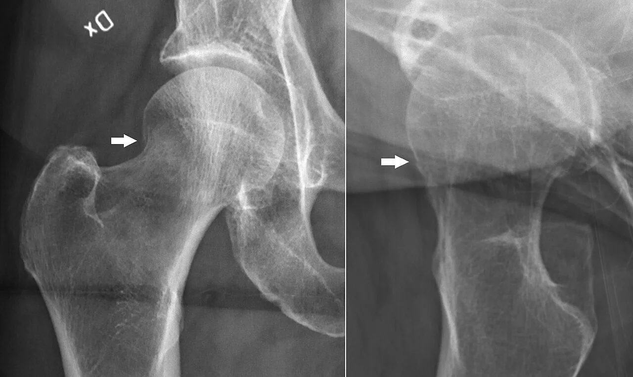
Plain radiographs are first-line: an AP view of the pelvis and a lateral view of the affected hip. The AP pelvis allows comparison with the contralateral, uninjured side, which is invaluable when a subtle fracture is suspected.
On the AP view, trace Shenton's line - the smooth, continuous arc formed by the medial border of the femoral neck and the superior border of the obturator foramen. A break or step in this line indicates a fracture or dislocation and is a key sign to actively look for, since a non-displaced or impacted intracapsular fracture can otherwise be easy to miss on a cursory look.5
Baseline bloods (FBC, U&Es, clotting, group and save), an ECG and a chest radiograph are performed as part of preoperative work-up, alongside a search for a precipitating medical cause of the fall as above.
Initial management
Hip fracture is a medical emergency in a frail patient, and initial management follows a structured pathway from the point of admission, run jointly by orthopaedics and orthogeriatrics.
- Analgesia early and adequately - paracetamol regularly, with opioids titrated as needed. A fascia iliaca compartment block is recommended and significantly reduces opioid requirement and its side effects; it should be offered on admission or in the emergency department.1
- Pressure area care - hip fracture patients are at high risk of pressure sores from prolonged time on hard surfaces (floor, trolley, theatre table) and reduced mobility; use pressure-relieving mattresses and reposition regularly
- VTE risk assessment and mechanical prophylaxis (and pharmacological once the bleeding risk of imminent surgery has passed)
- Urinary catheterisation only if clinically indicated (for example acute retention or to monitor output in a haemodynamically unstable patient) - not performed routinely, as it increases infection risk
- IV fluids if the patient is dehydrated or has been on the floor for a prolonged period (consider rhabdomyolysis and acute kidney injury with a long lie)
- Early orthogeriatric assessment, ideally within 72 hours and often on the day of admission, to identify and correct reversible comorbidities (anaemia, dehydration, electrolyte disturbance, uncontrolled AF, anticoagulation) before surgery
- Nutrition and early mobilisation planning started from admission
Definitive surgical management
The choice of operation is determined chiefly by the anatomical classification and the displacement of the fracture, since these dictate whether the femoral head's blood supply is likely to be intact.
| Fracture | Operation | Rationale |
|---|---|---|
| Intracapsular, undisplaced (Garden I-II) | Internal fixation with cannulated screws | Blood supply likely intact; preserves the native femoral head |
| Intracapsular, displaced (Garden III-IV) | Hemiarthroplasty or total hip replacement (see below) | Blood supply assumed disrupted; native head is at high risk of AVN |
| Extracapsular, intertrochanteric | Dynamic hip screw (DHS) | Stable extracapsular pattern amenable to a sliding screw-and-plate construct; blood supply preserved |
| Extracapsular, subtrochanteric | Intramedullary (IM) nail | High biomechanical stress at this level favours a load-sharing intramedullary implant over a DHS |
Undisplaced intracapsular fractures
Internal fixation, typically with parallel cannulated screws, is used because the blood supply to the femoral head is likely to be preserved. This avoids the morbidity of joint replacement and preserves the patient's own femoral head. There remains a real, if lower, risk of subsequent AVN or fixation failure, and patients should be counselled that a second operation (conversion to arthroplasty) may be needed if this occurs.
Displaced intracapsular fractures
Displaced intracapsular fractures are treated with arthroplasty rather than fixation, because the risk of AVN and non-union with fixation is unacceptably high. The choice between hemiarthroplasty (replacing only the femoral head) and total hip replacement (THR) (replacing both the femoral head and the acetabulum) is guided by NICE criteria balancing surgical durability against operative risk.1
NICE recommends offering a total hip replacement to patients who, before the fracture, were able to walk independently outdoors with no more than the use of a single stick, are not cognitively impaired, and are medically fit for the anaesthetic and the longer surgical procedure. THR gives better functional outcomes and a lower revision rate over time, at the cost of a longer operation, greater blood loss and a higher dislocation rate than hemiarthroplasty.
Hemiarthroplasty is preferred for patients who are older, less mobile before the fracture, cognitively impaired, or who have significant comorbidity that makes a longer procedure higher risk. It is a shorter, technically simpler operation with a lower dislocation rate, and is the appropriate choice for the majority of frail patients with displaced intracapsular fractures.
Non-operative management
A small minority of very frail patients with extremely limited life expectancy, or in whom the risks of any anaesthetic and surgery are judged to outweigh the benefits, may be managed non-operatively with a focus on analgesia, comfort and palliation. This decision should involve the patient (or their advocate), family and the multidisciplinary team, and is not taken lightly given that unrelieved pain from an unfixed fracture is itself a significant burden.
Secondary prevention
Every patient with a fragility hip fracture should be treated as having confirmed osteoporosis until proven otherwise, and secondary prevention should be started before discharge, not deferred to primary care.
- Falls risk assessment - review of medications (particularly sedatives and antihypertensives), vision, footwear, home hazards and gait, ideally with input from physiotherapy and occupational therapy
- Bone health assessment - DEXA scanning to quantify bone mineral density, although treatment is often started empirically without waiting for the result
- Vitamin D and calcium supplementation, correcting any deficiency
- Bone-protective therapy - a bisphosphonate (e.g. alendronic acid) first-line; alternatives such as denosumab, teriparatide or zoledronic acid where bisphosphonates are unsuitable or not tolerated
- Referral to a falls clinic or falls prevention service for ongoing multidisciplinary input
- Address the precipitant identified during the admission - cardiac, neurological or medication-related - rather than treating the fracture in isolation
Complications
Complications can be split into those specific to the fracture and fixation, and the systemic complications of surgery and immobility that dominate outcomes in this frail population.
- Avascular necrosis - specific to intracapsular fractures with disrupted blood supply; may present months to years later with collapse of the femoral head and pain
- Non-union or mal-union - failure of the fracture to heal, or healing in a poor position, more common after fixation of displaced or unstable fractures
- Implant failure - cut-out of screws, breakage of a DHS or nail, particularly in poor-quality osteoporotic bone
- Dislocation - of a hemiarthroplasty or THR, more common with the posterior surgical approach and in the early postoperative period
- Infection - superficial or deep surgical site infection, and periprosthetic joint infection, which is a serious complication often requiring revision surgery
- Leg length discrepancy, particularly after arthroplasty
- Systemic complications of surgery and immobility: hospital-acquired pneumonia, pressure sores, delirium, urinary tract infection, venous thromboembolism, and deconditioning with loss of independence
Prognosis
Hip fracture carries substantial mortality: approximately 8-10% of patients die within 30 days, and roughly 30% within one year. This reflects the burden of frailty, comorbidity and reduced physiological reserve in the population affected, rather than the fracture being intrinsically lethal - hip fracture is often described as a marker of, rather than solely the cause of, poor prognosis in a frail patient.1,6
Factors associated with worse outcome include increasing age, male sex, significant comorbidity, cognitive impairment, poor pre-fracture mobility, delay to surgery beyond the recommended window, and living in institutional care before the fracture.
Functional recovery is often incomplete. A substantial proportion of previously independent patients do not regain their pre-fracture level of mobility, and many who lived independently before the fracture require increased support or residential care afterwards. This is why the multidisciplinary, orthogeriatric-led pathway - covering surgery, medical optimisation, early mobilisation, rehabilitation and secondary prevention - is central to modern hip fracture care rather than the operation being viewed in isolation.
References
- NICE CG124. Hip fracture: management. 2011, updated 2023. Available here
- NICE QS16. Hip fracture in adults. Available here
- Lieberman JR, Berry DJ (eds). AAOS Comprehensive Orthopaedic Review - Hip fractures and blood supply of the femoral head. Available here
- Garden RS. Low-angle fixation in fractures of the femoral neck. Journal of Bone and Joint Surgery (Br). 1961. Available here
- Shenton EW. Disease in bone and its detection by X-rays. British Medical Journal. 1911. Available here
- Royal College of Physicians. National Hip Fracture Database Annual Report. Available here
- British Orthopaedic Association. BOAST - Care of the Older or Frail Orthopaedic Trauma Patient. Available here
- BNF. Bisphosphonates - osteoporosis treatment. Available here
This article is written for revision and education. It is not clinical guidance and must not be used to make decisions about the care of a patient. Always check current NICE guidance and local protocols.