
Pressure Sores
Key points
- Pressure sores (pressure ulcers/injuries): localised damage to skin and underlying tissue from sustained pressure, shear or friction, typically over a bony prominence, in patients with reduced mobility or sensation.
- Mechanism: sustained pressure exceeding capillary closing pressure causes local tissue ischaemia; shear forces further distort and occlude vessels, which is why damage can be worse than the visible skin surface suggests.
- Common sites: the sacrum, heels, ischial tuberosities, greater trochanters and occiput - anywhere bone lies close to the skin under sustained load.
- Staging: graded from stage 1 (non-blanching erythema with intact skin) to stage 4 (full-thickness loss with exposed bone, muscle or tendon), plus unstageable and deep tissue injury categories.
- Key risk factors: immobility, reduced sensation, malnutrition, incontinence/moisture, reduced consciousness and vascular disease - captured in structured risk assessment tools such as the Waterlow score.
- Prevention: the cornerstone of management - regular repositioning, pressure-relieving mattresses and cushions, skin inspection, moisture management and adequate nutrition, bundled together as the 'SSKIN' approach.
- Management: complete pressure relief from the affected area, dressing selection matched to stage and exudate, debridement of necrotic tissue, and surgical reconstruction for severe, non-healing stage 3-4 ulcers.
- Red flag: increasing pain, spreading erythema, malodour or systemic upset in a deep pressure sore should raise concern for osteomyelitis or sepsis rather than being managed as routine wound care.
Introduction
Pressure sores (also called pressure ulcers or pressure injuries) are areas of localised damage to the skin and underlying tissue caused by sustained pressure, shear or friction, almost always over a bony prominence. They occur in patients whose mobility, sensation or overall condition prevents them from naturally shifting their position to relieve pressure - critically ill patients, those with spinal cord injury, and frail older adults are all at particular risk.1
Pressure sores are largely preventable, and a very large proportion of NHS spending on wound care goes towards a condition that structured prevention programmes can substantially reduce. This makes prevention, rather than treatment after the fact, the central theme of this topic - and a marker of care quality that is now formally measured across NHS settings.2
Pathophysiology
Skin and subcutaneous tissue depend on capillary blood flow for oxygen and nutrient delivery. When external pressure on the tissue exceeds the capillary closing pressure (normally around 32 mmHg), local blood flow is occluded, causing tissue ischaemia. If this pressure is sustained for long enough - as happens when an immobile patient remains in one position - the ischaemic tissue progresses to necrosis.
Shear forces, generated when the skin is dragged over an underlying surface (for example when a patient slides down in a bed or chair), distort and stretch blood vessels at an angle, further impairing perfusion even at lower absolute pressures. Friction damages the superficial epidermis directly, and moisture (from incontinence or sweating) softens the skin and makes it more vulnerable to both friction and shear. All four - pressure, shear, friction and moisture - interact, which is why prevention strategies address them together rather than pressure alone.
Risk factors
- Immobility - critical illness, sedation, spinal cord injury, post-operative patients, and general frailty in older adults6
- Reduced sensation - peripheral neuropathy, spinal cord injury, or reduced consciousness, all of which blunt the normal discomfort that prompts a person to shift position
- Malnutrition and low body weight - impairs tissue resilience and wound healing capacity
- Incontinence and moisture - macerates and weakens the skin
- Vascular disease - reduces the tissue's tolerance of any additional ischaemic insult
- Older age, reflecting a combination of thinner skin, reduced subcutaneous fat over bony prominences, and comorbidity
- Previous pressure sore - a strong predictor of further episodes
Structured risk assessment tools, such as the Waterlow score, combine these factors into a single score used on admission and periodically thereafter to identify patients needing more intensive preventive measures.4
Clinical features and staging
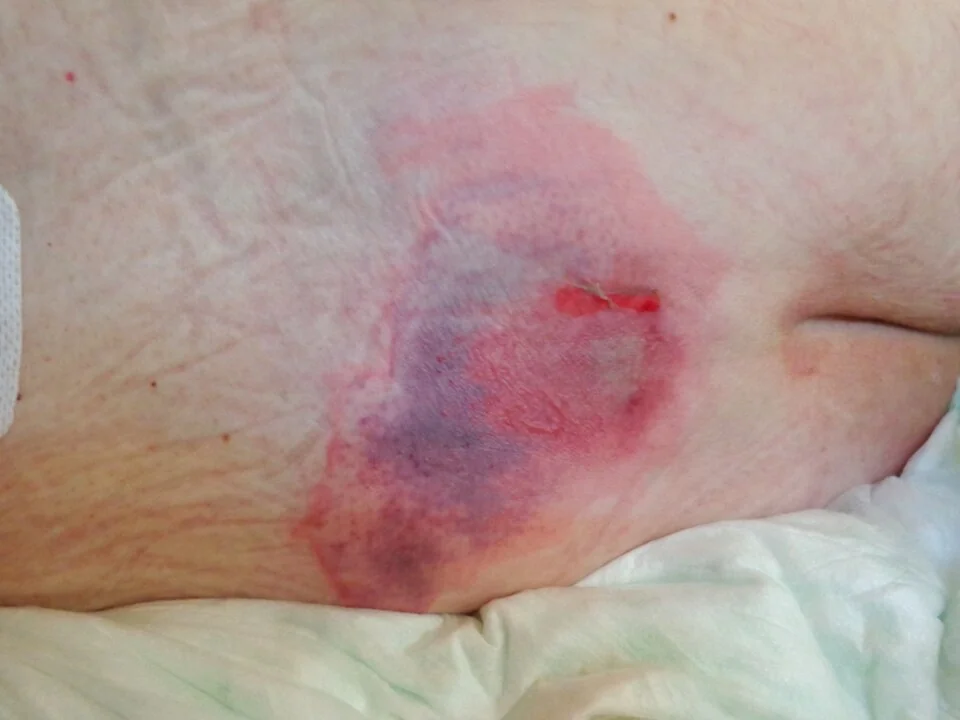
| Stage | Features |
|---|---|
| Stage 1 | Intact skin with non-blanching erythema over a bony prominence; may be painful, firm, warmer or cooler than surrounding skin |
| Stage 2 | Partial-thickness skin loss with exposed dermis, or an intact or ruptured serum-filled blister |
| Stage 3 | Full-thickness skin loss, with visible subcutaneous fat but no exposed bone, tendon or muscle; may include undermining and tunnelling |
| Stage 4 | Full-thickness skin and tissue loss with exposed or directly palpable bone, tendon or muscle |
| Unstageable | Full-thickness loss obscured by slough or eschar, so the true depth cannot be determined until debrided |
| Deep tissue pressure injury | Intact or non-intact skin with a localised area of persistent, non-blanchable deep red, maroon or purple discolouration, reflecting damage to underlying soft tissue that may not yet be visible at the surface |
Pressure sores occur almost exclusively over bony prominences - the sacrum and heels are the commonest sites, followed by the ischial tuberosities (particularly in seated patients), greater trochanters, elbows and occiput.
Clinical examination
- Regular, systematic skin inspection of all pressure-bearing areas, particularly the sacrum and heels, in any patient identified as at-risk
- Blanching test - press gently on reddened skin; skin that blanches (turns white) and refills is at an earlier, reversible stage than skin that remains persistently red (non-blanching)
- Depth, undermining and tunnelling - assessed once a wound has developed, to stage it accurately and guide dressing choice
- Signs of infection - increasing pain, spreading erythema, malodour, purulent discharge, or systemic signs (fever, tachycardia, confusion)
- Nutritional assessment - weight, appetite, and screening tools (for example MUST), since malnutrition both predisposes to and impairs healing of pressure sores
Differential diagnosis
- Moisture-associated skin damage (incontinence-associated dermatitis) - affects skin folds and perineal skin exposed to urine or faeces, generally more diffuse and following the pattern of moisture exposure rather than being confined to a bony prominence
- Skin tears - traumatic, often in fragile older skin, from friction or shearing injury rather than sustained pressure, and not necessarily over a bony prominence
- Arterial or venous leg ulcers - occur at characteristic lower leg sites rather than over pressure-bearing bony prominences; see Skin Ulcers
- Fungal intertrigo - affects skin folds rather than bony prominences, with associated erythema and satellite lesions
Investigations
Pressure sores are staged clinically. Further investigation is directed at identifying complications or contributing factors rather than confirming the diagnosis.
- Wound swab - only if clinical infection is suspected, since chronic wounds are normally colonised with bacteria and swabbing an uninfected wound adds little
- Blood cultures and inflammatory markers - if systemic infection is suspected
- Imaging (X-ray, MRI) and bone biopsy - if osteomyelitis is suspected in a deep (stage 3-4) sore, particularly one that fails to improve with standard wound care
- Nutritional bloods (albumin, prealbumin, vitamin and mineral levels) - to guide nutritional support in patients with poor healing
Management
Prevention - the SSKIN bundle
A structured approach used across NHS settings to reduce pressure sore incidence.5
- S - Surface - appropriate pressure-relieving mattresses and cushions for at-risk patients8
- S - Skin inspection - regular, systematic checking of vulnerable areas
- K - Keep moving - repositioning at regular intervals (commonly every 2-4 hours, individualised to risk), and encouraging mobility wherever possible
- I - Incontinence/moisture - prompt management of incontinence and skin protection to reduce moisture-related skin damage
- N - Nutrition/hydration - adequate nutrition to support tissue resilience and wound healing
Treatment of an established pressure sore
- Complete relief of pressure from the affected area - the single most important step, without which no dressing or treatment will allow healing
- Dressing selection matched to the wound - hydrocolloid or film dressings for stage 1-2, and more absorptive dressings (foam, alginate) for exudative stage 3-4 wounds
- Debridement of necrotic tissue or slough, surgical, sharp, enzymatic or autolytic, to allow accurate staging and healing
- Negative pressure wound therapy - can be used for selected deeper wounds to promote granulation
- Treat infection - systemic antibiotics for confirmed clinical infection or osteomyelitis, not for colonisation alone
- Nutritional optimisation, involving dietetic input for malnourished patients
- Surgical reconstruction (flap closure) - considered for severe, non-healing stage 3-4 ulcers, particularly in patients with spinal cord injury
Complications
- Local and spreading infection, including cellulitis
- Osteomyelitis - a significant risk with deep, longstanding stage 3-4 sores, particularly over the sacrum and ischial tuberosities
- Sepsis - a real risk from an infected, deep pressure sore, particularly in already frail patients
- Chronic non-healing wounds and sinus tracts
- Malignant transformation (Marjolin ulcer) - squamous cell carcinoma arising within a chronic, longstanding pressure sore, though uncommon7
- Prolonged hospital stay and reduced quality of life, and a recognised association with increased mortality in frail patients with severe pressure sores
Red flags
Prognosis
Stage 1 and 2 pressure sores generally heal well once pressure is relieved and appropriate wound care is provided, often within weeks. Stage 3 and 4 sores take considerably longer, sometimes months, and a proportion never fully heal, particularly in patients with ongoing risk factors (persistent immobility, malnutrition, vascular disease) that cannot be fully reversed.3
The outlook is closely tied to the patient's overall condition rather than the wound in isolation: pressure sores are frequently a marker of significant underlying frailty or critical illness, and their presence - particularly at a higher stage - is associated with worse overall outcomes in hospitalised and long-term care populations. This reinforces why structured prevention, rather than treatment after a sore has developed, remains the central priority in this area of care.
References
- NICE CG179. Pressure ulcers: prevention and management. 2014. Available here
- NICE QS89. Pressure ulcers. 2015. Available here
- European Pressure Ulcer Advisory Panel, National Pressure Injury Advisory Panel, Pan Pacific Pressure Injury Alliance. Prevention and Treatment of Pressure Ulcers/Injuries: Clinical Practice Guideline. 2019. Available here
- Waterlow J. Pressure sores: a risk assessment card. Nursing Times. 1985. Available here
- NHS England. Pan-UK pressure ulcer position statement. 2018. Available here
- Coleman S, Gorecki C, Nelson EA et al. Patient risk factors for pressure ulcer development: systematic review. International Journal of Nursing Studies. 2013. Available here
- Kerr-Valentic MA, Samimi K, Rohlen BH et al. Marjolin's ulcer: modern analysis of an ancient problem. Plastic and Reconstructive Surgery. 2009. Available here
- McInnes E, Jammali-Blasi A, Bell-Syer SE et al. Support surfaces for pressure ulcer prevention. Cochrane Database of Systematic Reviews. 2015. Available here
This article is written for revision and education. It is not clinical guidance and must not be used to make decisions about the care of a patient. Always check current NICE guidance and local protocols.