
Burns
Key points
- Burns: thermal, chemical, electrical or radiation injuries to the skin and sometimes deeper tissue, classified by depth (superficial through to full-thickness) and by the total body surface area (%TBSA) involved.
- Depth assessment: based on appearance, capillary refill/blanching and sensation - a burn that is painful and blanches has a good chance of healing without grafting, while a painless, non-blanching, leathery burn is full-thickness and will not.
- Estimating %TBSA: the Wallace rule of nines or, more accurately, the Lund-Browder chart (which accounts for the different body proportions of children); the patient's own palm represents roughly 1% of body surface area.
- Parkland formula: guides fluid resuscitation for burns over 15-20% TBSA in adults: 4mL x body weight (kg) x %TBSA of crystalloid over 24 hours, with half given in the first 8 hours from the time of injury, not from presentation.
- Referral to a specialist burns unit: based on %TBSA, depth, site (face, hands, feet, perineum, flexures, or circumferential), patient age, suspected inhalation injury, and mechanism (chemical, electrical, or suspected non-accidental injury).
- Initial management: stop the burning process, cool the burn with cool running water for 20 minutes (most effective within 3 hours of injury), remove constricting items before swelling develops, and cover with cling film.
- Circumferential burns: can restrict chest expansion or cause limb compartment syndrome as swelling develops beneath inelastic burnt skin, sometimes requiring urgent escharotomy.
- Red flag: facial burns, singed nasal hair, soot in the mouth or a hoarse voice suggest inhalation injury and airway compromise; a burn pattern inconsistent with the given history should prompt safeguarding assessment, especially in a child.
Introduction
Burns are injuries to the skin, and sometimes deeper tissue, caused by heat, chemicals, electricity or radiation. They range from minor, self-limiting injuries managed entirely in primary care to major, life-threatening trauma needing resuscitation and specialist burns unit care.1
Management follows a consistent structure regardless of cause: assess depth and extent, provide appropriate first aid and resuscitation, and identify the features - certain sites, depths, extents, mechanisms and patient factors - that mean a burn needs specialist rather than local management. Recognising inhalation injury and considering non-accidental injury in children are two further points that carry disproportionate clinical weight relative to how often they are actually present.
Classification by mechanism
- Thermal - the commonest cause, from flame, contact with a hot object, or scalds from hot liquid or steam; scalds are the commonest mechanism in young children
- Chemical - acids and alkalis; alkali burns tend to be deeper and more progressive than acid burns because of ongoing tissue liquefaction, and eye involvement needs urgent, prolonged irrigation
- Electrical - can cause deep tissue damage disproportionate to the visible skin injury, following the path of least electrical resistance through the body, and carries a risk of cardiac arrhythmia and rhabdomyolysis
- Radiation - including sunburn at the mild end of the spectrum, and radiotherapy-related skin injury
Classification by depth
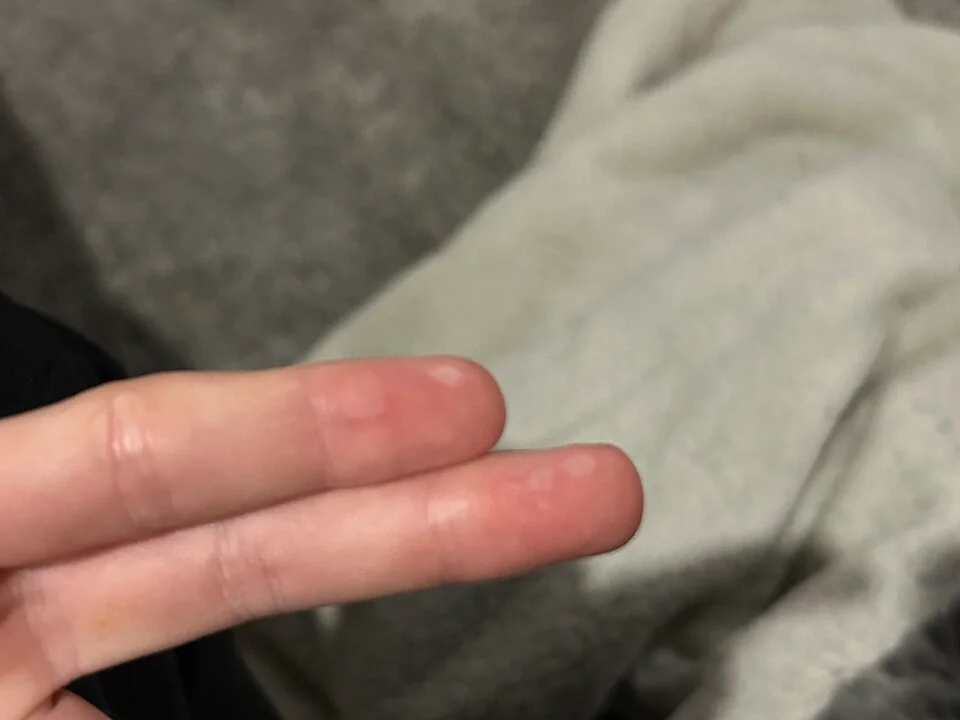
| Depth | Appearance | Sensation | Healing |
|---|---|---|---|
| Superficial (epidermal) | Red, dry, no blistering (e.g. simple sunburn) | Painful | Heals within about a week, no scarring |
| Superficial partial-thickness | Pale pink, blistered, moist, blanches with brisk capillary refill | Very painful | Usually heals within 2-3 weeks with minimal scarring |
| Deep partial-thickness | Blotchy red/white, may or may not blister, sluggish or absent blanching | Reduced sensation, less painful than superficial burns | Slow healing, often needs grafting, higher scarring risk |
| Full-thickness | White, waxy, leathery or charred; dry | Painless (nerve endings destroyed) | Will not heal without grafting; scarring and contracture likely |
Estimating extent - %TBSA
Only partial-thickness and full-thickness burns are included when calculating total body surface area (TBSA) affected - superficial (epidermal) burns, such as simple sunburn, are excluded from this calculation.
- Wallace rule of nines - each arm 9%, each leg 18%, the front and back of the trunk 18% each, the head and neck 9%, and the perineum 1%; a reasonable quick estimate in adults but inaccurate in children, whose head is proportionally larger
- Lund-Browder chart - a more accurate, age-adjusted chart that accounts for the changing body proportions of children, and is the preferred tool wherever available4
- Patient's palm (including fingers) - approximates 1% of total body surface area, useful for quickly estimating smaller or scattered burns
Clinical features suggesting inhalation injury
Inhalation injury should be actively considered in any burn from a fire in an enclosed space, and is suggested by:
- Burns to the face, singed eyebrows or nasal hair
- Soot around the mouth or nose, or carbonaceous sputum
- Hoarseness of voice, stridor, or a brassy cough
- History of being in an enclosed space with smoke or fire
- Any suggestion of reduced consciousness at the scene
Clinical examination
- Airway, breathing, circulation (ABCDE) approach first, since major burns are a form of trauma and airway compromise from inhalation injury can develop rapidly and unpredictably
- Depth and extent - assessed and documented using the tools above, ideally after any adherent clothing or jewellery has been carefully removed
- Circumferential burns - specifically assess any burn that encircles a limb, digit or the chest, given the risk of compartment syndrome or restricted ventilation as swelling develops
- Distal neurovascular status in any limb with a circumferential or deep burn
- Pattern and distribution - a burn pattern inconsistent with the history given, sharply demarcated 'glove and stocking' scalds, or old healed burns/other injuries in a child should prompt safeguarding consideration7
Differential diagnosis and mimics
- Toxic epidermal necrolysis - widespread skin detachment that can superficially resemble a burn, but with a drug-related history, mucosal involvement and a positive Nikolsky sign; see Stevens-Johnson Syndrome and Toxic Epidermal Necrolysis
- Staphylococcal scalded skin syndrome - toxin-mediated superficial skin detachment in young children, without a history of thermal exposure; see Impetigo
- Bullous impetigo or other blistering skin conditions - lack the clear history of heat, chemical or electrical exposure
Investigations
- Carboxyhaemoglobin level and arterial blood gas - if inhalation injury or carbon monoxide poisoning is suspected
- FBC, U&Es, glucose - baseline bloods, particularly important before and during large-volume fluid resuscitation
- Creatine kinase and urine myoglobin - in electrical burns, given the risk of significant deep tissue damage and rhabdomyolysis not reflected by the surface appearance
- Bronchoscopy - considered in suspected significant inhalation injury to directly assess the airway
- Cross-match - for extensive burns where surgical debridement or grafting is anticipated
Management
Immediate first aid
- Stop the burning process - remove the person from the source, extinguish flames, remove smouldering or chemical-soaked clothing
- Cool the burn with cool (not iced) running water for 20 minutes, most effective if started within 3 hours of injury; this reduces depth of injury and pain6
- Remove jewellery, watches and constricting clothing early, before swelling makes this difficult or dangerous
- Cover the burn with cling film (applied in layers rather than wrapped circumferentially) or a clean dressing, which reduces pain and contamination without adhering to the wound
- Avoid ice, butter or other home remedies, which can worsen tissue damage
- Analgesia, since burns - particularly partial-thickness burns - are extremely painful
Fluid resuscitation
Major burns (generally over 15-20% TBSA in adults, or a lower threshold in children) need formal intravenous fluid resuscitation, calculated using the Parkland formula:3
Total fluid in 24 hours (mL) = 4 x body weight (kg) x %TBSA burned, with half given in the first 8 hours from the time of injury (not from the time of arrival in hospital, which is a common and important source of error) and the remaining half over the following 16 hours. Fluid is titrated against urine output (a common target is 0.5mL/kg/hour in adults) rather than following the calculated volume rigidly.
Wound management
- Superficial and superficial partial-thickness burns - appropriate non-adherent dressings, with review to confirm healing progress
- Deep partial-thickness and full-thickness burns - typically need specialist assessment for debridement and skin grafting
- Escharotomy - surgical incision through full-thickness, inelastic burnt skin (eschar) encircling a limb or the chest, performed urgently where swelling beneath the eschar threatens the circulation to a limb or adequate chest wall movement
- Tetanus prophylaxis - reviewed and updated according to immunisation status and wound type
- Analgesia, nutrition and psychological support - major burns place a substantial metabolic demand on the body and carry significant psychological impact, both needing proactive management alongside the wound itself
Referral to a specialist burns service
Guided by national referral criteria, broadly including: burns over a threshold %TBSA (varying with age), full-thickness burns of any significant size, burns to the face, hands, feet, perineum, genitalia or major flexures, circumferential burns, chemical or electrical burns, suspected inhalation injury, burns in very young or very old patients, and any burn where non-accidental injury is suspected.2
Complications
- Hypovolaemic shock - from major fluid loss through the burn wound if resuscitation is inadequate or delayed
- Infection and sepsis - loss of the normal skin barrier over a large area is a major infection risk
- Inhalation injury and airway compromise - can progress over hours, so a normal initial airway examination does not exclude evolving injury
- Compartment syndrome - from circumferential limb burns and the swelling that follows
- Hypothermia - from major fluid and heat loss through damaged skin, particularly relevant during initial assessment and resuscitation
- Curling's ulcer - stress-related gastric ulceration associated with major burns
- Contractures and hypertrophic/keloid scarring - particularly over joints and flexures, sometimes needing later reconstructive surgery
- Significant psychological impact, including post-traumatic stress, particularly after major or disfiguring burns
Red flags
Prognosis
Outcome depends heavily on age, %TBSA burned, depth, and the presence of inhalation injury, which independently worsens prognosis for a given burn size. Small superficial and superficial partial-thickness burns generally heal well with minimal long-term consequence.5
Major burns, particularly full-thickness burns over a large %TBSA or with inhalation injury, carry significant mortality risk and, in survivors, a high burden of long-term physical (contractures, scarring, chronic pain) and psychological sequelae, which is why specialist burns unit care - combining surgical, medical, nursing, physiotherapy and psychological expertise - produces better outcomes than management outside a dedicated service for anyone meeting the referral criteria above.
References
- NICE Clinical Knowledge Summaries. Burns and scalds. Available here
- British Burn Association. National Burn Care Referral Guidance. 2012. Available here
- Baxter CR, Shires T. Physiological response to crystalloid resuscitation of severe burns. Annals of the New York Academy of Sciences. 1968. Available here
- MDCalc. Lund-Browder Chart for Burn Injuries. Available here
- ISBI Practice Guidelines Committee. ISBI Practice Guidelines for Burn Care. Burns. 2016. Available here
- Wood FM, Phillips M, Jovic T et al. Water first aid is beneficial in humans post-burn: evidence from a bi-national cohort study. PLoS One. 2016. Available here
- Maguire S, Moynihan S, Mann M et al. A systematic review of the features that indicate intentional scalds and burns in children. Burns. 2008. Available here
- Walker PF, Buehner MF, Wood LA et al. Diagnosis and management of inhalation injury: an updated review. Critical Care. 2015. Available here
This article is written for revision and education. It is not clinical guidance and must not be used to make decisions about the care of a patient. Always check current NICE guidance and local protocols.