
Lung Abscess: Cavitation, Foul Sputum and the Cause Behind It
Key points
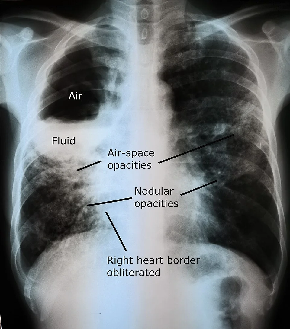
- Lung abscess: localised necrosis of lung parenchyma producing a cavity filled with pus, typically more than 2 cm across and containing an air-fluid level.
- Commonest mechanism: aspiration of oropharyngeal contents, which is why the classic patient has impaired consciousness, poor dentition or a swallowing disorder.
- Site: follows gravity - the posterior segment of the right upper lobe and the superior segment of the right lower lobe in a supine patient, and the right lower lobe when upright.
- Organisms: anaerobes above all, plus the Streptococcus anginosus group, Staphylococcus aureus and Klebsiella pneumoniae.
- Classic symptom: a subacute illness over weeks with fever, weight loss and copious foul-smelling or putrid sputum, which is highly suggestive of anaerobic infection.
- Imaging: a thick-walled cavity with an air-fluid level on the chest radiograph; CT distinguishes an abscess from an empyema and identifies an underlying obstruction.
- Treatment: prolonged antibiotics with anaerobic cover for 4 to 6 weeks or longer, guided by radiological resolution, together with postural drainage and dental treatment.
- The question always to ask: why did this happen? Exclude an obstructing bronchial carcinoma, especially in a smoker over 50 with no aspiration risk factors.
Introduction
A lung abscess is a localised collection of pus within a cavity formed by necrosis of lung parenchyma. By convention the term is applied to a single cavity greater than 2 cm; multiple smaller cavities are described as necrotising pneumonia, and the two lie on the same pathological spectrum.
Abscesses are conventionally divided into:
- Primary - arising in otherwise normal lung, almost always following aspiration. This accounts for the large majority.
- Secondary - arising because of an underlying problem: bronchial obstruction by tumour or foreign body, septic emboli from right-sided endocarditis, bronchiectasis, an infected bulla, immunosuppression, or direct spread from a subphrenic or hepatic abscess
The incidence has fallen substantially since the introduction of antibiotics, and it is now most often seen in people with alcohol dependence, poor dentition, neurological swallowing impairment, diabetes or immunosuppression. Because it is uncommon and the presentation is subacute, it is frequently misdiagnosed for weeks as unresolving pneumonia, tuberculosis or malignancy.
Aetiology
Why aspiration explains the anatomy
Aspirated material travels down the path of least resistance and then settles under gravity, which is why abscesses occur in predictable places. The right lung is affected more often because the right main bronchus is wider, shorter and more vertical than the left.
| Position when aspirating | Segment affected |
|---|---|
| Supine - the commonest, in an unconscious or anaesthetised patient | Posterior segment of the right upper lobe, and the superior (apical) segment of the right lower lobe |
| Upright | Basal segments of the right lower lobe |
| Lying on one side | Dependent segments of that side |
Risk factors
- Reduced conscious level - alcohol intoxication, drug misuse, seizures, general anaesthesia, head injury, sedation
- Impaired swallow - stroke, bulbar and pseudobulbar palsy, motor neurone disease, Parkinson's disease, dementia
- Poor dental hygiene and periodontal disease - the reservoir of anaerobic organisms, and the reason abscesses are rare in edentulous patients unless there is an obstructing tumour
- Oesophageal disease - achalasia, strictures, reflux, pharyngeal pouch, tracheo-oesophageal fistula
- Bronchial obstruction - carcinoma, an inhaled foreign body, or extrinsic compression by nodes
- Immunosuppression - HIV, chemotherapy, corticosteroids, transplantation, poorly controlled diabetes
- Intravenous drug use - through right-sided infective endocarditis producing septic pulmonary emboli, which characteristically cause multiple peripheral cavitating nodules
- Pre-existing lung disease - bronchiectasis, cystic fibrosis, emphysematous bullae
Organisms
- Anaerobes - Peptostreptococcus, Prevotella, Bacteroides and Fusobacterium. The classic cause of the primary aspiration abscess and the source of the putrid sputum.
- Streptococcus anginosus (milleri) group - strongly associated with abscess and empyema formation
- Staphylococcus aureus - including Panton-Valentine leukocidin-producing strains, which cause a fulminant necrotising cavitating pneumonia in young previously healthy people, often after influenza, and carry a very high mortality
- Klebsiella pneumoniae - upper lobe cavitation with redcurrant jelly sputum, classically in alcohol dependence and diabetes
- Pseudomonas aeruginosa and other Gram-negatives in hospital-acquired disease
- Mycobacterium tuberculosis - always in the differential of any cavity
- Nocardia and Actinomyces - subacute, in the immunosuppressed and after dental disease respectively; actinomycosis classically crosses tissue planes and produces sulphur granules
- Fungi - Aspergillus and, in the profoundly immunosuppressed, others
- Infection is often polymicrobial, particularly after aspiration
Clinical features
The presentation is characteristically subacute, evolving over one to several weeks, in contrast to the abrupt onset of pneumococcal pneumonia. This indolent course is itself a diagnostic clue, and is why patients often present having already had one or more unsuccessful courses of antibiotics.
Symptoms
- Fever, rigors and night sweats
- Cough productive of large volumes of sputum. Once the abscess ruptures into a bronchus, the patient may expectorate a large quantity of pus at once (vomica), which is dramatic and clinically important because it establishes bronchial communication.
- Foul-smelling or putrid sputum - strongly suggests anaerobic infection, and is one of the few genuinely specific clinical signs in respiratory medicine
- Weight loss, anorexia and profound malaise over weeks
- Pleuritic chest pain if the pleura is involved
- Haemoptysis - usually mild, but occasionally massive if the cavity erodes a vessel
Signs
- Fever and tachycardia
- Signs of consolidation over the affected area - dull percussion, bronchial breathing, coarse crackles
- Amphoric or cavernous breath sounds over a large superficial cavity - hollow and blowing, like air across the mouth of a bottle. Uncommon but classic.
- Finger clubbing - developing over weeks in a chronic abscess
- Poor dentition and gingival disease - examine the mouth, as it is both diagnostic and a treatment target
- Cachexia and anaemia in chronic disease
- Signs of the underlying cause - a swallowing abnormality, needle marks and a murmur suggesting endocarditis, or lymphadenopathy and cachexia suggesting malignancy
Investigations
Imaging
- Chest radiograph - a thick-walled cavity with an air-fluid level, usually surrounded by consolidation. The air-fluid level only appears once the abscess has ruptured into a bronchus, and is best seen on an erect film.
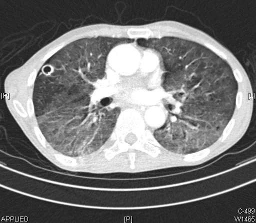
- CT thorax with contrast - more sensitive and the key investigation. It confirms the cavity, defines its size and site, identifies multiple lesions, and above all distinguishes an abscess from an empyema and reveals an underlying obstructing lesion.
| Feature | Lung abscess | Empyema |
|---|---|---|
| Shape | Spherical | Lenticular (biconvex) |
| Angle with the chest wall | Acute | Obtuse |
| Wall | Thick and irregular, with an indistinct outer margin | Thin, smooth, with the split pleura sign |
| Effect on adjacent lung | Destroys it; vessels and bronchi end abruptly at the cavity | Compresses it, with vessels displaced around the collection |
| Treatment | Antibiotics, usually without drainage | Chest drain is mandatory |
Microbiology and blood tests
- Sputum culture - including anaerobic culture, though yield is limited because sputum is contaminated by oral flora, which are the same organisms
- Blood cultures - and repeat sets if endocarditis is suspected
- Sputum for AFB smear and culture - in every cavitating lesion, since tuberculosis is the most important infective mimic
- Bronchoscopy with washings or protected specimen brush - for microbiology where the diagnosis is unclear, and essential where bronchial obstruction is suspected or the abscess is not responding
- Percutaneous or transbronchial aspiration - occasionally used in specialist centres to obtain uncontaminated material
- FBC, CRP, U&Es, LFTs and albumin - a neutrophilia with anaemia of chronic disease and a low albumin is typical
- HIV test, and consider immunodeficiency or diabetes screening
- Echocardiogram if septic emboli or endocarditis are suspected
Differential diagnosis of a lung cavity
- Cavitating bronchial carcinoma - especially squamous cell carcinoma, which cavitates most readily. A thick, irregular, nodular wall in an older smoker with fewer infective features.
- Tuberculosis - upper lobe, with fibrosis, calcification and volume loss, and a longer history of weight loss and night sweats
- Granulomatosis with polyangiitis - multiple cavitating nodules with upper airway and renal involvement, and positive c-ANCA
- Septic pulmonary emboli - multiple peripheral cavitating nodules, in intravenous drug use or with an infected line
- Pulmonary infarction - which may cavitate
- Rheumatoid nodules
- Infected emphysematous bulla - a thin wall with little surrounding consolidation
- Fungal infection - including an aspergilloma, in which a fungal ball sits within a pre-existing cavity
Management
Antibiotics
- Prolonged antibiotic treatment is the mainstay, and around 80 to 90% of abscesses resolve on antibiotics alone
- Anaerobic cover is essential. First line is usually co-amoxiclav, or clindamycin in penicillin allergy; a combination such as a cephalosporin with metronidazole is an alternative. Follow local microbiology guidance.
- Start intravenously until the patient is afebrile and improving, then switch to oral
- Continue for 4 to 6 weeks, and often longer - the endpoint is radiological resolution or a stable residual scar, not simply clinical improvement. Stopping early is the commonest cause of relapse.
- Note that metronidazole alone is inadequate - it does not cover the microaerophilic streptococci that are frequently co-pathogens
Drainage and clearance
- Postural drainage and chest physiotherapy - to help the abscess empty through the bronchus
- Percutaneous catheter drainage under image guidance - considered for large abscesses (typically above 6 to 8 cm), or where there is no response after 5 to 7 days of appropriate antibiotics
- Bronchoscopic drainage - occasionally used, and also allows the exclusion of an obstructing lesion
- Surgical resection (lobectomy or segmentectomy) - required in fewer than 10%. Indications are failure of medical treatment, massive haemoptysis, a very large abscess, suspected underlying malignancy, or a bronchopleural fistula.
- Nutritional support, correction of anaemia, and VTE prophylaxis
Treating the cause
Follow-up
- Serial chest radiographs to confirm resolution - typically at 4 to 6 weeks and then until stable. A residual thin-walled cavity or scar may persist and is acceptable if the patient is clinically well.
- Any cavity that fails to resolve requires further investigation for malignancy or tuberculosis
- Monitor CRP and clinical progress to guide the duration of treatment
Complications
- Rupture into the pleural space - producing an empyema or a pyopneumothorax, and requiring chest drainage
- Bronchopleural fistula
- Massive haemoptysis - from erosion of a bronchial artery within the cavity wall; a life-threatening emergency treated with airway protection, tranexamic acid and bronchial artery embolisation
- Metastatic infection - especially brain abscess, and also osteomyelitis and septic arthritis. New neurological symptoms in a patient with a lung abscess warrant urgent brain imaging.
- Sepsis and septic shock
- Chronic abscess with persistent cavitation, recurrent infection and clubbing
- Bronchiectasis in the surrounding lung
- Secondary AA amyloidosis - rare, from prolonged chronic suppuration
- Respiratory failure, particularly in necrotising pneumonia
Red flags
Prognosis
With appropriate prolonged antibiotic treatment, the outlook for a primary aspiration abscess in an otherwise reasonably well patient is good: 80 to 90% resolve on medical treatment alone, and overall mortality is around 5 to 10%.
The picture is considerably worse in secondary abscess, where mortality rises above 20% or more. The factors that predict a poor outcome are an underlying bronchial carcinoma, immunosuppression, large abscesses over 6 cm, multiple abscesses, hospital-acquired and Gram-negative organisms, low serum albumin and advanced age.
Radiological resolution is slow, taking weeks to months, and a residual thin-walled cavity or area of scarring is common and not in itself concerning if the patient is well. What always matters is whether the underlying cause was found and treated - a patient whose teeth are fixed and whose swallow is assessed is unlikely to return with a second abscess, while one who is simply given antibiotics frequently does.
References
- NICE Clinical Knowledge Summaries. Chest infections - adult. Available here
- British Thoracic Society. Guidelines for the management of community acquired pneumonia in adults. Available here
- Kuhajda I, Zarogoulidis K, Tsirgogianni K et al. Lung abscess: etiology, diagnostic and treatment options. Annals of Translational Medicine. 2015. Available here
- NICE NG12. Suspected cancer: recognition and referral. 2015, updated 2023. Available here
- NICE NG33. Tuberculosis. 2016, updated 2019. Available here
- BNF. Co-amoxiclav and clindamycin - indications and dosing. Available here
- Cerevisae, CC BY-SA 4.0, via Wikimedia Commons. Available here
- Yale Rosen, CC BY-SA 2.0, via Wikimedia Commons. Available here
This article is written for revision and education. It is not clinical guidance and must not be used to make decisions about the care of a patient. Always check current NICE guidance and local protocols.