
Head Injury
Key points
- Primary vs secondary injury: primary injury happens at the moment of impact and cannot be undone; secondary injury from hypoxia, hypotension or raised ICP develops afterwards and is what management aims to prevent.
- GCS 8 or below: defines severe head injury and means the airway is assumed unprotected - involve anaesthetics immediately.
- NICE CT head rules: specific red-flag criteria determine who needs a CT within 1 hour and who needs one within 8 hours - anticoagulation is an automatic trigger for the 1-hour pathway.
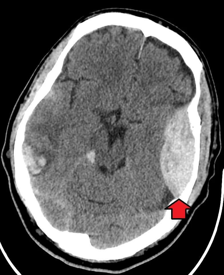
- Extradural haematoma: classically a lucid interval after initial loss of consciousness, then rapid deterioration - a neurosurgical emergency, often from a temporal bone fracture tearing the middle meningeal artery.
- Subdural haematoma: from tearing of bridging veins; more common in the elderly, on anticoagulants, or after alcohol excess, and can present acutely or, in the chronic form, over weeks.
- C-spine protection: assume cervical spine injury in any significant head injury until clinically or radiologically cleared.
- Anticoagulated patients: need an urgent CT head and, if bleeding is confirmed, immediate reversal of their anticoagulant - do not wait for deterioration.
- Cushing's triad: hypertension, bradycardia and irregular breathing - a late, pre-terminal sign of critically raised intracranial pressure.
Introduction
Head injury is one of the commonest reasons for attendance at UK emergency departments - well over a million attendances a year - ranging from a trivial bump to a devastating traumatic brain injury. The central task of assessment is separating the very large number of low-risk presentations from the small number harbouring a surgically important intracranial bleed, using a validated set of clinical criteria rather than gut feeling or scanning everyone. Falls and road traffic collisions account for most cases, with alcohol a frequent contributing factor in adults and sport a common cause in younger patients.
Brain injury is conventionally split into primary and secondary injury. Primary injury is the direct mechanical damage at the moment of impact - contusion, diffuse axonal injury, laceration - and is fixed by the time the patient is assessed. Secondary injury develops afterwards, driven by hypoxia, hypotension, raised intracranial pressure, seizures, hyperglycaemia or fever, and is largely preventable. Every element of emergency management is aimed at limiting secondary injury, since primary injury cannot be reversed.1
Diffuse axonal injury deserves specific mention: it results from rotational or shearing forces (typically rapid deceleration, as in a high-speed road traffic collision) that stretch and damage axons diffusely throughout the brain, rather than from a focal blow. It often produces a conscious level far worse than the initial CT appearance suggests, since the damage is microscopic and only becomes visible as diffuse swelling over subsequent imaging or on MRI.
Classification of intracranial haemorrhage
The pattern of bleeding on CT, and the clinical course that goes with it, is heavily examined and worth knowing cold.
| Type | Source | CT appearance | Classic course |
|---|---|---|---|
| Extradural (epidural) | Middle meningeal artery, usually with a temporal/temporoparietal skull fracture | Biconvex (lentiform), does not cross suture lines | Lucid interval after initial brief loss of consciousness, then rapid deterioration as the arterial bleed expands |
| Subdural | Tearing of bridging veins between cortex and dural sinuses | Crescentic (concave), can cross suture lines | More insidious; acute form after significant trauma, chronic form over weeks in the elderly, alcohol excess or anticoagulation, sometimes after trivial or forgotten trauma |
| Subarachnoid (traumatic) | Disruption of surface vessels in the subarachnoid space | Hyperdense blood outlining the sulci and basal cisterns | Headache, meningism; distinguish from spontaneous (aneurysmal) subarachnoid haemorrhage by the trauma history |
| Intracerebral/contusion | Direct parenchymal injury, often coup-contrecoup | Hyperdense focus within brain tissue, often with surrounding oedema | Variable; can evolve and enlarge over the following 24-48 hours |
Initial assessment
Assess using ABCDE, with cervical spine protection assumed throughout for any significant mechanism until it is clinically or radiologically cleared.2
- Airway with in-line cervical spine immobilisation if there is any suspicion of spinal injury
- Breathing - hypoxia is one of the strongest predictors of secondary brain injury and must be corrected immediately
- Circulation - hypotension is equally damaging to an injured brain; identify and treat any other source of bleeding, since isolated head injury rarely causes hypotension in adults except pre-terminally or in infants
- Disability - GCS (trended, not a single reading), pupils, blood glucose, and a focused neurological examination
- Exposure - look for scalp lacerations, other injuries, and signs of a base of skull fracture
| Sign | Location |
|---|---|
| Battle's sign | Bruising over the mastoid process, appearing 1-3 days after injury |
| Periorbital ecchymosis ('panda/racoon eyes') | Bruising around both orbits, not explained by direct orbital trauma |
| CSF rhinorrhoea or otorrhoea | Clear fluid leaking from the nose or ear; test for beta-2 transferrin if uncertain |
| Haemotympanum | Blood visible behind the tympanic membrane on otoscopy |
When to CT the head
NICE CG176 sets out specific criteria for urgent CT imaging, designed to identify the small proportion of head injuries with a clinically important bleed without scanning every minor bump.1
CT head within 1 hour
- GCS below 13 on initial assessment, or below 15 two hours after injury
- Suspected open or depressed skull fracture, or signs of a base of skull fracture
- Post-traumatic seizure
- Focal neurological deficit
- More than one episode of vomiting
- Taking an anticoagulant (for example warfarin or a DOAC) - this alone triggers the 1-hour pathway regardless of other findings
CT head within 8 hours
- Age 65 or over, with any loss of consciousness or amnesia since the injury
- A dangerous mechanism (pedestrian or cyclist struck by a vehicle, occupant ejected from a vehicle, a fall from over 1 metre or five stairs)
- More than 30 minutes of retrograde amnesia of events immediately before the injury
- Any loss of consciousness or amnesia with a history of bleeding or clotting disorder (not on anticoagulants, which triggers the 1-hour pathway instead)
Preventing secondary brain injury
Once primary injury has occurred, management is entirely about protecting the brain from further insult while any surgically remediable lesion is identified and treated.
- Oxygenation - maintain saturations at least 94-98%; avoid hypoxia
- Ventilation - maintain normocapnia; both hypercapnia (cerebral vasodilatation, raised ICP) and excessive hypocapnia (cerebral vasoconstriction, worsening ischaemia) are harmful
- Blood pressure - avoid hypotension, which reduces cerebral perfusion pressure; a systolic target is often set higher than in other trauma to protect the injured brain
- Glucose - maintain normoglycaemia; both hypo- and hyperglycaemia worsen outcome
- Temperature - avoid fever, which increases cerebral metabolic demand
- Seizure control - treat promptly, since seizures sharply increase cerebral oxygen demand
- Positioning - head-up tilt to around 30° once the spine is cleared or adequately immobilised, to aid venous drainage
Managing suspected raised intracranial pressure
- Head-up positioning and a neutral neck position (a tight cervical collar can obstruct venous drainage and should be loosened if not needed for spinal protection)
- Osmotic therapy - hypertonic saline or mannitol as a temporising measure while arranging definitive treatment
- Brief hyperventilation may be used as a short-term measure to reduce ICP in extremis while awaiting surgery, but is not sustained, since the resulting vasoconstriction can itself cause ischaemia
- Urgent neurosurgical referral for evacuation of a significant haematoma or decompressive craniectomy where indicated
Anticoagulation reversal
Any anticoagulated patient with a confirmed traumatic intracranial bleed needs immediate reversal, in parallel with neurosurgical discussion, not sequentially after it.
| Anticoagulant | Reversal |
|---|---|
| Warfarin | IV vitamin K plus prothrombin complex concentrate (PCC) for immediate correction |
| Dabigatran | Idarucizumab |
| Apixaban / rivaroxaban | Andexanet alfa where available, or PCC |
| Antiplatelet agents (aspirin, clopidogrel) | No specific reversal agent in routine use; platelet transfusion is not generally recommended and management is largely supportive |
Children
NICE publishes separate, more conservative CT criteria for children under 16, reflecting both a higher sensitivity to certain mechanisms and a strong desire to limit ionising radiation exposure to a developing brain. Suspicion of non-accidental injury - a mechanism inconsistent with the injury, delayed presentation, or other unexplained injuries - should be actively considered and escalated via local safeguarding processes in any child presenting with head injury, particularly infants.
Discharge and safety-netting
Most patients with a minor head injury and a normal CT (or no CT indication) can be safely discharged, provided there is a responsible adult to observe them and clear written advice on when to return.
- Written head injury advice covering worsening headache, repeated vomiting, increasing drowsiness, seizure, weakness, or visual disturbance
- Avoid alcohol and sedating medication for the first 48 hours
- No driving, contact sport, or unsupervised return to work until symptoms have settled and, where relevant, occupational or DVLA guidance has been checked
- Post-concussion syndrome - headache, dizziness, poor concentration, irritability and fatigue can persist for weeks after apparently minor head injury; advise a graded return to activity and safety-net for symptoms that fail to improve
Red flags
Prognosis
The great majority of head injuries presenting to UK emergency departments are minor and resolve without sequelae. Outcome after moderate to severe traumatic brain injury depends on the severity of the primary injury, how effectively secondary injury is prevented, and how quickly a surgically remediable lesion such as an extradural haematoma is evacuated - which is why rapid, protocol-driven assessment exists in the first place.
References
- NICE CG176. Head injury: assessment and early management. Available here
- Resuscitation Council UK. The ABCDE approach. Available here
This article is written for revision and education. It is not clinical guidance and must not be used to make decisions about the care of a patient. Always check current NICE guidance and local protocols.