
Malignant Spinal Cord Compression
Key points
- Definition: compression of the dural sac and its contents - spinal cord or cauda equina - by an extradural mass, with radiological indentation of the theca.
- Scale: affects around 5% of patients with cancer; breast, lung and prostate primaries account for the majority, followed by myeloma, renal and lymphoma.
- First symptom: back pain in over 90%, typically preceding any neurology by weeks - progressive, band-like, worse lying flat and on coughing.
- The determining factor: neurological function when treatment starts. Patients who are walking usually keep walking; those already paraplegic rarely recover.
- Imaging: MRI of the whole spine within 24 hours, because a third of patients have compression at more than one level.
- Immediate treatment: dexamethasone 16 mg daily with gastric protection, analgesia, and lie flat with log-rolling until the spine is known to be stable.
- Definitive treatment: surgical decompression and stabilisation where the patient is fit and the disease is limited, otherwise radiotherapy - both within 24 hours.
- Prevention: every patient with known bone metastases must be told to report new or worsening back pain immediately, before neurology develops.
Introduction
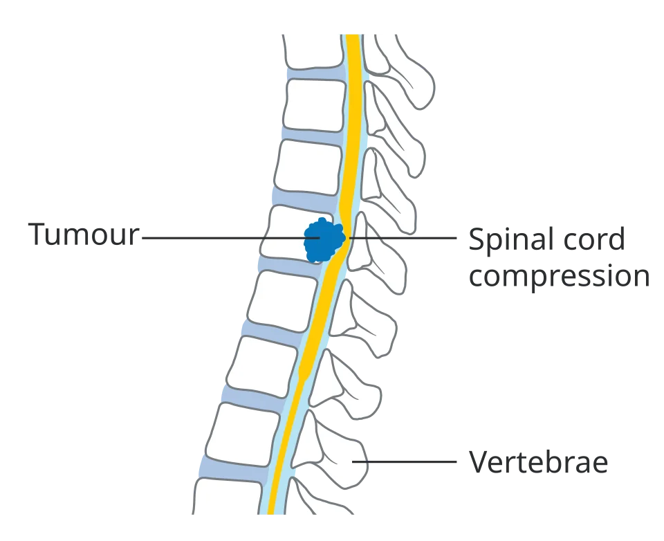
Metastatic spinal cord compression (MSCC) is compression of the dural sac and its contents by an extradural tumour mass. It affects around 5% of people with cancer, and in about a fifth of cases it is the presentation that leads to the cancer diagnosis in the first place.1
It earns its place as an emergency for a specific reason: the neurological function a patient has when treatment begins is broadly the function they will keep. Around 80% of patients who are walking at diagnosis remain able to walk; fewer than 10% of those who are paraplegic regain useful function.2 The clinical skill being tested is therefore not managing established paraplegia but recognising the back pain that comes weeks before it.
That is why the message given to patients with known bone metastases matters as much as anything done in hospital. New, progressive or unusual back pain in someone with cancer is an indication to contact the oncology team the same day, not to wait and see.
Aetiology and pathophysiology
In around 85% of cases the mechanism is a metastasis in the vertebral body that expands backwards into the epidural space. Less commonly, a paravertebral mass grows through the intervertebral foramen - the classic route in lymphoma and, in children, neuroblastoma - or the vertebral body collapses and retropulses bone into the canal. Intramedullary and leptomeningeal metastases cause a similar clinical picture but are much rarer.
Compression first obstructs the epidural venous plexus, producing vasogenic cord oedema. If it is not relieved, oedema progresses to venous infarction and then to irreversible axonal loss. This sequence explains two things you will be asked about: why dexamethasone helps (it reduces the oedema and can improve function within hours), and why time matters so much - the transition from reversible oedema to infarction can occur over hours once neurology begins.
Which cancers, and where
| Detail | |
|---|---|
| Commonest primaries | Breast, lung and prostate together account for roughly 60%; then myeloma, renal cell carcinoma, lymphoma, thyroid, gastrointestinal tumours and melanoma |
| Cancer not previously known | About 20% of cases - MSCC is the presenting feature, most often of lung, myeloma or an unknown primary |
| Thoracic spine | 60 to 70% of cases - the narrowest canal relative to the cord, and the largest number of vertebrae |
| Lumbosacral spine | 20 to 30%; below L1 to L2 the cord has ended, so this produces a cauda equina rather than a cord syndrome |
| Cervical spine | About 10%, but the most dangerous level because of the risk to respiratory function |
| Multiple levels | Present in around a third of patients - the reason imaging must cover the whole spine, not the painful segment |
Clinical features
Back pain - the warning symptom
Pain is the first symptom in more than 90% of patients and typically precedes neurological signs by several weeks. Recognising its character is the single most useful thing in this topic.
- Progressive and unremitting, unlike mechanical back pain which fluctuates with activity and settles with rest
- Worse on lying flat, and waking the patient at night - the opposite of degenerative pain
- Worse on coughing, sneezing or straining, which raises epidural venous pressure
- Band-like or radicular, radiating around the chest or abdomen in a dermatomal distribution as nerve roots are involved
- Localised spinal tenderness on percussion over the affected vertebra
- Thoracic back pain in any adult is itself a red flag, since mechanical thoracic pain is uncommon
Neurological features
- Motor - limb weakness, described as heaviness, dragging a leg, difficulty climbing stairs or rising from a chair, or unsteadiness. Weakness is usually bilateral and symmetrical, though it may start asymmetrically.
- Sensory - paraesthesiae and numbness, ascending from the feet, with a sensory level on the trunk. Ask the patient to tell you where sensation changes as you move a cold object up the trunk.
- Sphincter dysfunction - urinary hesitancy, retention with overflow incontinence, constipation and loss of the urge to defaecate. These are late features, and their presence indicates advanced compression with a poor outlook for recovery.
- Gait disturbance - often the earliest objective sign; watch the patient walk if they can
- Autonomic - postural hypotension and, in high cervical lesions, respiratory compromise
Examination
Examine tone, power, reflexes and sensation in all four limbs, look for a sensory level, assess perianal sensation and anal tone, palpate for a distended bladder, and percuss the spine for focal tenderness. Document the findings precisely with a power grade for each muscle group and the level of any sensory change, because this is the baseline against which deterioration is judged - and deterioration is what triggers urgent surgery.
Differential diagnosis
- Degenerative disease and disc prolapse - much commoner, but the pain pattern differs and it does not usually produce a sensory level
- Osteoporotic vertebral collapse - can coexist with malignancy and can be difficult to distinguish; MRI signal characteristics usually separate them
- Spinal epidural abscess - fever, raised inflammatory markers, intravenous drug use or bacteraemia; a surgical emergency in its own right
- Spinal epidural haematoma - anticoagulation, recent procedure, abrupt onset
- Leptomeningeal metastasis (carcinomatous meningitis) - patchy, asymmetrical, multi-level deficits with cranial nerve involvement
- Radiation myelopathy - months to years after spinal irradiation, and a diagnosis of exclusion
- Paraneoplastic myelopathy or transverse myelitis
- Brain metastasis or stroke - can mimic with limb weakness, but there is no sensory level and no spinal pain
Investigations
MRI of the whole spine is the investigation of choice and should be performed within 24 hours of suspected MSCC - sooner if there is neurological deterioration, and it should never be delayed to the next working day.1 Imaging the whole spine rather than the symptomatic level is essential because a third of patients have disease at multiple levels, which changes the radiotherapy field or the surgical plan.
- MRI whole spine with contrast - confirms the level, the number of levels, the degree of compression, and whether there is a paravertebral mass
- CT spine - if MRI is contraindicated (pacemaker, severe claustrophobia) or unavailable; also used to assess bone stability for surgical planning
- Plain radiographs - insufficient to exclude MSCC and should not be used for this purpose. Around 30 to 50% of vertebral bone must be lost before a lesion is visible.
- Bloods - FBC, U&Es, LFTs, corrected calcium (hypercalcaemia frequently coexists), and a myeloma screen where there are lytic lesions
- PSA in men and staging CT chest, abdomen and pelvis where the primary is unknown
- Biopsy - if there is no known malignancy, tissue is needed before treatment wherever it can be obtained without delay, since infection, lymphoma and myeloma are all managed differently
Management
Immediate measures
- Dexamethasone 16 mg daily, orally or intravenously, started as soon as MSCC is suspected and before imaging if there is neurological deficit. Prescribe a proton pump inhibitor, monitor capillary glucose, give it in the morning to avoid insomnia, and plan a reducing regimen once definitive treatment has begun.
- Analgesia - these patients are usually in severe pain. Follow the analgesic ladder and use strong opioids early, with adjuvants for neuropathic pain.
- Nurse flat with neutral spine alignment and log-rolling until the spine has been assessed for stability by the MSCC coordinator or spinal service. Do not sit the patient up until stability is confirmed.
- Venous thromboembolism prophylaxis - mechanical initially where surgery is being considered, since these patients are immobile and have a very high VTE risk
- Bladder and bowel care - assess for retention with a bladder scan and catheterise as needed; start a bowel regimen
- Pressure area care - a patient nursed flat with sensory loss is at high risk of pressure damage within hours
- Contact the MSCC coordinator - every cancer network in England has a 24-hour service to coordinate imaging and definitive treatment
Surgery or radiotherapy
Definitive treatment should begin within 24 hours of diagnosis. The choice between decompressive surgery and radiotherapy is made jointly by the spinal surgeons and oncologists, and the key evidence is the Patchell trial, which found that direct decompressive surgery followed by radiotherapy preserved and restored the ability to walk substantially better than radiotherapy alone in selected patients.3
| Favours surgical decompression and stabilisation | Favours radiotherapy alone |
|---|---|
| Spinal instability or bone retropulsed into the canal | Multiple levels of compression |
| Compression at a single level | Poor performance status or extensive systemic disease |
| Good performance status and life expectancy of at least 3 months | Expected survival of weeks |
| Radioresistant tumour - renal cell, melanoma, sarcoma | Radiosensitive tumour - myeloma, lymphoma, small cell lung, germ cell, breast, prostate |
| Previously irradiated to spinal cord tolerance at that level | No previous radiotherapy to that site |
| No tissue diagnosis, so a specimen is needed | Known histology |
| Deterioration during or after radiotherapy | Established complete paraplegia for more than 24 to 48 hours, where recovery is very unlikely |
Radiotherapy is usually given as a single 8 Gy fraction or 20 Gy in 5 fractions; the SCORAD III trial found single-fraction treatment non-inferior for ambulatory status in patients with poor prognosis, which is relevant given how much time in hospital matters to this group.4 Post-operative radiotherapy is given after surgery once the wound has healed sufficiently.
Systemic and supportive treatment
- Chemotherapy may be the primary treatment where the tumour is highly chemosensitive - lymphoma, myeloma, germ cell tumours and small cell lung cancer
- Androgen deprivation therapy in newly diagnosed metastatic prostate cancer can produce a rapid response, though radiotherapy is still usually given
- Bisphosphonates or denosumab for bone metastases, reducing further skeletal-related events
- Vertebroplasty or kyphoplasty for painful vertebral collapse without cord compression
- Rehabilitation - early physiotherapy and occupational therapy, spinal cord injury rehabilitation where appropriate, wheelchair and home assessment, and continence management
- Palliative care involvement - the majority of these patients have advanced disease, and this is often the point at which prognosis and preferred place of care should be discussed
Complications
The dominant complication is permanent paraplegia or tetraplegia with sphincter dysfunction, with all the consequences that follow: loss of independence, need for long-term catheterisation and bowel management, and a substantial impact on where and how the patient can be cared for at the end of life.
- Venous thromboembolism - very high risk in an immobile patient with cancer
- Pressure ulceration - develops rapidly in a patient nursed flat with sensory loss and poor nutrition
- Spinal instability and further collapse, with worsening deficit or intractable mechanical pain
- Urinary tract infection and urosepsis from long-term catheterisation
- Hypercalcaemia, which coexists in a significant minority and compounds confusion and constipation
- Corticosteroid toxicity - hyperglycaemia, proximal myopathy (which itself impairs mobility), agitation, insomnia, gastric ulceration and candidiasis. This is why the dexamethasone is reduced promptly once definitive treatment is under way.
- Recurrent compression at the same or a different level
Red flags
Pain alone in a patient with known cancer warrants MRI within a week; pain with any neurological symptom or sign warrants MRI within 24 hours and dexamethasone now.
Prognosis
Median survival after an episode of MSCC is in the region of 3 to 6 months, reflecting the fact that it occurs in advanced disease, though it varies greatly by primary - patients with myeloma, lymphoma or hormone-sensitive prostate and breast cancer may live considerably longer.2,5
The strongest predictor of functional outcome is ambulatory status at the start of treatment, followed by the speed of onset of the deficit (a deficit developing over days does worse than one over weeks), the primary tumour type, and the number of levels involved. Patients who are still walking when treated overwhelmingly continue to walk; those with established complete paraplegia for more than 48 hours almost never recover.
The practical conclusion is that outcome is decided largely before the patient reaches the scanner. Systems that shorten the interval from symptom to treatment - patient education about back pain, direct-access MRI, and a 24-hour MSCC coordinator - do more for these patients than any refinement of the treatment itself. When you counsel a patient starting treatment for bone metastases, telling them what back pain to report and who to ring is a concrete intervention, not a formality.
References
- NICE NG234. Spinal metastases and metastatic spinal cord compression. 2023. Available here
- Loblaw DA, Laperriere NJ, Mackillop WJ. A population-based study of malignant spinal cord compression in Ontario. Clinical Oncology. 2003. Available here
- Patchell RA, Tibbs PA, Regine WF et al. Direct decompressive surgical resection in the treatment of spinal cord compression caused by metastatic cancer: a randomised trial. The Lancet. 2005. Available here
- Hoskin PJ, Hopkins K, Misra V et al. Effect of single-fraction vs multifraction radiotherapy on ambulatory status among patients with spinal canal compression (SCORAD): a randomised clinical trial. JAMA. 2019. Available here
- NICE Clinical Knowledge Summaries. Metastatic spinal cord compression. Available here
This article is written for revision and education. It is not clinical guidance and must not be used to make decisions about the care of a patient. Always check current NICE guidance and local protocols.