
Intervertebral Disc Prolapse
Key points
- Definition: displacement of nucleus pulposus through a defect in the annulus fibrosus, compressing or chemically irritating a nerve root.
- Where: over 90% of lumbar prolapses occur at L4/5 or L5/S1; the cervical spine most often at C5/6 and C6/7.
- Which root: a posterolateral lumbar herniation compresses the traversing root - so an L4/5 disc affects the L5 root. A far lateral herniation compresses the exiting root instead.
- Cardinal symptom: leg pain worse than back pain, in a dermatomal distribution, aggravated by coughing, sneezing and sitting.
- Imaging: not routine. MRI is for red flags, severe or progressive deficit, or persisting symptoms where an intervention is being considered.
- MRI findings are common in normal people: disc degeneration is present in around a third of asymptomatic 20-year-olds - the scan must match the clinical picture.
- Natural history: around 90% improve within 6 to 12 weeks, and extruded fragments frequently resorb spontaneously.
- Surgery: emergency for cauda equina syndrome, urgent for severe or progressive motor deficit, otherwise for radicular pain failing 6 to 12 weeks of conservative care.
Introduction
Intervertebral disc prolapse is displacement of nucleus pulposus material through a tear in the annulus fibrosus. When the displaced material contacts a nerve root it causes radiculopathy - pain, sensory disturbance and weakness in that root's distribution - which in the lumbar spine is the commonest cause of true sciatica.
It is worth being clear at the outset that this is a different entity from non-specific low back pain, which is far more common and is not caused by an identifiable structural lesion, and from cauda equina syndrome, which is a surgical emergency. Those are covered separately; this article concerns the disc itself and the radiculopathy it causes.
The examinable core is anatomical: knowing which root a given disc compresses, and being able to identify that root from the pattern of pain, weakness, sensory loss and reflex change. Get that right and the rest of the topic - imaging, management, surgical indications - follows logically.
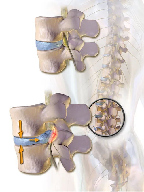
Anatomy and pathophysiology
The intervertebral disc has three parts. The nucleus pulposus is a proteoglycan-rich gel, around 80% water in youth, which acts hydrostatically to distribute load. The annulus fibrosus is a series of concentric lamellae of collagen fibres running obliquely in alternating directions, which contains that pressure. Cartilaginous endplates separate the disc from the vertebral bodies.
The adult disc is avascular, receiving nutrition by diffusion through the endplates - which is why it heals poorly, why smoking (which impairs endplate perfusion) is a risk factor, and why degeneration is essentially universal with age. As proteoglycan content falls, the nucleus dehydrates and loses height, load transfers to the annulus, and radial fissures develop through which nuclear material can extrude.
Herniations are posterolateral in the great majority of cases, for two anatomical reasons: the annulus is thinnest posteriorly, and the posterior longitudinal ligament, which reinforces the midline, narrows considerably in the lower lumbar spine, leaving the posterolateral corner unsupported.
Why it hurts
Radicular pain is not purely mechanical. Nuclear material is antigenic and provokes an intense local inflammatory response, with release of phospholipase A2, tumour necrosis factor alpha and other mediators that sensitise the nerve root. This explains two clinical observations: a small herniation can cause severe pain while a large one may be asymptomatic, and symptoms often improve before any change is visible on imaging.
Which root is compressed
This is the single most examined point in the topic and it depends on the geometry of the lumbar nerve roots. Each lumbar root exits below its correspondingly numbered pedicle, so the L4 root exits through the L4/5 foramen. It has already left the thecal sac and moved laterally by the time it reaches that level, while the L5 root is still descending medially, passing the L4/5 disc en route to its own foramen at L5/S1.
| Herniation type | Root compressed | Example |
|---|---|---|
| Paracentral (posterolateral) - the common one | The traversing root, that is the root numbered one below the level | An L4/5 disc compresses the L5 root; an L5/S1 disc compresses the S1 root |
| Foraminal or far lateral | The exiting root, numbered the same as the upper vertebra | An L4/5 far lateral disc compresses the L4 root |
| Large central | Multiple roots bilaterally | Can produce cauda equina syndrome - a surgical emergency |
In the cervical spine the arithmetic is different, because cervical roots C1 to C7 exit above their correspondingly numbered vertebra (there are eight cervical roots but seven cervical vertebrae). A C5/6 disc therefore compresses the C6 root, and a C6/7 disc compresses C7 - the root number matching the lower vertebra.
| Root | Sensory territory | Motor weakness | Reflex |
|---|---|---|---|
| L3 | Anterior thigh, down to the medial knee | Hip flexion and knee extension | Knee jerk reduced |
| L4 | Medial shin and medial malleolus | Knee extension; ankle dorsiflexion and inversion (tibialis anterior) | Knee jerk reduced |
| L5 | Lateral lower leg and dorsum of the foot, including the great toe | Extension of the great toe (extensor hallucis longus); ankle dorsiflexion; hip abduction (gluteus medius, so a positive Trendelenburg) | Normal - no reflex tests L5 reliably |
| S1 | Posterior calf, lateral border of the foot and the sole | Ankle plantarflexion and eversion; unable to stand on tiptoe | Ankle jerk reduced |
Risk factors
- Age 30 to 50 - the peak, when the nucleus is still hydrated enough to extrude but the annulus has begun to fissure. Prolapse is less common in the elderly, in whom the desiccated disc herniates less readily.
- Male sex - roughly twice as common
- Heavy manual work, particularly lifting with twisting, and repetitive bending
- Whole-body vibration - professional drivers
- Obesity and a sedentary lifestyle
- Smoking - impairs endplate diffusion and disc nutrition, and is associated with poorer outcomes after surgery
- Genetic predisposition - heritability of disc degeneration is substantial, and a family history is a stronger predictor than occupation in several studies
- Tall stature and previous spinal injury
Clinical features
- Leg pain worse than back pain - the defining feature. Sharp, burning or electric, radiating below the knee in a dermatomal pattern.
- Onset - may follow a lifting or twisting incident, but frequently there is no clear precipitant
- Aggravated by raised intrathecal pressure - coughing, sneezing, straining at stool, and by sitting and forward flexion
- Relieved by lying down, and often by standing or walking - the opposite of spinal stenosis, in which walking provokes symptoms
- Paraesthesia and numbness in the same dermatome
- Weakness - the patient may report catching their toe, tripping, or difficulty on stairs
- Cervical prolapse presents with neck pain and brachialgia radiating into the arm, aggravated by neck extension and rotation towards the affected side, and often relieved by placing the hand on the head (the shoulder abduction relief sign)
Examination
- Observe gait - a foot drop with a high-stepping gait, or inability to heel-walk (L5) or toe-walk (S1)
- Posture - a sciatic list or scoliosis away from the side of the herniation is common
- Spinal movements - restricted forward flexion, with reproduction of leg pain
- Full neurological examination of the lower limbs - tone, power in each myotome, reflexes, plantar responses and sensation, mapped to dermatomes
- Straight leg raise (Lasegue's test) - positive when passive elevation of the straight leg to between about 30 and 70 degrees reproduces the patient's radicular pain below the knee. Pain in the back or hamstring alone does not count. Sensitive but not very specific.
- Crossed straight leg raise - raising the unaffected leg reproduces pain in the affected leg. Much more specific for a herniation, though insensitive.
- Femoral stretch test - the patient prone, knee flexed and hip extended, reproducing anterior thigh pain in upper lumbar (L2 to L4) radiculopathy
- Spurling's test for cervical radiculopathy - extension, lateral flexion and rotation towards the affected side with axial compression reproduces the arm pain
- Always assess for cauda equina - perianal sensation, anal tone, and a post-void bladder scan; and for cervical myelopathy - hyperreflexia, clonus, upgoing plantars, Hoffmann's sign and gait ataxia
Differential diagnosis
- Non-specific low back pain - back pain dominant, no dermatomal radiation, normal neurology
- Lumbar spinal stenosis - older patient, bilateral buttock and leg pain on walking (neurogenic claudication), relieved by sitting or leaning forward, with preserved pedal pulses
- Spondylolisthesis and facet joint arthropathy
- Sacroiliac joint dysfunction and greater trochanteric pain syndrome, both of which refer to the buttock and lateral thigh but not below the knee
- Piriformis syndrome - sciatic irritation in the buttock, with pain on resisted external rotation
- Common peroneal nerve palsy at the fibular head - foot drop without back pain, often after prolonged squatting, a plaster cast or weight loss5
- Diabetic amyotrophy - proximal thigh pain and quadriceps wasting in diabetes
- Vascular claudication - calf pain on walking with a fixed claudication distance, absent pulses, relieved by standing still rather than by sitting
- Hip osteoarthritis - groin pain, worse on weight bearing, with restricted internal rotation
- Serious pathology - malignancy, discitis or vertebral osteomyelitis, fracture, inflammatory spondyloarthropathy, herpes zoster before the rash appears
Investigations
Disc prolapse is a clinical diagnosis, and NICE is explicit that imaging should not be requested routinely in a non-specialist setting - it does not change management in most patients and generates findings that alarm without informing.1
- MRI is the investigation of choice where imaging is needed. Indications are: suspected cauda equina syndrome (emergency, same day), severe or progressive neurological deficit, suspected malignancy, infection or fracture, and radicular symptoms persisting beyond about 4 to 6 weeks where surgery or an epidural injection is being considered.
- CT - if MRI is contraindicated; better for bony detail but poorer for soft tissue and neural structures
- Plain radiographs - of little value in disc prolapse, though used to assess alignment, spondylolisthesis and instability
- Nerve conduction studies and EMG - occasionally used where the diagnosis is uncertain, particularly to distinguish radiculopathy from a peripheral nerve entrapment
- Bloods - FBC, CRP, ESR if infection or malignancy is suspected; myeloma screen and PSA where relevant
Management
Natural history and explanation
Around 90% of patients improve substantially within 6 to 12 weeks without surgery, and MRI follow-up studies show that extruded and sequestrated fragments frequently resorb spontaneously through a macrophage-mediated inflammatory process - with, counter-intuitively, the largest herniations often resorbing most completely. Explaining this changes how patients cope with the pain, and is itself part of the treatment.
Conservative treatment
- Stay active. Bed rest is harmful and delays recovery. Encourage continuation of normal activity, including work where possible, within the limits of pain.
- NSAIDs at the lowest effective dose for the shortest period, with gastroprotection where indicated. NICE recommends these as the analgesic of first choice for sciatica.1
- Weak opioids may be considered only if NSAIDs are contraindicated or ineffective, and only short-term. Do not use opioids for chronic sciatica.
- A structured exercise programme and physiotherapy, tailored to the individual, with manual therapy only as part of a package that includes exercise
- Psychological support as part of a combined physical and psychological programme where there are significant psychosocial obstacles to recovery
- Return to work planning, which is a clinical outcome and not an administrative one
Interventional and surgical treatment
- Epidural injection of local anaesthetic and steroid - considered for acute and severe sciatica; gives useful short-term relief of leg pain, with little effect on long-term outcome or on the eventual need for surgery
- Microdiscectomy - the standard operation, removing the herniated fragment through a small incision, usually as a day case or overnight stay
- Cervical prolapse - anterior cervical discectomy and fusion, or posterior foraminotomy; surgery is urgent where there is myelopathy rather than radiculopathy alone
| Urgency | Indication |
|---|---|
| Emergency | Cauda equina syndrome - decompression as soon as possible, and certainly within hours rather than days |
| Urgent | Severe or progressive motor deficit, such as a new foot drop or a rapidly progressing weakness |
| Elective | Radicular pain that is disabling, concordant with imaging, and has failed 6 to 12 weeks of appropriate conservative treatment |
| Not an indication | Back pain without radiculopathy; an incidental disc abnormality on MRI; pain that does not match the imaged level |
The evidence on elective surgery is worth knowing. The SPORT trial and the Dutch trial by Peul and colleagues both found that surgery produces faster relief of leg pain - a matter of weeks rather than months - but that outcomes in the surgical and conservative groups converge by one to two years.3,6 The honest way to present this to a patient is therefore that discectomy buys earlier recovery rather than a better final result, which allows them to weigh the operative risk against how much the next few months matter to them.
Complications
- Persistent neurological deficit - a foot drop that does not recover, or lasting sensory loss, particularly where decompression was delayed
- Cauda equina syndrome with permanent bladder, bowel and sexual dysfunction
- Chronic pain and disability, with the associated loss of work and psychological consequences
- Recurrent herniation at the same level after discectomy, in around 5 to 10%
- Surgical complications - dural tear with CSF leak, nerve root injury, epidural haematoma, wound infection, and discitis, which presents with escalating back pain and raised inflammatory markers some weeks postoperatively
- Failed back surgery syndrome - persistent pain after technically successful surgery, more likely where the indication was back pain rather than radiculopathy
- Adjacent segment degeneration after cervical fusion
Prognosis
The outlook for a first episode is good. The majority of patients are substantially better by 6 to 12 weeks, and most recover fully without surgery. Sensory symptoms often settle before motor recovery is complete, and residual numbness in a dermatome may persist without functional consequence.
Recurrence of some form of back or leg pain over subsequent years is common, and around 5 to 10% of patients who undergo discectomy will herniate again at the same level. Predictors of a poorer outcome are prolonged symptoms before treatment, smoking, obesity, significant psychosocial distress, low expectations of recovery, and involvement in compensation or litigation - which is why explanation, activity and early return to work matter as much as any drug.
Neurological recovery depends heavily on the duration and severity of compression. A foot drop present for a few days usually recovers well after decompression; one present for months often does not. That principle - that the deficit at the time of treatment largely determines the deficit afterwards - is the same one that governs cauda equina syndrome and malignant spinal cord compression, and it is why the urgency categories exist.
References
- NICE NG59. Low back pain and sciatica in over 16s: assessment and management. 2016, updated 2020. Available here
- Fardon DF, Williams AL, Dohring EJ et al. Lumbar disc nomenclature: version 2.0. The Spine Journal. 2014. Available here
- Weinstein JN, Tosteson TD, Lurie JD et al. Surgical vs nonoperative treatment for lumbar disk herniation: the Spine Patient Outcomes Research Trial (SPORT). JAMA. 2006. Available here
- Brinjikji W, Luetmer PH, Comstock B et al. Systematic literature review of imaging features of spinal degeneration in asymptomatic populations. American Journal of Neuroradiology. 2015. Available here
- NICE Clinical Knowledge Summaries. Sciatica (lumbar radiculopathy). Available here
- Peul WC, van Houwelingen HC, van den Hout WB et al. Surgery versus prolonged conservative treatment for sciatica. New England Journal of Medicine. 2007. Available here
This article is written for revision and education. It is not clinical guidance and must not be used to make decisions about the care of a patient. Always check current NICE guidance and local protocols.