
Cervical Cancer
Key points
- Cervical cancer: almost always caused by persistent infection with high-risk human papillomavirus (hrHPV), most commonly types 16 and 18.
- Histology: squamous cell carcinoma (~70-80%) is most common; adenocarcinoma (~20%) arises from the endocervical glandular epithelium and is harder to detect on screening.
- Precursor: cervical intraepithelial neoplasia (CIN) is the pre-invasive stage, detected and treated through the national screening programme before it becomes cancer - see the cervical screening article.
- Presentation: postcoital, intermenstrual or postmenopausal bleeding, and offensive vaginal discharge; many early cases are asymptomatic and found on screening.
- Staging: FIGO clinical/radiological staging (I-IV), based on tumour size and spread rather than surgical pathology alone.
- Management: fertility-sparing local excision for the earliest stages; radical hysterectomy with lymphadenectomy for early invasive disease; chemoradiotherapy for locally advanced or unresectable disease.
- Prevention: HPV vaccination and cervical screening together are the two pillars of prevention; smoking is a major independent risk factor for progression.
- Prognosis: strongly stage-dependent - early-stage disease has excellent survival, underscoring why screening and prompt referral for red-flag bleeding both matter.
Introduction
Cervical cancer arises from the epithelium of the cervix and is, with very rare exceptions, caused by persistent infection with high-risk human papillomavirus (hrHPV) - making it one of the few cancers with a clearly identified, preventable infective cause.1 This has driven the two major public health interventions covered in more detail elsewhere: HPV vaccination (primary prevention) and the national cervical screening programme (secondary prevention, detecting and treating pre-cancerous change before it becomes invasive - see the cervical screening article).
Globally, cervical cancer remains a major cause of cancer death in women, particularly where screening and vaccination programmes are less established; in the UK, incidence has fallen substantially since the introduction of screening and is expected to continue falling as vaccinated cohorts age into the screened population.
Aetiology and risk factors
HPV types 16 and 18 together account for the majority of cervical cancers; HPV integrates its DNA into cervical epithelial cells, and viral oncoproteins (E6 and E7) inactivate the tumour suppressor genes p53 and Rb respectively, disabling normal cell cycle control and allowing progressive dysplasia (CIN) to develop into invasive cancer if the infection persists rather than clears.2
- Persistent high-risk HPV infection (the necessary cause)
- Smoking - an independent risk factor, thought to impair local immune clearance of HPV and to have direct carcinogenic effects on cervical cells via tobacco metabolites concentrated in cervical mucus
- Immunosuppression (HIV, transplant recipients, long-term immunosuppressant use) - impairs viral clearance
- Early age at first intercourse and multiple sexual partners - increase cumulative HPV exposure
- Long-term combined oral contraceptive use (weak, likely modest, independent association)
- High parity
- Non-attendance at cervical screening
Histology
| Subtype | Proportion | Notes |
|---|---|---|
| Squamous cell carcinoma | ~70-80% | Arises from the ectocervical squamous epithelium/transformation zone; well detected by cytology |
| Adenocarcinoma | ~20% | Arises from endocervical glandular epithelium, higher in the canal; harder to sample and detect on screening, so a rising relative proportion of screen-detected cancers |
| Adenosquamous and other rare subtypes | <10% | Mixed or rarer histological patterns |
Clinical features
Early-stage cervical cancer is often asymptomatic and detected through screening, which is precisely the point of the programme - catching disease before symptoms develop.3 Symptomatic presentation usually reflects more established disease:
- Postcoital bleeding: the classic presenting symptom and should never be attributed to a benign cause (e.g. ectropion) without adequate assessment
- Intermenstrual bleeding
- Postmenopausal bleeding
- Offensive or blood-stained vaginal discharge
- Pelvic pain or dyspareunia: suggests more advanced local disease
- Symptoms of advanced disease: haematuria or urinary symptoms (bladder invasion), rectal bleeding or altered bowel habit (rectal invasion), leg swelling or pain (pelvic sidewall/lymphatic involvement), and flank pain (ureteric obstruction)
On speculum examination, an invasive tumour may appear as an irregular, friable, ulcerated or exophytic mass on the cervix that bleeds readily on contact - a very different appearance from the smooth, symmetrical red ring of a benign ectropion (see that article for the comparison).
Investigations
Colposcopy and biopsy
Direct visualisation with acetic acid/iodine application and biopsy of any suspicious area provides histological diagnosis - the essential first step whenever cervical cancer is suspected clinically, whether or not it was screening that raised concern.
Staging investigations
Once cancer is histologically confirmed, imaging defines local, nodal and distant spread to guide staging and treatment planning: MRI pelvis for local tumour size and parametrial/pelvic sidewall invasion, CT chest/abdomen/pelvis or PET-CT for nodal and distant metastatic disease, and examination under anaesthesia in some cases to assess local extent clinically.
Staging
Cervical cancer uses the FIGO staging system, which is based on clinical and radiological assessment of tumour size, depth of invasion, and spread to adjacent structures, lymph nodes and distant sites - distinct from purely surgical/pathological staging used in some other cancers.4
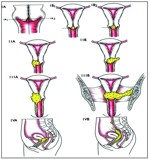
| Stage | Extent |
|---|---|
| I | Confined to the cervix |
| II | Extends beyond the cervix/uterus but not to the pelvic sidewall or lower third of the vagina |
| III | Extends to the pelvic sidewall, lower third of the vagina, causes hydronephrosis, or involves regional lymph nodes |
| IV | Extends beyond the true pelvis, or involves the bladder/rectal mucosa (IVA), or distant metastasis (IVB) |
Management
Treatment is guided by stage, tumour size, and the woman's wish to preserve fertility, and is planned via a specialist gynaecological oncology multidisciplinary team.1
| Stage | Typical approach |
|---|---|
| Very early (IA1), fertility desired | Cone biopsy / large loop excision alone may be sufficient |
| Early invasive (IA2-IB), fertility desired and tumour suitable | Radical trachelectomy (removal of the cervix, preserving the uterine body) with pelvic lymphadenectomy |
| Early invasive (IA2-IIA), fertility not a priority | Radical hysterectomy with pelvic lymphadenectomy |
| Locally advanced (IIB-IVA) | Chemoradiotherapy (concurrent cisplatin-based chemotherapy with external beam radiotherapy and brachytherapy) - generally not primarily surgical at this stage |
| Metastatic (IVB) / recurrent | Systemic chemotherapy ± targeted therapy/immunotherapy, with palliative intent and symptom-focused supportive care |
Complications
- Ureteric obstruction and renal impairment from local tumour spread
- Fistula formation (vesicovaginal or rectovaginal) in advanced or treated disease
- Lymphoedema following pelvic lymphadenectomy or radiotherapy
- Premature ovarian insufficiency if radiotherapy or chemotherapy affects ovarian function (ovarian transposition can be performed before pelvic radiotherapy to preserve function where fertility/hormonal preservation matters)
- Vaginal stenosis after radiotherapy, affecting sexual function
- Psychological impact of a cancer diagnosis and, where relevant, loss of fertility
Prognosis
Prognosis is strongly dependent on stage at diagnosis: early-stage disease (confined to the cervix) has a very good prognosis with high five-year survival, while locally advanced and metastatic disease carry a substantially poorer outlook despite chemoradiotherapy. This stage-dependence is the central argument for both HPV vaccination and consistent cervical screening uptake, and for taking postcoital and postmenopausal bleeding seriously rather than assuming a benign cause without adequate assessment.
References
- NICE NG12. Suspected cancer: recognition and referral - cervical cancer. 2021. Available here
- Cancer Research UK. Cervical cancer statistics and information. Available here
- British Gynaecological Cancer Society. Cervical cancer guidelines. Available here
- Bhatla N, Aoki D, Sharma DN, Sankaranarayanan R. Cancer of the cervix uteri. FIGO Cancer Report 2021. Available here
This article is written for revision and education. It is not clinical guidance and must not be used to make decisions about the care of a patient. Always check current NICE guidance and local protocols.