
Endometrial Cancer
Key points
- Endometrial cancer: the most common gynaecological cancer in the UK, arising from the endometrial lining; most cases are diagnosed early because postmenopausal bleeding prompts rapid investigation.
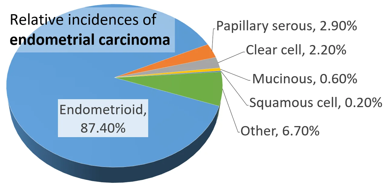
- Type 1 (endometrioid): the great majority of cases (~87%); oestrogen-driven, arises via endometrial hyperplasia, typically lower grade with a better prognosis.
- Type 2 (non-endometrioid): serous, clear cell and other subtypes; not oestrogen-driven, arises in atrophic endometrium, higher grade, and a worse prognosis despite being less common.
- Key risk factor: unopposed oestrogen exposure - obesity (peripheral aromatisation), nulliparity, early menarche/late menopause, unopposed HRT, PCOS, and tamoxifen.
- Presentation: postmenopausal bleeding is the classic and most useful red flag; in premenopausal women, persistent intermenstrual bleeding is the equivalent warning sign.
- First-line investigation: transvaginal ultrasound measuring endometrial thickness - 4 mm or less has a high negative predictive value in postmenopausal women.
- Diagnosis: endometrial biopsy (pipelle or hysteroscopic) for histological confirmation if ultrasound is abnormal or bleeding persists.
- Management: total hysterectomy with bilateral salpingo-oophorectomy for most early-stage disease; adjuvant radiotherapy/chemotherapy for higher-risk or advanced disease.
Introduction
Endometrial cancer arises from the glandular epithelium lining the uterine cavity and is the most common gynaecological cancer in the UK, more common than ovarian or cervical cancer.1 It predominantly affects postmenopausal women, with a peak incidence around 65-75 years, though it can occur in younger women, particularly those with obesity or PCOS.
Endometrial cancer has a comparatively favourable prognosis overall compared with other gynaecological cancers, largely because it tends to present early - postmenopausal bleeding is a highly visible symptom that prompts rapid investigation, so most cases are diagnosed while still confined to the uterus.
Classification: type 1 versus type 2
Endometrial cancer is traditionally divided into two broad types with different pathogenesis, histology and prognosis, which is one of the most examinable frameworks in this topic.2
| Feature | Type 1 (endometrioid) | Type 2 (non-endometrioid) |
|---|---|---|
| Proportion | ~80-90% of cases | ~10-20% of cases |
| Driver | Oestrogen-dependent | Not oestrogen-dependent |
| Background endometrium | Arises from endometrial hyperplasia | Arises in an atrophic endometrium |
| Typical histology | Endometrioid adenocarcinoma | Serous, clear cell, carcinosarcoma |
| Grade | Usually lower grade | Usually higher grade |
| Typical patient | Obesity, PCOS, unopposed oestrogen exposure | Older, thinner women; can arise without classic hormonal risk factors |
| Prognosis | Generally favourable | Poorer, even at an equivalent stage |
Risk factors
Because type 1 disease dominates, most recognised risk factors relate to cumulative or unopposed oestrogen exposure - oestrogen stimulates endometrial proliferation, and without progestogen to oppose this effect, hyperplasia and eventually malignant change can develop.3
Factors that increase oestrogen exposure
- Obesity - peripheral aromatisation of androgens to oestrogen in adipose tissue is the single most important modifiable risk factor
- Nulliparity
- Early menarche and late menopause (more lifetime ovulatory/oestrogen-exposed cycles)
- Polycystic ovary syndrome (chronic anovulation causes unopposed oestrogen)
- Unopposed oestrogen HRT (oestrogen without progestogen in a woman with a uterus)
- Tamoxifen (has a paradoxical oestrogenic effect on the endometrium despite being an anti-oestrogen in breast tissue)
Other risk factors
- Type 2 diabetes mellitus (independent of obesity, via insulin's proliferative effect)
- Lynch syndrome (hereditary non-polyposis colorectal cancer) - carries a high lifetime endometrial cancer risk, alongside colorectal and other cancers; a strong family history should prompt genetic counselling and consideration of risk-reducing surgery
- Increasing age
Protective factors
- Combined oral contraceptive pill use (progestogen component opposes endometrial proliferation)
- Multiparity
- Smoking (paradoxically reduces risk, likely via anti-oestrogenic effects, though this is never a reason to smoke given its far greater harms)
- LNG-IUS use
Clinical features
Postmenopausal bleeding is the classic and most useful presenting symptom - any bleeding 12 months or more after the last period should be regarded as endometrial cancer until proven otherwise and investigated urgently, even though the majority of such bleeding turns out to have a benign cause (see the abnormal uterine bleeding article).4
In premenopausal women, endometrial cancer is less common but should be considered with persistent intermenstrual bleeding, particularly in women with risk factors (obesity, PCOS). Other features include abnormal or blood-stained vaginal discharge, and, in more advanced disease, pelvic pain or a palpable mass.
Investigations
Transvaginal ultrasound
First-line investigation, measuring endometrial thickness. In postmenopausal women, an endometrial thickness of 4 mm or less has a high negative predictive value for endometrial cancer, and further investigation may be deferred if bleeding is a single episode and thickness is reassuring; a thicker endometrium, or persistent/recurrent bleeding despite a thin endometrium, warrants biopsy.5
Endometrial biopsy
Provides histological diagnosis. Can be performed as an outpatient pipelle biopsy, or via hysteroscopy (allowing direct visualisation and targeted biopsy of any focal lesion), which is preferred if ultrasound suggests a focal abnormality or pipelle sampling is inadequate/inconclusive.
Staging investigations
Once cancer is confirmed, MRI pelvis assesses depth of myometrial invasion and cervical involvement, and CT chest/abdomen/pelvis assesses for nodal and distant spread, to guide surgical and adjuvant treatment planning.
Endometrial hyperplasia
Endometrial hyperplasia is the histological precursor to type 1 endometrial cancer, resulting from the same unopposed oestrogen exposure, and is classified as hyperplasia without atypia or atypical hyperplasia (endometrial intraepithelial neoplasia) - the latter carries a substantial risk of progression to, or coexistence with, endometrial cancer.3
| Type | Management |
|---|---|
| Without atypia | LNG-IUS (preferred) or oral progestogens, with repeat biopsy to confirm regression; low progression risk |
| Atypical (EIN) | Hysterectomy is generally recommended given the significant risk of coexisting or progressing to cancer; high-dose progestogen with close surveillance is an option if fertility preservation is essential |
Staging and management
Endometrial cancer is staged using the FIGO system (surgical-pathological, based on depth of myometrial invasion, cervical involvement, and spread beyond the uterus), and management is planned by a specialist gynaecological oncology multidisciplinary team.1
Surgery
Total hysterectomy with bilateral salpingo-oophorectomy (removal of the uterus, cervix, fallopian tubes and ovaries) is the mainstay of treatment for most early-stage disease, usually performed laparoscopically or robotically where possible. Pelvic (± para-aortic) lymph node assessment is performed for higher-risk histology or more advanced disease to guide adjuvant treatment.
Adjuvant therapy
Adjuvant radiotherapy (external beam and/or vaginal brachytherapy) is used for higher-risk features (deep myometrial invasion, higher grade, lymphovascular space invasion) to reduce local recurrence. Chemotherapy is added for high-risk histology (including all type 2 disease) or advanced-stage cancer.
Fertility-sparing management
In carefully selected young women with early, low-grade, superficial disease who strongly wish to preserve fertility, high-dose progestogen therapy with very close surveillance (regular biopsy) is sometimes used instead of immediate hysterectomy, with definitive surgery recommended once childbearing is complete - this is a specialist decision made with full understanding of the trade-off against a small increase in progression risk.
Complications
- Anaemia from chronic abnormal bleeding
- Local spread to the cervix, vagina, bladder or bowel in advanced disease
- Lymphoedema following pelvic lymphadenectomy
- Surgical menopause (if premenopausal at the time of bilateral salpingo-oophorectomy) with associated symptoms and long-term bone/cardiovascular implications
- Psychological impact of cancer diagnosis and loss of fertility
Prognosis
Overall prognosis for endometrial cancer is comparatively good, largely reflecting early diagnosis driven by postmenopausal bleeding as a highly visible warning symptom, with the great majority of type 1 disease diagnosed at an early, surgically curable stage. Type 2 disease and higher-grade tumours carry a substantially worse prognosis even at an equivalent stage, which is why histological subtype, not just stage, drives decisions about adjuvant treatment.
References
- NICE NG12. Suspected cancer: recognition and referral - endometrial cancer. 2021. Available here
- Cancer Research UK. Uterine (womb) cancer statistics and information. Available here
- Royal College of Obstetricians and Gynaecologists. Green-top Guideline No. 67: Management of Endometrial Hyperplasia. Available here
- NICE Clinical Knowledge Summaries (CKS). Menopause and postmenopausal bleeding. Available here
- British Gynaecological Cancer Society. Endometrial cancer guidelines. Available here
- NICE DG42. Testing strategies for Lynch syndrome in people with endometrial cancer. 2020. Available here
This article is written for revision and education. It is not clinical guidance and must not be used to make decisions about the care of a patient. Always check current NICE guidance and local protocols.