
Brain Tumours: Diagnosis and Management
Key points
- Brain tumours: may be primary (arising from brain tissue or its coverings) or metastatic - and metastases are considerably more common than primary brain tumours in adults.
- Four presenting syndromes: raised intracranial pressure, seizures, progressive focal neurological deficit, and personality or cognitive change - often in combination.
- Red flag headache: progressive, worse on waking or lying flat, worse with coughing or straining, and associated with vomiting or papilloedema.
- Commonest primary malignant tumour in adults: glioblastoma (a grade 4 glioma), which carries a poor prognosis despite maximal treatment.
- Commonest primary tumour overall: meningioma - usually benign, extra-axial, arising from the arachnoid, and often found incidentally.
- Commonest sources of brain metastases: lung, breast, melanoma, renal and colorectal cancer.
- First-line investigation: MRI brain with contrast; CT is used acutely where MRI is not immediately available.
- Dexamethasone: reduces vasogenic oedema surrounding a tumour and can rapidly improve symptoms, but is symptomatic treatment and not definitive.
Introduction
Brain tumours encompass a wide range of pathologies, from benign, slow-growing meningiomas found incidentally to aggressive, rapidly fatal glioblastomas. In adults, metastatic disease is considerably more common than primary brain tumour, which is why any new intracranial mass should prompt consideration of a systemic primary.1
Whatever the histology, brain tumours produce symptoms through a limited number of mechanisms: mass effect and raised intracranial pressure, irritation of cortex causing seizures, destruction or compression of functional brain tissue causing focal deficit, and sometimes endocrine disturbance where the pituitary or hypothalamus is involved. Reasoning from these mechanisms, rather than memorising tumour types in isolation, is what makes the clinical picture predictable.
The examinable core is recognising the red flag headache pattern that warrants urgent imaging, and distinguishing it from the far commoner primary headache disorders.
Primary brain tumours account for a small proportion of all cancers - around 2-3% - but a disproportionate share of cancer-related years of life lost, because they affect a relatively young population and carry poor survival in their malignant forms. Brain metastases are considerably more common, developing in perhaps a fifth of patients with systemic cancer, and their incidence is rising as systemic treatments extend survival and patients live long enough to develop intracranial disease.
Classification
Primary brain tumours
| Tumour | Origin and behaviour | Key features |
|---|---|---|
| Glioblastoma (grade 4 glioma) | Astrocytic glial cells; highly malignant and infiltrative | The commonest primary malignant brain tumour in adults; rapid progression, characteristic irregular ring enhancement with central necrosis and marked surrounding oedema on MRI |
| Lower-grade gliomas (grades 2-3) | Glial cells; infiltrative but slower growing | Often present with seizures in younger adults; tend to transform to higher grade over time |
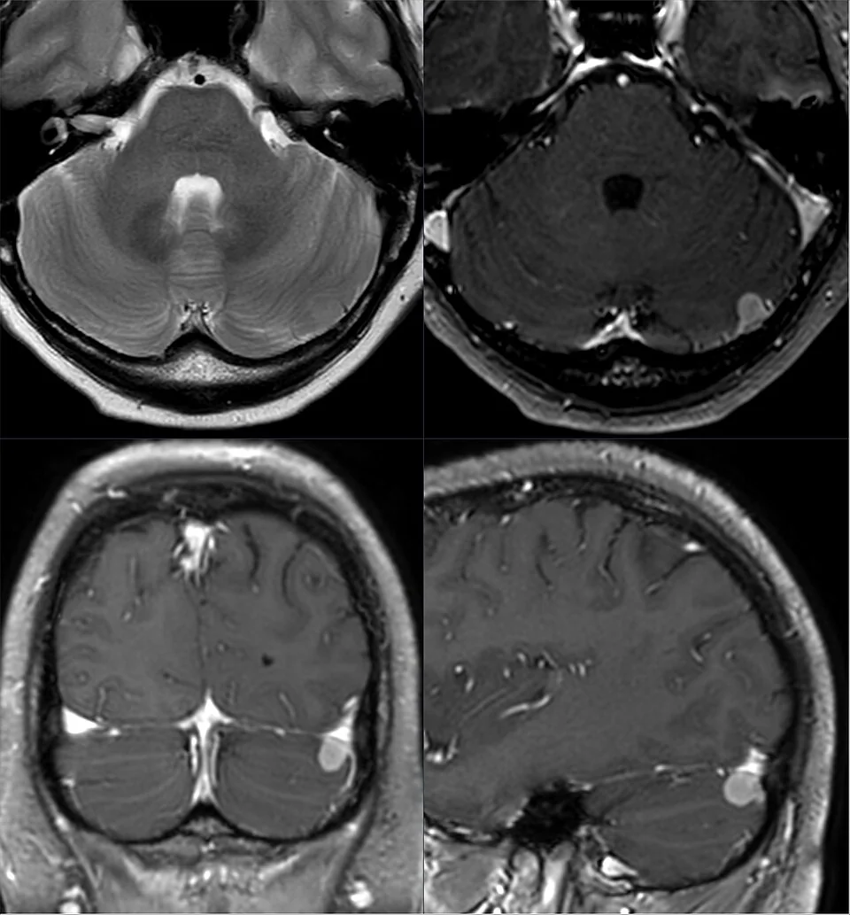
| Meningioma | Arachnoid cap cells - extra-axial, arising from the meninges | The commonest primary intracranial tumour overall; usually benign and slow-growing, often incidental; classically shows a 'dural tail' on contrast MRI |
| Pituitary adenoma | Anterior pituitary | Bitemporal hemianopia from optic chiasm compression, plus endocrine effects (prolactinoma, acromegaly, Cushing's disease) or hypopituitarism |
| Vestibular schwannoma | Schwann cells of the vestibulocochlear nerve | Unilateral sensorineural hearing loss and tinnitus; covered in its own article |
| CNS lymphoma | Lymphoid cells | More common in the immunocompromised, particularly advanced HIV; often periventricular and avidly enhancing |
Metastatic tumours
Brain metastases are more common than primary brain tumours in adults, and are typically multiple, located at the grey-white matter junction (reflecting haematogenous spread where vessel calibre narrows), and surrounded by disproportionately marked vasogenic oedema. The commonest primary sources are lung, breast, melanoma, renal and colorectal cancer - melanoma has a particular tendency to metastasise to brain and to bleed.
Molecular classification has become central rather than supplementary in recent years, and the 2021 WHO classification formally integrates it with histology. The practical point for a student is that two tumours that look identical down a microscope can behave completely differently depending on their molecular profile - IDH-mutant gliomas carry a substantially better prognosis than IDH-wildtype tumours of the same grade, and MGMT promoter methylation predicts a better response to temozolomide.
This is why tissue diagnosis retains its importance even when imaging appears characteristic, and why biopsy may be undertaken in lesions that will not be resected: the molecular result changes both the treatment offered and the prognosis discussed with the patient.
Clinical features
Raised intracranial pressure
- Headache - classically progressive over weeks, worse on waking or lying flat, and aggravated by coughing, straining or bending forward
- Vomiting - often without preceding nausea
- Papilloedema on fundoscopy
- Reduced conscious level in more advanced disease
- Sixth nerve palsy - a false localising sign of raised pressure rather than a lesion in the nerve's path
Seizures
A new-onset seizure in an adult - particularly focal seizures, or a first seizure over the age of 40 - should always prompt consideration of an underlying structural lesion and warrants brain imaging. Seizures are a particularly common presenting feature of lower-grade gliomas in younger adults.
An important principle underlies the difference between fast- and slow-growing tumours. A slowly enlarging lesion such as a low-grade glioma or meningioma allows the brain to accommodate it gradually, so it may reach a considerable size before causing anything other than seizures. A rapidly expanding lesion such as a glioblastoma, or a metastasis with marked surrounding oedema, exhausts the compensatory reserve quickly and presents with raised intracranial pressure and focal deficit over weeks.
This is why the rate of change in a patient's symptoms is often more informative than their absolute severity, and why a history of subtly worsening headache or personality change over two months should be taken as seriously as a more dramatic acute presentation.
Focal neurological deficit
- Progressive hemiparesis, dysphasia, visual field defect or ataxia, depending on tumour location - the gradual, worsening course distinguishes this from the sudden onset of stroke
- Frontal lobe tumours - personality change, disinhibition, apathy and executive dysfunction, which may be attributed to depression or dementia before the tumour is found
- Cerebellar tumours - ataxia, nystagmus, and early hydrocephalus from fourth ventricle obstruction
- Pituitary/suprasellar tumours - bitemporal hemianopia from chiasmal compression, plus endocrine features
Clinical examination
- Fundoscopy for papilloedema - essential in any suspected raised intracranial pressure
- Full neurological examination, including visual fields to confrontation (bitemporal hemianopia in pituitary lesions, homonymous defects in hemispheric lesions)
- Cognitive and behavioural assessment, particularly for frontal lobe involvement
- Cranial nerve examination, including hearing where a cerebellopontine angle lesion is suspected
- Cerebellar signs and gait assessment
- Systemic examination for a primary malignancy - breasts, skin (melanoma), lymph nodes, chest and abdomen
- Vital signs for Cushing's triad if raised intracranial pressure is severe
Differential diagnosis
- Brain abscess - also produces a ring-enhancing lesion; distinguished by infective context and restricted diffusion on diffusion-weighted MRI
- Stroke - sudden rather than progressive onset, and follows a vascular territory
- Multiple sclerosis (tumefactive demyelination) - can produce a large enhancing lesion mimicking tumour, particularly in younger patients
- Primary headache disorders - by far the commoner explanation for headache, but lack the red flag features above
- Idiopathic intracranial hypertension - raised pressure and papilloedema without a mass lesion on imaging
- Cerebral venous sinus thrombosis - headache with raised pressure, excluded by venography
- Neurocysticercosis - multiple cystic lesions in patients from endemic areas
Investigations
- MRI brain with contrast - the investigation of choice, characterising the lesion, its enhancement pattern, surrounding oedema and mass effect, and distinguishing intra-axial from extra-axial lesions
- CT head with contrast - used acutely where MRI is unavailable or the patient is too unwell, and to exclude haemorrhage or hydrocephalus urgently
- CT chest, abdomen and pelvis - to identify a primary malignancy where metastatic disease is suspected5
- Biopsy or surgical resection specimen - provides definitive histological and molecular diagnosis, which increasingly determines treatment and prognosis (for example IDH mutation and MGMT methylation status in gliomas)
- Bloods - including tumour markers and HIV testing where CNS lymphoma is a consideration
- Endocrine profile - for suspected pituitary lesions, assessing both hypersecretion and hypopituitarism
- Avoid lumbar puncture where a mass lesion with raised intracranial pressure is suspected, because of the risk of herniation
Management
Immediate symptomatic management
- Dexamethasone - reduces vasogenic oedema surrounding the tumour and can produce rapid, sometimes dramatic symptomatic improvement in headache and focal deficit. This is one of the specific settings where steroids genuinely help raised intracranial pressure, unlike in traumatic brain injury or stroke
- Antiepileptic drugs - for patients who have had a seizure; prophylactic use in those who have not seized is not routine
- Urgent neurosurgical referral for significant mass effect, hydrocephalus, or declining conscious level
- Analgesia and antiemetics
Definitive treatment
- Surgery - maximal safe resection where the lesion is accessible and the patient fit; for gliomas this improves survival and provides tissue for molecular diagnosis, while for meningiomas complete resection can be curative
- Radiotherapy - used adjuvantly after resection of high-grade gliomas, and for metastases (either whole-brain radiotherapy or, increasingly, stereotactic radiosurgery for limited numbers of metastases)
- Chemotherapy - temozolomide alongside radiotherapy is standard adjuvant treatment for glioblastoma, with greatest benefit in tumours showing MGMT promoter methylation
- Systemic therapy for the primary - in metastatic disease, alongside targeted CNS treatment; some targeted agents and immunotherapies have meaningful intracranial activity
- Observation - appropriate for small, asymptomatic, incidentally found meningiomas, with serial imaging rather than immediate intervention
- Multidisciplinary team discussion - neuro-oncology MDT input determines management for essentially all cases
Supportive and palliative care
- Early palliative care involvement for high-grade tumours, alongside active treatment rather than only at the end of life
- Neurorehabilitation - physiotherapy, occupational therapy and speech and language therapy for focal deficits
- Advance care planning, initiated while capacity and communication are preserved
- Psychological support for patient and family
- Driving - patients must inform the DVLA of a brain tumour diagnosis, and will be required to stop driving for a period determined by tumour type, treatment and seizure history
Complications
- Raised intracranial pressure and herniation
- Seizures and post-treatment epilepsy
- Hydrocephalus, particularly with posterior fossa or intraventricular tumours
- Progressive focal neurological deficit and loss of independence
- Cognitive impairment and personality change, from the tumour itself, from surgery, or from radiotherapy
- Haemorrhage into the tumour, particularly with melanoma and renal cell metastases
- Venous thromboembolism - patients with brain tumours, especially gliomas, are at markedly increased risk
- Complications of corticosteroid treatment - hyperglycaemia, proximal myopathy, infection risk, neuropsychiatric effects
Red flags
Prognosis
Prognosis varies enormously with histology. Meningiomas are usually benign, and complete surgical resection is often curative, with many small incidental lesions safely observed indefinitely. Lower-grade gliomas may follow an indolent course over years, though most eventually transform to higher grade. Glioblastoma carries a poor prognosis despite maximal surgery, radiotherapy and temozolomide, with median survival typically quoted at around 12-15 months, though molecular features such as MGMT methylation and IDH mutation identify subgroups with better outcomes.2,3
Prognosis in brain metastases depends primarily on the behaviour of the primary tumour, the number of intracranial lesions, and the patient's performance status. Historically poor, outcomes have improved meaningfully with stereotactic radiosurgery and with targeted agents and immunotherapies that achieve useful intracranial activity in specific tumour types.
Supportive management deserves as much attention as tumour-directed treatment, particularly in high-grade disease. Seizure control, corticosteroid dose minimisation to limit the substantial side-effect burden, venous thromboembolism prophylaxis given the markedly elevated risk in glioma, fatigue management and mood support all materially affect how a patient actually feels. Early palliative care involvement alongside oncological treatment improves quality of life and should be framed as additional expertise rather than a withdrawal of active care.
References
- NICE NG99. Brain tumours (primary) and brain metastases in over 16s. 2018, updated 2021. Available here
- Stupp R, Mason WP, van den Bent MJ et al. Radiotherapy plus concomitant and adjuvant temozolomide for glioblastoma. New England Journal of Medicine. 2005. Available here
- Louis DN, Perry A, Wesseling P et al. The 2021 WHO Classification of Tumors of the Central Nervous System. Neuro-Oncology. 2021. Available here
- DVLA. Assessing fitness to drive: a guide for medical professionals. Available here
- NICE NG12. Suspected cancer: recognition and referral. 2015, updated 2023. Available here
This article is written for revision and education. It is not clinical guidance and must not be used to make decisions about the care of a patient. Always check current NICE guidance and local protocols.