
Vestibular Schwannoma: Diagnosis and Management
Key points
- Vestibular schwannoma: a benign tumour arising from Schwann cells of the vestibular division of the eighth cranial nerve, usually within the internal auditory canal - also called acoustic neuroma, though this is a misnomer.
- Cardinal presentation: progressive unilateral sensorineural hearing loss with tinnitus - asymmetry is the key feature that should trigger investigation.
- Why balance is often preserved: the tumour grows slowly enough for central compensation of the vestibular deficit, so vertigo is often mild or absent despite the vestibular nerve being the site of origin.
- Cranial nerve sequence: as the tumour enlarges into the cerebellopontine angle it affects CN VIII first, then CN V (reduced corneal reflex, facial numbness), then CN VII (facial weakness) - a late feature.
- Investigation of choice: MRI of the internal auditory meati with gadolinium - the gold standard, detecting even small intracanalicular tumours.
- Bilateral tumours: are effectively diagnostic of neurofibromatosis type 2 and warrant genetic referral.
- Management options: serial imaging observation for small tumours, stereotactic radiosurgery for small-to-medium tumours, and microsurgical resection for large or growing tumours.
- Key surgical risk: facial nerve injury, given the intimate relationship of CN VII to the tumour in the internal auditory canal.
Introduction
Vestibular schwannoma is a benign, slow-growing tumour arising from the Schwann cells that myelinate the vestibular division of the vestibulocochlear nerve (CN VIII), typically originating within the internal auditory canal and growing medially into the cerebellopontine angle.1
It is often still called an acoustic neuroma, but both halves of that name are inaccurate: it arises from the vestibular, not the acoustic (cochlear), division, and it is a schwannoma, not a neuroma. Despite arising from the vestibular nerve, its dominant symptoms are cochlear - hearing loss and tinnitus - because the slow growth allows the brain to compensate centrally for a gradually failing vestibular input.3
It accounts for the large majority of cerebellopontine angle tumours and is the single most important diagnosis to exclude in unilateral (asymmetric) sensorineural hearing loss.2 That referral trigger is the highest-yield point in this topic.
Vestibular schwannomas account for roughly 8% of intracranial tumours and around 80% of cerebellopontine angle lesions, with an incidence of a few per 100,000 per year that has risen substantially with the widespread availability of MRI. Much of that rise reflects detection of small, asymptomatic tumours that would previously have gone unrecognised - which is part of why conservative management has become a more prominent option than it was when tumours were typically diagnosed only once large.
Aetiology
- Sporadic - the great majority of cases, unilateral, typically presenting in middle age with no identifiable cause; many involve somatic mutation of the NF2 gene within the tumour itself
- Neurofibromatosis type 2 (NF2) - an autosomal dominant condition caused by germline NF2 mutation, in which bilateral vestibular schwannomas are the defining feature; presents at a younger age and is often accompanied by meningiomas, other schwannomas and ependymomas4
- Previous radiation exposure to the head and neck - a recognised but uncommon risk factor
Clinical features
Symptoms reflect which structures the enlarging tumour affects, and they appear in a fairly predictable anatomical sequence as it grows from the internal auditory canal into the cerebellopontine angle.
The pattern of hearing loss deserves particular attention because it is what distinguishes a retrocochlear lesion from far commoner cochlear causes. Vestibular schwannomas typically impair speech discrimination out of proportion to the pure-tone thresholds - a patient may hear that speech is present but be unable to make out words, particularly on the telephone or in background noise. This disproportion is a genuinely useful clinical sign, because it points to disordered transmission along the nerve rather than simple loss of sensitivity in the cochlea.
A minority present with sudden sensorineural hearing loss rather than gradual decline, thought to result from compromise of the labyrinthine artery. This is an important trap: sudden hearing loss is often treated empirically with steroids as idiopathic, and unless MRI is arranged the underlying tumour is missed.
- Unilateral sensorineural hearing loss - the commonest presenting symptom, typically gradual and progressive, though a minority present with sudden sensorineural hearing loss
- Unilateral tinnitus - frequently accompanies the hearing loss and may be the symptom the patient notices first
- Reduced speech discrimination out of proportion to the pure-tone audiogram - a characteristic finding suggesting a retrocochlear (nerve) rather than cochlear cause
- Balance disturbance or unsteadiness - usually mild, chronic imbalance rather than true rotational vertigo, because slow growth permits central vestibular compensation
- Facial numbness and reduced corneal reflex (CN V) - occurs as the tumour enlarges into the cerebellopontine angle; the corneal reflex is often the earliest trigeminal sign
- Facial weakness (CN VII) - a relatively late feature despite the facial nerve running alongside CN VIII, because the facial nerve is remarkably resistant to slow compression
- Cerebellar signs and ataxia - with large tumours compressing the cerebellum or brainstem
- Raised intracranial pressure and hydrocephalus - with very large tumours obstructing the fourth ventricle
Clinical examination
- Otoscopy - should be normal, excluding conductive causes such as wax, effusion or perforation
- Rinne and Weber tests - Weber lateralises to the contralateral (better) ear and Rinne remains positive bilaterally, confirming a sensorineural pattern
- Corneal reflex - reduced or absent on the affected side is an important early sign of trigeminal involvement
- Facial sensation in all three trigeminal divisions
- Facial nerve function - assess for any weakness, noting this is typically a late feature
- Cerebellar examination and gait - for larger tumours
- Fundoscopy for papilloedema if the tumour is large enough to cause hydrocephalus
- Full cranial nerve examination, since lower cranial nerves (IX, X, XI) may be involved with very large lesions
Differential diagnosis
- Meningioma of the cerebellopontine angle - the next commonest CPA tumour; usually broader-based against the petrous bone with a dural tail, and less likely to extend into the internal auditory canal
- Epidermoid cyst - a CPA lesion characteristically showing restricted diffusion on diffusion-weighted MRI
- Facial nerve schwannoma - causes facial weakness disproportionately early relative to hearing loss
- Presbycusis - age-related hearing loss, but characteristically symmetric; asymmetry is what distinguishes a schwannoma
- Noise-induced hearing loss - typically bilateral with a notch at 4 kHz, though occupational exposure can be asymmetric
- Meniere's disease - episodic vertigo, fluctuating low-frequency hearing loss and aural fullness, quite different from progressive unilateral loss
- Sudden sensorineural hearing loss of other cause - still warrants MRI, since a minority of vestibular schwannomas present this way
- Metastatic disease to the CPA or internal auditory canal
Investigations
- Pure-tone audiometry - the first-line test, confirming and quantifying asymmetric sensorineural hearing loss; any significant asymmetry warrants imaging
- Speech audiometry - reduced speech discrimination disproportionate to the pure-tone thresholds supports a retrocochlear lesion
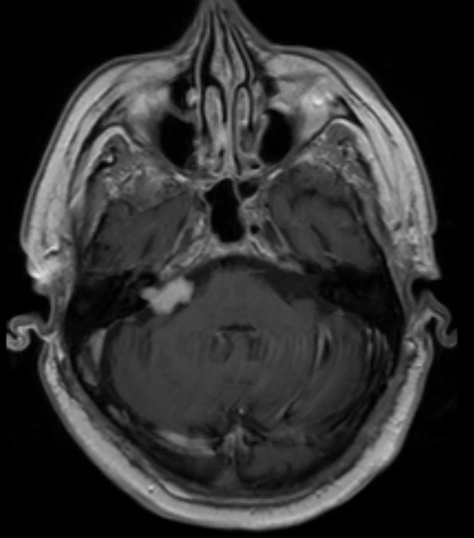
- MRI internal auditory meati with gadolinium - the gold standard investigation, detecting even small intracanalicular tumours as an avidly enhancing lesion, classically with an 'ice cream cone' appearance where the intracanalicular portion (the cone) extends into a CPA component (the scoop)
- CT - inferior to MRI and may miss small tumours entirely, but used where MRI is contraindicated, and shows widening of the internal auditory canal in larger lesions
- Auditory brainstem response testing - historically used as a screening test but largely superseded by MRI, which is far more sensitive
- Vestibular function testing - not usually needed diagnostically, but may inform surgical planning
- Genetic testing and referral - where bilateral tumours or a suggestive family history raise the possibility of NF2
Management
Because these tumours are benign and often very slow-growing, treatment is individualised to tumour size, growth rate on serial imaging, symptoms, hearing status, patient age and comorbidity, and is decided within a skull base multidisciplinary team.
Growth rate, rather than size at diagnosis, is often the decisive factor. Many vestibular schwannomas grow at only 1-2 mm per year, and a proportion do not grow at all, so the first scan establishes a baseline rather than a mandate to intervene. A repeat scan at six to twelve months, and then at lengthening intervals if stable, allows the tumour's own behaviour to guide management.
The counterpoint is that hearing tends to decline over time whether or not the tumour grows, so a patient with useful hearing who is hoping to preserve it may prefer earlier intervention. This trade-off - between avoiding the risks of treatment and preserving hearing that may be lost while waiting - is the central conversation to have with the patient, and there is rarely a single right answer.
| Option | Best suited to | Key considerations |
|---|---|---|
| Observation ('watch, wait and rescan') | Small, asymptomatic or minimally symptomatic tumours, and older or comorbid patients | Serial MRI, initially annually; many tumours grow very slowly or not at all, so intervention may never be needed |
| Stereotactic radiosurgery (e.g. Gamma Knife) | Small-to-medium tumours (generally under ~3 cm), or patients unfit for open surgery | Aims to arrest growth rather than remove the tumour; good facial nerve preservation rates, though hearing often declines over time |
| Microsurgical resection | Large tumours, those causing brainstem compression or hydrocephalus, and documented significant growth | Definitive removal, but carries risks of facial nerve injury, hearing loss and CSF leak; approach chosen partly by whether hearing preservation is realistic |
Supportive management
- Hearing rehabilitation - conventional hearing aids where useful hearing remains, and bone-anchored hearing aids or a CROS aid for single-sided deafness
- Vestibular rehabilitation - physiotherapy to promote central compensation for imbalance
- Eye protection - essential if there is combined facial weakness and reduced corneal sensation, since the eye is at high risk of exposure keratopathy; lubricants, taping at night and ophthalmology input
- Tinnitus management - counselling, sound therapy and psychological support
- Genetic counselling and surveillance for patients with NF2 and their families
Complications
- Progressive or complete unilateral hearing loss, with the practical difficulties of single-sided deafness in noisy environments
- Persistent tinnitus
- Chronic imbalance
- Facial nerve palsy - from tumour growth or, more often, as a complication of treatment
- Exposure keratopathy where facial weakness and corneal anaesthesia coexist
- Trigeminal sensory loss and, occasionally, neuropathic facial pain
- Brainstem compression, cerebellar dysfunction and obstructive hydrocephalus with very large tumours
- Surgical complications - CSF leak, meningitis, and (rarely) intracranial haemorrhage
Red flags
Prognosis
Vestibular schwannomas are benign and do not metastasise, so prognosis for survival is excellent. Many small tumours grow extremely slowly, or not at all, and a substantial proportion of patients managed with observation never require intervention - which is why 'watch, wait and rescan' is a legitimate active management strategy rather than simply doing nothing.
The main determinants of long-term quality of life are hearing outcome, facial nerve function and balance, rather than tumour recurrence. Hearing tends to decline over time regardless of the approach taken, while facial nerve outcomes are generally good with radiosurgery and with microsurgery for smaller tumours, but less predictable when resecting large lesions. Patients with NF2 face a more complex course, with bilateral disease, a higher likelihood of eventual bilateral deafness, and the burden of associated tumours requiring lifelong surveillance.
Patients frequently find the term acoustic neuroma more alarming than the reality warrants, particularly when it is first mentioned alongside the word tumour. Explaining clearly that these lesions are benign, do not spread, frequently grow very slowly or not at all, and that observation is an active and legitimate management strategy rather than neglect, addresses a great deal of anxiety at the point of diagnosis and helps patients engage with a surveillance plan rather than pressing for immediate intervention.
References
- NICE CKS. Hearing loss in adults. Available here
- NICE NG98. Hearing loss in adults: assessment and management. 2018. Available here
- Carlson ML, Link MJ. Vestibular schwannomas. New England Journal of Medicine. 2021. Available here
- Evans DG. Neurofibromatosis type 2: genetics, clinical features and management. Orphanet Journal of Rare Diseases. 2009. Available here
This article is written for revision and education. It is not clinical guidance and must not be used to make decisions about the care of a patient. Always check current NICE guidance and local protocols.