
Cranial Nerve Examination: A Practical Guide
Key points
- Purpose: the cranial nerve examination localises lesions - the specific combination of nerves affected tells you where the problem is, often more precisely than imaging alone.
- Structure: work through the nerves in numerical order (I-XII); this prevents omissions under exam pressure and produces a clear, reproducible presentation.
- Pupillary pathway: the light reflex tests CN II (afferent) and CN III (efferent) - so a lesion in either produces an abnormal response, but in different ways.
- Facial nerve: forehead sparing indicates an upper motor neurone (central) lesion; forehead involvement indicates a lower motor neurone lesion such as Bell's palsy.
- Third nerve palsy: ptosis, a 'down and out' eye, and pupil involvement suggesting a compressive cause (posterior communicating artery aneurysm) rather than a medical one.
- Rinne and Weber: together distinguish conductive from sensorineural hearing loss at the bedside, and should be interpreted as a pair, never in isolation.
- Cerebellopontine angle: lesions here characteristically affect CN VIII, then V, then VII - the pattern that suggests a vestibular schwannoma.
- Localisation principle: multiple adjacent cranial nerves affected together suggests a single lesion at a shared anatomical site, such as the brainstem, cavernous sinus or skull base.
Introduction
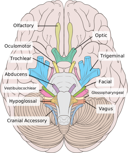
The cranial nerve examination is one of the most commonly assessed OSCE stations in medical finals, and one of the most clinically useful examinations in neurology. Its power lies in localisation: because the twelve cranial nerves have precisely known courses through the brainstem, skull base and orbit, the specific combination of nerves affected tells you where a lesion must be, often before any imaging is performed.1
This article covers how to perform each component, what the classic abnormal findings mean, and - most importantly - how to reason from a pattern of findings to an anatomical site. It complements the articles on Bell's palsy, vestibular schwannoma and stroke, where these signs frequently appear.
Before you begin
- Introduce yourself, confirm the patient's identity, explain what you would like to do and obtain consent
- Position the patient sitting comfortably, roughly at your eye level and about an arm's length away
- Expose appropriately - the head and neck need to be visible
- General inspection first - look from the end of the bed for facial asymmetry, ptosis, obvious wasting, abnormal posture, hearing aids, walking aids, or a nasogastric tube (suggesting swallowing difficulty)
- Ask about pain before touching the patient
CN I - Olfactory
Test: ask whether the patient has noticed any change in their sense of smell or taste. Formal testing uses standardised odours presented to each nostril separately with the other occluded, though in most OSCE settings the question alone is accepted.
- Anosmia - causes include upper respiratory tract infection (much the commonest), head trauma shearing the olfactory fibres at the cribriform plate, nasal disease, a frontal lobe or olfactory groove meningioma, and Parkinson's disease, in which hyposmia can precede motor symptoms by years
- Kallmann syndrome - anosmia with hypogonadotropic hypogonadism, a classic association
CN II - Optic
The optic nerve is assessed in five parts, conventionally remembered as acuity, fields, reflexes, colour and fundoscopy.
- Visual acuity - Snellen chart at the correct distance, testing each eye separately, with the patient's usual glasses on (correcting refractive error, which is not a neurological problem)
- Visual fields - confrontation testing, comparing the patient's field with your own, quadrant by quadrant, each eye separately. Also test for visual inattention by presenting stimuli in both fields simultaneously
- Pupillary reflexes - direct and consensual responses to light, the swinging light test for a relative afferent pupillary defect, and accommodation
- Colour vision - Ishihara plates; red desaturation (asking whether a red object looks less vivid in one eye) is a sensitive early sign of optic nerve disease
- Fundoscopy - looking for papilloedema, optic atrophy, and retinal pathology
| Defect | Site of lesion |
|---|---|
| Monocular visual loss | Optic nerve on that side (anterior to the chiasm) |
| Bitemporal hemianopia | Optic chiasm - classically compression from a pituitary adenoma or craniopharyngioma |
| Homonymous hemianopia | Optic tract or radiation, contralateral to the field loss (i.e. behind the chiasm) |
| Homonymous quadrantanopia | Superior quadrant loss - temporal lobe (Meyer's loop); inferior quadrant loss - parietal lobe |
| Homonymous hemianopia with macular sparing | Occipital cortex, typically posterior cerebral artery territory infarction |
CN III, IV and VI - Oculomotor, trochlear and abducens
These three are tested together because they act as a unit to move the eye. Test: inspect for ptosis and resting eye position, then ask the patient to follow your finger through an 'H' pattern, keeping their head still, and ask about diplopia at each position. Check for nystagmus at the extremes of gaze.
| Nerve | Supplies | Palsy produces |
|---|---|---|
| CN III (oculomotor) | All extraocular muscles except lateral rectus and superior oblique; levator palpebrae superioris; parasympathetic supply to the pupil | Ptosis, eye deviated 'down and out', and (if the parasympathetic fibres are involved) a fixed, dilated pupil |
| CN IV (trochlear) | Superior oblique | Vertical diplopia, worse on looking down and inward - patients characteristically tilt the head away from the affected side to compensate; classically noticed on descending stairs |
| CN VI (abducens) | Lateral rectus | Failure of abduction, with horizontal diplopia worse on looking towards the affected side |
CN V - Trigeminal
Test: sensation in all three divisions (ophthalmic V1, maxillary V2, mandibular V3) bilaterally to light touch and, if indicated, pinprick and temperature; motor function by palpating masseter and temporalis while the patient clenches, and testing jaw opening against resistance; and the corneal reflex (afferent V1, efferent CN VII) with a wisp of cotton wool - though this is often only mentioned rather than performed in OSCEs.
- Sensory loss in a trigeminal distribution - consider a cerebellopontine angle lesion, cavernous sinus pathology, or a brainstem lesion
- Reduced or absent corneal reflex - often the earliest trigeminal sign, and an important finding in vestibular schwannoma
- Jaw deviation towards the weak side on opening, from unopposed action of the healthy pterygoid
- Trigeminal neuralgia - severe paroxysmal facial pain, but with a characteristically normal examination; sensory loss should prompt imaging for a secondary cause
CN VII - Facial
Test: inspect for facial asymmetry at rest, then ask the patient to raise their eyebrows, screw their eyes tightly shut (and try to open them against resistance), puff out their cheeks, show their teeth, and purse their lips. Ask about taste on the anterior two-thirds of the tongue and about hyperacusis.
- Lower motor neurone facial palsy - Bell's palsy, Ramsay Hunt syndrome (with a vesicular rash in the ear), parotid tumour, cerebellopontine angle lesion, or Lyme disease (which can cause bilateral palsy)
- Upper motor neurone facial weakness - stroke or other contralateral hemispheric lesion, with forehead sparing
- Inability to close the eye - risks exposure keratopathy and needs active eye protection
CN VIII - Vestibulocochlear
Test: gross hearing by whispering a number in each ear while masking the other, then perform Rinne's and Weber's tests with a 512 Hz tuning fork. Assess balance and, where vertigo is the complaint, consider positional testing.
| Pattern | Rinne | Weber | Interpretation |
|---|---|---|---|
| Normal | Air > bone conduction, both ears | Central (no lateralisation) | Normal hearing, or symmetrical loss |
| Conductive loss (left) | Bone > air on the left | Lateralises to the LEFT (affected ear) | Wax, effusion, perforation, otosclerosis |
| Sensorineural loss (left) | Air > bone both ears | Lateralises to the RIGHT (better ear) | Presbycusis, noise damage, vestibular schwannoma |
CN IX and X - Glossopharyngeal and vagus
Test: listen to the patient's voice for hoarseness or a nasal quality, ask them to cough (a 'bovine' cough without the normal explosive onset suggests recurrent laryngeal nerve palsy), ask them to open their mouth and say 'aah' while observing palatal movement, and assess swallow if clinically indicated. The gag reflex (afferent IX, efferent X) is unpleasant and usually only mentioned rather than performed.
- Uvula deviates AWAY from the side of the lesion - the healthy side pulls it across, so the uvula points away from the weak side
- Hoarseness or bovine cough - recurrent laryngeal nerve palsy; consider lung cancer, thyroid surgery, or an aortic arch lesion given the left recurrent laryngeal nerve's course through the chest
- Dysphagia and nasal regurgitation - bulbar or pseudobulbar palsy
- Bulbar palsy (lower motor neurone) - a wasted, fasciculating tongue with a nasal voice, seen in motor neurone disease and Guillain-Barre syndrome; pseudobulbar palsy (upper motor neurone) - a spastic tongue, brisk jaw jerk and emotional lability
CN XI - Accessory
Test: ask the patient to shrug their shoulders against resistance (trapezius) and to turn their head to each side against resistance while you palpate the opposite sternocleidomastoid.
- Note the crossed innervation: the right sternocleidomastoid turns the head to the left, so weakness turning the head to the left indicates a right sternocleidomastoid (and therefore right accessory nerve) problem
- Causes of palsy: iatrogenic injury during posterior triangle neck surgery or lymph node biopsy is the commonest, plus trauma, and lesions at the jugular foramen or brainstem
CN XII - Hypoglossal
Test: inspect the tongue in the floor of the mouth at rest for wasting and fasciculation (fasciculations are unreliable to assess in a protruded tongue), then ask the patient to protrude it and move it side to side, and to push it into each cheek against your finger.
- Tongue deviates TOWARDS the side of the lesion in a lower motor neurone palsy - the healthy genioglossus pushes it across
- Wasting and fasciculation - a lower motor neurone sign; a wasted, fasciculating tongue is a classic and important finding in motor neurone disease
- A spastic, slow-moving tongue without wasting suggests an upper motor neurone (pseudobulbar) lesion
Localising from the pattern
The real diagnostic power comes from recognising combinations of affected nerves, because cranial nerves that travel together are affected together by a single lesion.
| Pattern | Site | Typical causes |
|---|---|---|
| CN VIII, then V, then VII | Cerebellopontine angle | Vestibular schwannoma, meningioma |
| CN III, IV, V1, V2, VI | Cavernous sinus | Cavernous sinus thrombosis, pituitary apoplexy, carotid aneurysm, tumour |
| CN IX, X, XI | Jugular foramen | Glomus tumour, skull base metastasis, basal skull fracture |
| Ipsilateral cranial nerve palsy with contralateral limb weakness | Brainstem ('crossed' signs) | Brainstem stroke, demyelination, tumour |
| Multiple lower cranial nerves, progressive | Skull base | Nasopharyngeal carcinoma, metastasis, chronic meningitis, granulomatous disease |
Completing the examination
- Thank the patient and ensure they are comfortable and appropriately covered
- State what you would do to complete the examination: a full peripheral neurological examination of the upper and lower limbs, cerebellar examination, fundoscopy if not already performed, otoscopy, blood pressure, and a swallow assessment where bulbar function is impaired
- Summarise your findings clearly, stating the abnormal nerves, whether the lesion is upper or lower motor neurone, and your proposed anatomical localisation
- Suggest relevant investigations - typically MRI brain for a structural cause, alongside bloods and any condition-specific tests
References
- Fuller G. Neurological Examination Made Easy. Elsevier. Available here
- NICE CKS. Bell's palsy. Available here
- NICE NG98. Hearing loss in adults: assessment and management. 2018. Available here
- NICE NG128. Stroke and transient ischaemic attack in over 16s. 2019, updated 2022. Available here
This article is written for revision and education. It is not clinical guidance and must not be used to make decisions about the care of a patient. Always check current NICE guidance and local protocols.