
Myasthenia Gravis: Diagnosis and Management
Key points
- Myasthenia gravis (MG): an autoimmune disease in which antibodies attack the postsynaptic neuromuscular junction, most often targeting the acetylcholine receptor.
- Cardinal feature: fatigable weakness - muscle strength worsens with sustained or repeated use and improves with rest, classically affecting the eyes first.
- Typical early presentation: ptosis and diplopia, often asymmetric and variable through the day, worse in the evening.
- Key antibody: anti-acetylcholine receptor (anti-AChR) antibodies in the majority; anti-MuSK antibodies in a further subset with a distinct, often more severe phenotype.
- Thymus association: thymic hyperplasia or thymoma is present in a significant proportion - CT chest is part of the standard work-up.
- First-line symptomatic treatment: pyridostigmine, an acetylcholinesterase inhibitor.
- Immunosuppression: corticosteroids and steroid-sparing agents for disease not controlled by pyridostigmine alone.
- Myasthenic crisis: life-threatening respiratory failure from severe weakness - a medical emergency treated with IVIG or plasma exchange and, if needed, ventilatory support.
Introduction
Myasthenia gravis (MG) is an autoimmune disease of the neuromuscular junction, in which antibodies impair transmission at the postsynaptic membrane, most commonly by targeting the nicotinic acetylcholine receptor (AChR).1 The result is fatigable weakness: muscle strength that is relatively preserved at rest but declines with sustained or repetitive activity, recovering again after rest - a pattern that, once recognised, is highly distinctive.
MG classically affects the extraocular, bulbar and proximal limb muscles, sparing sensation entirely, and follows a bimodal age distribution: younger women (often with thymic hyperplasia) and older men (more often with thymoma or no thymic abnormality at all). It is a compact, high-yield topic combining immunology, a specific and testable clinical sign (fatigability), and a genuine medical emergency (myasthenic crisis).
Prevalence is roughly 15-20 per 100,000 and has risen over recent decades, partly through better recognition and partly through improved survival. The classic bimodal distribution matters clinically: early-onset disease predominantly affects women in their twenties and thirties and is more often associated with thymic hyperplasia, while late-onset disease predominantly affects men over 50 and carries a higher likelihood of thymoma - which is why the CT chest is not optional in either group.
Pathophysiology
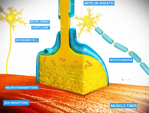
In the majority of patients, IgG antibodies bind the postsynaptic acetylcholine receptor, causing complement-mediated destruction of the receptor-rich folds of the postsynaptic membrane and accelerated receptor turnover. This reduces the safety margin for neuromuscular transmission, so that with repeated stimulation - as acetylcholine release itself normally declines slightly with each successive impulse - the fewer available receptors are progressively unable to trigger an action potential, producing fatigable weakness.
A smaller but clinically important subset of patients instead have antibodies against muscle-specific kinase (MuSK), a protein essential for clustering acetylcholine receptors during neuromuscular junction formation and maintenance; this group tends to have more prominent bulbar and respiratory involvement and responds differently to some treatments.2
Aetiology and associations
- Thymic hyperplasia - present in a substantial proportion, particularly younger women with AChR-antibody-positive disease
- Thymoma - present in around 10-15% of patients, more often in older patients; MG can also occur as a paraneoplastic phenomenon associated with thymoma
- Other autoimmune disease - increased association with autoimmune thyroid disease, rheumatoid arthritis, and other organ-specific autoimmune conditions
- Drug-induced/precipitated myasthenic syndromes - penicillamine can induce a myasthenic syndrome; several drugs (see Red flags) can unmask or worsen existing MG
Clinical features
The defining feature is fatigability: weakness that worsens with sustained or repeated activity through the day and with exercise, and improves with rest - classically worse in the evening and after prolonged use of the affected muscles.
Understanding why the eyes are affected first, and so disproportionately, is worth a moment. Extraocular muscles fire at very high frequencies, have a lower safety margin for neuromuscular transmission than limb muscles, and have relatively few acetylcholine receptors per junction to begin with. They are therefore the first to fail when receptor numbers fall. In addition, the visual system detects even slight misalignment of the two eyes immediately as diplopia, so a small degree of weakness that would be imperceptible in a limb produces an obvious and disabling symptom.
The same logic explains why bulbar and respiratory muscles matter clinically out of proportion to limb strength. A patient can have near-normal power in the arms and legs while their diaphragm and swallowing muscles are failing, which is precisely why assessment must include forced vital capacity and a swallow assessment rather than relying on limb testing to gauge severity.
- Ocular symptoms - the commonest presenting feature: ptosis (often asymmetric, worsening as the day goes on or with sustained upward gaze) and diplopia from extraocular muscle weakness; pupillary responses are always spared, which helps distinguish MG from a third nerve palsy
- Bulbar symptoms - fatigable chewing (worse towards the end of a meal), dysphagia, and a nasal, quiet, or slurred voice that worsens with prolonged speech
- Facial weakness - a 'myasthenic snarl' on smiling, from weakness of orbicularis oris
- Limb weakness - typically proximal and symmetric, affecting shoulder abduction and hip flexion more than distal muscles
- Respiratory muscle weakness - can progress to life-threatening respiratory failure (myasthenic crisis)
- No sensory loss and reflexes are preserved - important negatives that support the diagnosis and distinguish it from other neuromuscular conditions
- Ocular myasthenia - disease confined to the eyes; a significant proportion of these patients go on to generalise to other muscle groups within 2 years
Clinical examination
- Ptosis - assess at rest and after sustained upgaze (worsens); the 'curtain sign' - lifting one drooping eyelid manually can unmask or worsen ptosis on the other side
- Eye movements - test for diplopia and extraocular weakness; pupils should be normal
- Facial power and 'myasthenic snarl'
- Repetitive/sustained testing - shoulder abduction held or repeated to demonstrate fatigable weakness, rather than a single maximal-effort test
- Bulbar function - count aloud, or ask the patient to read for several minutes and listen for a progressively nasal, weakening voice
- Reflexes and sensation - both should be entirely normal
- Respiratory function - observe respiratory effort; assess forced vital capacity if there is any concern about respiratory involvement
Differential diagnosis
- Lambert-Eaton myasthenic syndrome (LEMS) - a presynaptic disorder (antibodies against voltage-gated calcium channels), often paraneoplastic (classically small cell lung cancer); weakness characteristically improves with sustained contraction (post-tetanic facilitation) rather than worsening, and autonomic symptoms (dry mouth, erectile dysfunction) are prominent
- Third nerve palsy - ptosis and diplopia, but with a dilated, unreactive pupil (in a compressive lesion) and no fatigability
- Chronic progressive external ophthalmoplegia - a mitochondrial disorder causing slowly progressive, non-fatigable ptosis and ophthalmoplegia
- Guillain-Barré syndrome - ascending weakness with areflexia, but not classically fatigable, and often with sensory symptoms
- Botulism - descending paralysis with autonomic involvement, from toxin-mediated presynaptic blockade
- Thyroid eye disease - can cause diplopia and lid abnormalities but typically with proptosis and lid retraction rather than ptosis
Investigations
- Anti-acetylcholine receptor (AChR) antibodies - positive in the majority of patients with generalised MG and a smaller proportion with purely ocular disease; highly specific when positive
- Anti-MuSK antibodies - tested if AChR antibodies are negative; associated with more prominent bulbar/respiratory involvement and a different treatment response profile
- Anti-LRP4 antibodies - a further, smaller subgroup; tested in specialist settings when both of the above are negative but suspicion remains high
- Edrophonium (Tensilon) test - historically used, showing transient improvement in weakness after IV edrophonium (a short-acting acetylcholinesterase inhibitor); now rarely used because of cardiac arrhythmia risk and superseded by antibody testing and neurophysiology
- Repetitive nerve stimulation - shows a decremental response in compound muscle action potential amplitude with repeated stimulation
- Single-fibre electromyography - the most sensitive test, showing increased 'jitter' between muscle fibres in the same motor unit; used when antibody testing is negative but clinical suspicion remains
- CT chest - performed in all patients with confirmed MG to look for thymoma or thymic hyperplasia, since it changes management
- Pulmonary function tests (forced vital capacity) - to assess and monitor respiratory muscle involvement, particularly if crisis is a concern
Management
Symptomatic treatment
Pyridostigmine, an acetylcholinesterase inhibitor, is first-line symptomatic treatment. By slowing the breakdown of acetylcholine in the synaptic cleft, it increases the chance that the reduced number of available receptors will still be activated, improving strength - though it treats the symptom, not the underlying autoimmune process.
Immunosuppression
- Corticosteroids - for disease not adequately controlled by pyridostigmine alone; started cautiously, sometimes with initial in-patient monitoring, since high-dose steroids can transiently worsen myasthenic weakness before improving it
- Steroid-sparing immunosuppressants - azathioprine, mycophenolate mofetil, or others, used to reduce long-term steroid exposure
- Biologic therapies (e.g. rituximab, complement inhibitors, FcRn antagonists) - increasingly used in refractory disease, particularly MuSK-antibody-positive MG which often responds well to rituximab specifically
Thymectomy
Thymectomy is indicated for all patients with a thymoma, regardless of age or disease severity, since the tumour itself needs treating. It is also considered in AChR-antibody-positive generalised MG without thymoma, particularly in younger patients, since trial evidence shows it improves clinical outcomes and reduces immunosuppressant requirements even without a demonstrable thymoma.3
Myasthenic crisis
Complications
- Myasthenic crisis with respiratory failure
- Aspiration pneumonia from bulbar weakness
- Progression from ocular to generalised disease
- Falls and injury from limb weakness
- Side effects of long-term immunosuppression (infection risk, osteoporosis with steroids)
- Cholinergic side effects, or rarely cholinergic crisis, from acetylcholinesterase inhibitor treatment
Red flags
Prognosis
With modern treatment, most patients with MG achieve good symptom control and a normal life expectancy, though the disease typically requires long-term management and treatment adjustment. Ocular disease that has not generalised within about 2 years is less likely to do so subsequently. Thymectomy and early, appropriately escalated immunosuppression have improved long-term outcomes considerably. Myasthenic crisis carries significant morbidity but is usually survivable with prompt recognition, respiratory support and rapid-acting immunotherapy.
Myasthenia has important implications in two specific situations that are easily forgotten. In pregnancy, the course is unpredictable and may worsen in the first trimester and puerperium; magnesium sulfate, used for eclampsia, blocks neuromuscular transmission and is relatively contraindicated. Maternal antibodies can cross the placenta to cause transient neonatal myasthenia, which resolves as maternal antibody is cleared but requires the neonatal team to be forewarned.
In the perioperative period, patients are sensitive to non-depolarising neuromuscular blockers and may need markedly reduced doses, with a real risk of prolonged post-operative ventilation. Anaesthetic teams should always be informed of the diagnosis in advance, and elective surgery is best undertaken when the disease is well controlled.
Finally, patients should be given clear guidance on recognising deterioration and on which drugs to avoid, ideally in written form and with an alert card. Because myasthenic crisis is frequently precipitated by an intercurrent infection or by a newly prescribed antibiotic, patients who understand to seek help early with breathlessness, weak cough or worsening swallowing - and to flag their diagnosis before any new prescription - are considerably safer than those relying on clinicians to remember the interactions.
References
- NICE CKS. Myasthenia gravis. Available here
- Gilhus NE, Verschuuren JJ. Myasthenia gravis: subgroup classification and therapeutic strategies. The Lancet Neurology. 2015. Available here
- Wolfe GI, Kaminski HJ, Aban IB et al. Randomized trial of thymectomy in myasthenia gravis. New England Journal of Medicine. 2016. Available here
This article is written for revision and education. It is not clinical guidance and must not be used to make decisions about the care of a patient. Always check current NICE guidance and local protocols.