
Principles of Cancer Diagnosis and Staging
Key points
- Tissue is the issue: with very few exceptions, a cancer diagnosis requires histological or cytological confirmation before treatment begins.
- Grade: how abnormal the cells look down the microscope - a property of the tumour cells themselves.
- Stage: how far the tumour has spread anatomically - a property of the disease in the patient. Grade and stage are not the same thing.
- TNM: T is local tumour extent, N is regional lymph node involvement, M is distant metastasis. These combine into an overall stage of I to IV.
- Staging investigations: usually CT of the chest, abdomen and pelvis, with PET-CT, MRI or bone scan added where they change the plan.
- Performance status: the ECOG/WHO scale of 0 to 4 predicts tolerance of systemic anti-cancer therapy and is often the deciding factor in treatment.
- Tumour markers: are used for monitoring and prognosis, not for making a diagnosis - the important exceptions are AFP and hCG in germ cell tumours.
- MDT: every new cancer diagnosis in the NHS is discussed at a site-specific multidisciplinary team meeting, which records the stage and the intent of treatment.
Introduction
Around 375,000 people are diagnosed with cancer in the UK each year, and roughly one in two people born after 1960 will develop cancer at some point in their lives.3 Whichever specialty you end up in, you will diagnose cancer, break the news, and follow patients through treatment. The UKMLA expects you to understand the shared framework that sits behind every tumour site rather than the details of any one of them.
That framework has three questions in a fixed order. What is it? - answered by tissue diagnosis. How far has it spread? - answered by staging. What can this particular patient tolerate? - answered by performance status and comorbidity assessment. Only when all three are known can a multidisciplinary team decide whether treatment is given with curative or palliative intent, and that single decision shapes everything that follows.
Getting the order right matters clinically. Treating before tissue confirmation risks giving toxic therapy for a benign condition or for the wrong malignancy; staging after treatment has started makes the result uninterpretable. The main exception is an oncological emergency, where treatment such as dexamethasone for suspected cord compression begins before the diagnostic work-up is complete.
How cancer comes to attention
Recognising the route by which a patient presents tells you something about the likely stage and prognosis before any investigation is done. Emergency presentations carry markedly worse outcomes than screen-detected disease, largely because the disease is more advanced by the time it declares itself.
- Symptomatic presentation to primary care - the commonest route. NICE NG12 sets symptom-based thresholds for urgent suspected cancer referral, most of which correspond to a positive predictive value of around 3%.1
- Screening - asymptomatic detection through one of the three national adult programmes, generally identifying earlier-stage disease
- Incidental finding - a lesion found on imaging or blood tests requested for another reason, an increasingly common route as cross-sectional imaging use rises
- Emergency presentation - bowel obstruction, pathological fracture, seizure from a brain metastasis, malignant hypercalcaemia or spinal cord compression. Around one in five cancers in the UK is still diagnosed this way.
- Paraneoplastic syndrome - hyponatraemia from SIADH, Cushing's syndrome from ectopic ACTH, or Lambert-Eaton myasthenic syndrome, sometimes preceding the tumour by months
The national screening programmes
| Programme | Population | Test | Interval |
|---|---|---|---|
| Bowel | Age 50 to 74, following a phased extension downwards from 60 | Faecal immunochemical test (FIT) posted to the home | Every 2 years |
| Breast | Age 50 to 71 | Two-view mammography | Every 3 years |
| Cervical | Age 25 to 64 | Primary high-risk HPV testing, with cytology only if HPV positive | Every 3 years to age 49, then every 5 years |
Two points are examined repeatedly. First, screening is offered to people without symptoms - a patient with rectal bleeding needs a suspected cancer referral, not a screening kit, and telling them to wait for their next round is a serious error. Second, cervical screening moved to primary HPV testing because it is more sensitive for the lesions that matter, and a negative HPV test allows a longer interval safely.5
Making the diagnosis: obtaining tissue
Imaging can be highly suggestive but it cannot distinguish a lymphoma from a carcinoma, establish receptor status, or exclude a benign mimic such as tuberculosis or sarcoidosis. Histology remains the reference standard, and the practical question is which technique will yield enough tissue with the least risk.
| Technique | What it gives | Typical use and limitation |
|---|---|---|
| Fine needle aspiration (FNA) | Cytology - individual cells only, no architecture | Thyroid nodules and neck lumps. Quick and low risk, but cannot distinguish follicular adenoma from carcinoma, and is poor for lymphoma. |
| Core biopsy | Histology - a cylinder of tissue with preserved architecture | The workhorse for breast, prostate, liver and lung lesions. Allows grading, immunohistochemistry and molecular testing. |
| Incisional biopsy | A sample of a larger lesion | Used when a core is non-diagnostic. In suspected sarcoma the biopsy tract must be planned by the sarcoma centre so that it can be excised later. |
| Excisional biopsy | The whole lesion | Lymph nodes in suspected lymphoma, and skin lesions in suspected melanoma, where architecture and full depth are essential. |
| Endoscopic biopsy | Direct visualisation plus histology | Upper GI endoscopy, colonoscopy, bronchoscopy, cystoscopy. Also allows tattooing of a lesion for later surgery. |
| Image-guided biopsy | Access to deep lesions | Ultrasound or CT-guided. Carries a small risk of bleeding, pneumothorax and, rarely, seeding along the needle tract. |
| Cytology of fluid | Malignant cells in an effusion or in urine | Pleural fluid, ascites, urine. A positive result confirms malignancy; a negative result excludes nothing, as sensitivity in pleural fluid is only around 60%. |
When tissue is not obtained first
- Testicular tumours - a suspicious testicular mass is removed by radical inguinal orchidectomy, which is both diagnostic and therapeutic. Trans-scrotal biopsy risks seeding and alters the lymphatic drainage.
- Some hepatocellular carcinomas - a lesion in a cirrhotic liver with characteristic arterial enhancement and venous washout on multiphase imaging is diagnostic without biopsy
- Brain lesions - where the location makes biopsy hazardous, treatment may proceed on radiological grounds after neuro-oncology MDT discussion
- Oncological emergencies - dexamethasone for suspected cord compression, or rasburicase where tumour lysis risk is high, is given before histology is available
What the pathologist reports
A histopathology report is more than a name. Royal College of Pathologists cancer datasets specify a minimum set of items for each tumour site, because those items are exactly what the MDT needs to make a decision.6
Tumour type
The broad lineage is established first: carcinoma (epithelial, subdivided into adenocarcinoma, squamous cell carcinoma and others), sarcoma (mesenchymal), lymphoma or leukaemia (haematopoietic), melanoma (melanocytic), or germ cell tumour. This is then refined using the current WHO classification for that organ, which increasingly incorporates molecular features alongside morphology.10
Grade
Grade describes how closely the tumour resembles the tissue it arose from - its degree of differentiation. A well-differentiated tumour retains recognisable architecture and generally behaves less aggressively; a poorly differentiated or anaplastic tumour has lost it. Grading is usually reported as G1 (well differentiated) to G4 (undifferentiated), but several sites use their own system.
- Prostate - the Gleason score, adding the two commonest patterns (each scored 3 to 5), now reported as Grade Groups 1 to 5
- Breast - the Nottingham (modified Bloom-Richardson) grade, scoring tubule formation, nuclear pleomorphism and mitotic count
- Sarcoma and glioma - grade is one of the strongest predictors of behaviour and drives treatment directly
- Cervical and other squamous sites - grade adds relatively little once stage is known, and is not used to select treatment
Immunohistochemistry and molecular testing
Immunohistochemistry uses labelled antibodies to detect proteins in tissue, which both confirms lineage and identifies therapeutic targets. Cytokeratins indicate epithelial origin, CD45 (leucocyte common antigen) indicates lymphoid origin, S100 and SOX10 indicate melanocytic origin, and TTF-1 suggests a lung or thyroid primary. Oestrogen receptor, progesterone receptor and HER2 status in breast cancer determine whether endocrine therapy and trastuzumab are used at all.
Molecular testing has moved from research into routine care. The examples that recur in finals are EGFR mutation and ALK rearrangement in non-small cell lung cancer, RAS and BRAF status in colorectal cancer (a RAS mutation predicts a lack of response to cetuximab), BRAF V600E in melanoma, and mismatch repair or microsatellite instability testing, which flags possible Lynch syndrome and predicts response to immunotherapy.
Staging
Staging answers the question of anatomical extent, and it is the single most important determinant of both treatment intent and prognosis. The international standard is the TNM classification, maintained by the UICC and now in its eighth edition.2,9
| Component | What it describes | Typical values |
|---|---|---|
| T - tumour | Size and, more often, depth of local invasion through the layers of the organ | Tis (carcinoma in situ), then T1 to T4, with T4 usually meaning invasion of adjacent structures |
| N - nodes | Involvement of regional lymph nodes, by number or by anatomical station | N0 (none) to N3. Nodes outside the regional field count as metastatic disease, not as N. |
| M - metastasis | Distant spread beyond the regional nodes | M0 or M1. There is no M2 - the presence of any distant metastasis is what matters. |
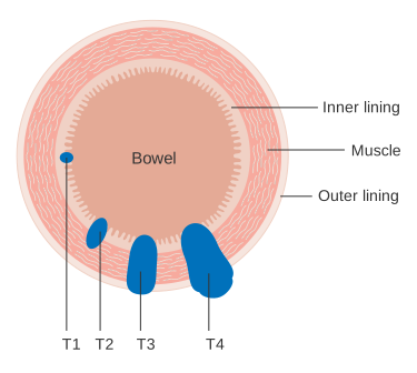
Prefixes carry meaning and are worth recognising on a report. c denotes clinical staging based on examination and imaging (cT3N1M0); p denotes pathological staging after resection (pT2N0); y denotes staging after neoadjuvant therapy (ypT0N0 meaning a complete pathological response); and r denotes recurrent disease. Pathological staging is more accurate, but it is only available if the tumour is resected.
From TNM to overall stage
The T, N and M categories are combined into an overall stage group of I to IV using site-specific rules. Although the groupings differ between tumours, the underlying logic is consistent:
| Stage | General meaning | Usual treatment intent |
|---|---|---|
| 0 | Carcinoma in situ - has not breached the basement membrane | Curative, often local excision alone |
| I | Small tumour confined to the organ of origin, no nodal involvement | Curative, usually surgery alone |
| II | Larger or more deeply invasive, still node-negative | Curative, with adjuvant therapy considered |
| III | Regional lymph node involvement | Curative but multimodal - surgery plus chemotherapy and/or radiotherapy |
| IV | Distant metastasis | Usually palliative, with important exceptions such as germ cell tumours, lymphoma and oligometastatic colorectal disease |
Site-specific systems you still need to know
- Ann Arbor with the Lugano modification for lymphoma - stages I to IV by the number of nodal regions involved and whether they lie on one or both sides of the diaphragm, with the suffix B for weight loss, fever and night sweats
- FIGO for gynaecological cancers - cervical, endometrial, ovarian and vulval, broadly parallel to TNM
- Breslow thickness for melanoma - depth of invasion in millimetres, the strongest single prognostic factor, feeding directly into the T category
- Rai and Binet for chronic lymphocytic leukaemia, which stage by blood counts and organ involvement rather than by imaging
- Dukes for colorectal cancer - A confined to the bowel wall, B through the wall, C nodal involvement, D distant metastasis. Superseded by TNM but still quoted on wards and in exams.
Staging investigations
The principle is to request the investigations that will change the plan, and no more. A patient who is clearly unfit for any anti-cancer treatment gains nothing from a full staging work-up, and subjecting them to it is a failure of judgement rather than a display of thoroughness.
- CT of the chest, abdomen and pelvis with contrast - the default staging scan for almost all solid tumours, assessing nodal disease and the common metastatic sites of lung, liver, adrenal and bone
- MRI - superior soft tissue contrast. The standard for local staging in rectal cancer (predicting the circumferential resection margin), prostate cancer (multiparametric MRI before biopsy), gynaecological tumours, sarcoma, and any suspected brain or spinal involvement.
- PET-CT using 18F-fluorodeoxyglucose - detects metabolically active disease. Used for lymphoma staging and response assessment, before radical treatment of lung and oesophageal cancer, and to search for an occult primary. Beware false positives from infection, inflammation and sarcoidosis, and false negatives in low-grade or mucinous tumours.
- Isotope bone scan - whole-body detection of osteoblastic metastases in breast and prostate cancer, though MRI is more sensitive for early marrow disease and for cord compression
- Endoscopic ultrasound - the most accurate assessment of T and N stage in oesophageal, gastric and pancreatic tumours, and allows fine needle aspiration at the same sitting
- Sentinel lymph node biopsy - the first draining node is identified with radioisotope and blue dye and removed. If it is clear, the rest of the nodal basin is spared, avoiding the lymphoedema of a full axillary or groin clearance. Standard in clinically node-negative breast cancer and melanoma.
- Bone marrow aspirate and trephine - required for staging in lymphoma, myeloma and the leukaemias
Assessing the patient, not just the tumour
Two patients with identical stage IV disease may be offered completely different treatment because of their baseline function. Performance status is the standard shorthand for this, and the ECOG scale (also called the WHO or Zubrod scale) is used throughout UK oncology.4
| Grade | Description |
|---|---|
| 0 | Fully active, able to carry on all pre-disease activity without restriction |
| 1 | Restricted in physically strenuous activity but ambulatory and able to do light work |
| 2 | Ambulatory and self-caring but unable to work; up and about more than 50% of waking hours |
| 3 | Capable of only limited self-care; confined to bed or chair for more than 50% of waking hours |
| 4 | Completely disabled; no self-care; totally confined to bed or chair |
As a rule of thumb, most trials of systemic anti-cancer therapy recruit only patients with performance status 0 to 1, and patients at 3 or 4 are generally managed with supportive and palliative care rather than chemotherapy, because the risk of toxicity outweighs any realistic benefit. Performance status 2 is the difficult middle ground where individual judgement and patient preference dominate.
Alongside performance status, the assessment covers comorbidity, renal and hepatic function (which determine drug dosing), cardiac function before anthracyclines or trastuzumab, nutritional state and weight loss, frailty, and the patient's own priorities. Comprehensive geriatric assessment is increasingly used in patients over 70, because chronological age alone is a poor guide to who will tolerate treatment - and older patients are systematically under-treated when it is not done.
Tumour markers
Tumour markers are substances, usually proteins, that can be measured in blood and are elevated in some malignancies. Their most important limitation is that almost none are specific enough to diagnose cancer: they rise in benign disease, and a normal level does not exclude malignancy.8 Their real value is in monitoring a known cancer, where a rising level after treatment suggests recurrence.
| Marker | Associated malignancy | Important caveats |
|---|---|---|
| PSA | Prostate cancer | Also raised by benign prostatic hyperplasia, prostatitis, urinary infection, catheterisation, recent ejaculation and vigorous exercise |
| CA 125 | Ovarian cancer | Raised in endometriosis, fibroids, pelvic infection, menstruation, pregnancy, ascites and heart failure. Combined with ultrasound in the risk of malignancy index. |
| CA 19-9 | Pancreatic and biliary cancer | Raised in cholestasis of any cause, and undetectable in the 5-10% of people who are Lewis antigen negative |
| CEA | Colorectal cancer | Not a diagnostic test. Used for surveillance after curative resection, where a rising level prompts imaging. Also raised by smoking. |
| AFP | Hepatocellular carcinoma; non-seminomatous germ cell tumours | Also raised in cirrhosis, hepatitis and pregnancy. A raised AFP excludes pure seminoma. |
| beta-hCG | Germ cell tumours; gestational trophoblastic disease | Raised in pregnancy - always exclude this first in a woman of childbearing age |
| LDH | Non-specific marker of cell turnover | Used in germ cell tumour and lymphoma prognostic scores; also raised in haemolysis and tissue injury |
| Calcitonin | Medullary thyroid carcinoma | Also used to screen relatives in MEN 2 |
The multidisciplinary team and treatment intent
Every new cancer diagnosis in the NHS is discussed at a site-specific MDT meeting attended by surgeons, oncologists, radiologists, pathologists, clinical nurse specialists and palliative care. The meeting confirms the diagnosis and stage, records performance status, and agrees a recommendation - which is then discussed with the patient, whose decision is the one that counts.
That recommendation is framed by intent, and using this vocabulary correctly is expected of you:
- Curative (radical) intent - treatment aimed at eliminating the cancer, accepting greater toxicity in exchange for the chance of cure
- Neoadjuvant - systemic therapy or radiotherapy given before definitive surgery, to shrink the tumour, improve resectability and test the biology of the disease in vivo
- Adjuvant - treatment given after apparently curative surgery to eradicate micrometastatic disease and reduce the risk of relapse
- Palliative intent - active anti-cancer treatment aimed at controlling symptoms and prolonging life where cure is not achievable
- Best supportive care - symptom control without anti-cancer treatment, appropriate where the burden of therapy would exceed any benefit
Red flags
Alongside the site-specific NG12 criteria, certain presentations demand action the same day rather than a suspected cancer referral.1
Equally, remember the referral triggers that do not depend on finding a lump: unexplained weight loss with a new symptom, unexplained iron deficiency anaemia in a man or a postmenopausal woman, visible haematuria over the age of 45, and any postmenopausal bleeding.
Prognosis and outcomes
Cancer survival in the UK has roughly doubled over the last 50 years, and around half of people diagnosed now survive ten years or more, but the average conceals enormous variation.3 Ten-year survival exceeds 95% for testicular cancer and melanoma, yet remains under 10% for pancreatic cancer, and stage at diagnosis explains much of that gap.
Survival is conventionally expressed as five-year survival, which for many tumours approximates cure, and as disease-free or progression-free survival, which measure time to relapse rather than time to death. Be careful interpreting improvements in five-year survival after a screening programme is introduced: lead time bias (diagnosing the same disease earlier without changing the date of death) and length time bias (screening preferentially detects slow-growing tumours) can both inflate apparent survival without anyone living longer. Disease-specific mortality is the more reliable measure.
For the individual patient in front of you, population statistics are a starting point rather than an answer. Prognosis depends on stage, grade, molecular features, performance status, comorbidity and how the disease responds to first-line treatment. When asked "how long have I got", the honest and useful reply gives a range in units of time - days to short weeks, weeks to months, months to years - acknowledges the uncertainty openly, and first checks what the patient actually wants to know.
References
- NICE NG12. Suspected cancer: recognition and referral. 2015, updated 2023. Available here
- Brierley JD, Gospodarowicz MK, Wittekind C (eds). TNM Classification of Malignant Tumours, 8th edition. UICC. 2017. Available here
- Cancer Research UK. Cancer statistics for the UK. Available here
- Oken MM, Creech RH, Tormey DC et al. Toxicity and response criteria of the Eastern Cooperative Oncology Group. American Journal of Clinical Oncology. 1982. Available here
- UK National Screening Committee. NHS population screening explained. Available here
- Royal College of Pathologists. Cancer datasets and tissue pathways. Available here
- NHS England. Faster Diagnosis Standard for cancer. Available here
- Sturgeon CM, Duffy MJ, Stenman UH et al. National Academy of Clinical Biochemistry laboratory medicine practice guidelines for use of tumor markers. Clinical Chemistry. 2008. Available here
- Amin MB, Greene FL, Edge SB et al. The Eighth Edition AJCC Cancer Staging Manual. CA: A Cancer Journal for Clinicians. 2017. Available here
- WHO Classification of Tumours (the WHO Blue Books). International Agency for Research on Cancer. Available here
This article is written for revision and education. It is not clinical guidance and must not be used to make decisions about the care of a patient. Always check current NICE guidance and local protocols.