
Metastatic Cancer of Unknown Primary
Key points
- Definition: histologically confirmed metastatic malignancy in which the primary site remains unidentified after a defined set of investigations.
- Terminology: malignancy of undefined primary origin (MUO) becomes provisional CUP after initial tests, and confirmed CUP after specialist assessment.
- Scale: accounts for roughly 3% of UK cancer diagnoses, and is one of the commonest causes of cancer death because it presents late.
- Histology: around 60% are adenocarcinoma and 30% poorly differentiated carcinoma; immunohistochemistry on the biopsy is the single most informative test.
- Core work-up: history, full examination including breast, testes, skin and rectum, bloods with site-directed markers, CT chest, abdomen and pelvis, and a biopsy.
- Favourable subsets: a minority of patients fit a pattern that can be treated as a known cancer with curative or near-curative intent - identifying these is the whole point of the work-up.
- Acute oncology: NICE requires every trust to have a CUP team and a named key worker, so these patients are not passed between specialties.
- Prognosis: median survival for unfavourable CUP is around 3 to 9 months, but favourable subsets do far better and must not be missed.
Introduction
Cancer of unknown primary (CUP) is metastatic malignancy that has been confirmed on histology or cytology, but in which no primary tumour can be found despite appropriate investigation. It is not a single disease; it is a clinical situation, and the patients within it range from a young man with a curable extragonadal germ cell tumour to a frail patient with widespread adenocarcinoma and days to live.1
It accounts for around 3% of all cancer diagnoses in the UK, and because it is by definition metastatic at presentation it contributes disproportionately to cancer mortality.2 Historically these patients did badly not only because of the biology but because no specialty owned them, and they were referred back and forth while their performance status declined. NICE CG104 exists largely to fix that organisational problem.
The examinable core is a sequence of judgements: confirm that this really is metastatic malignancy, take a targeted rather than exhaustive look for the primary, decide whether the patient fits one of the treatable subsets, and know when further searching stops serving the patient. That last judgement - when to stop looking - is what distinguishes good management here.
Terminology and definitions
NICE defines three stages along the pathway, and using the terms precisely matters because they determine which team is responsible.1
| Term | Definition | Who is responsible |
|---|---|---|
| Malignancy of undefined primary origin (MUO) | Metastatic malignancy identified on the basis of a limited work-up, without an obvious primary, before a full assessment | The admitting or referring team, supported by the acute oncology service |
| Provisional carcinoma of unknown primary (provisional CUP) | Metastatic epithelial or neuroendocrine malignancy with no primary found after the specified initial screen, and before specialist review | The CUP team |
| Confirmed carcinoma of unknown primary (confirmed CUP) | Metastatic epithelial or neuroendocrine malignancy with no primary identified after all appropriate specialist investigations are complete | The CUP team, with oncology and palliative care |
Note that the terms carcinoma of unknown primary and cancer of unknown primary are both used. Strictly, CUP describes epithelial and neuroendocrine tumours; a metastasis that turns out to be lymphoma, sarcoma, melanoma or germ cell tumour on immunohistochemistry leaves the CUP pathway immediately, because each of those has its own effective treatment.
Why the primary is not found
There are several explanations, and they are not mutually exclusive:
- The primary is too small to see - a sub-centimetre pancreatic or lung tumour can seed the liver long before it is radiologically visible
- The primary has regressed - well described in melanoma, where the cutaneous lesion involutes after the metastases are established, and suspected in some carcinomas
- The primary was removed or destroyed - a previously excised skin lesion, or a tumour obliterated by its own necrosis
- The biology genuinely differs - CUP tumours show a distinctive pattern of early, widespread and often atypical metastasis, with chromosomal instability and overexpression of angiogenesis and metastasis genes. This is why CUP metastases behave differently from metastases of a known primary of the same histology.
- The work-up was incomplete or the patient too unwell to complete it
The practical consequence of the biological explanation is important: identifying a probable tissue of origin does not always translate into better outcomes. Randomised evidence has not shown that gene-expression profiling to predict the site of origin, and then treating accordingly, improves survival compared with empirical chemotherapy. This is why molecular profiling is not routinely commissioned in the NHS.
Clinical features
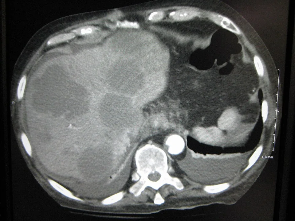
Most patients present with symptoms from the metastases rather than from the primary, and non-specific systemic symptoms are the rule. The commonest presentations are with liver metastases, malignant lymphadenopathy, bone metastases and malignant effusions.
- Constitutional - unexplained weight loss, anorexia, fatigue, night sweats. Weight loss with no localising symptom is one of the classic routes into this diagnosis.
- Hepatic - right upper quadrant pain, hepatomegaly, jaundice, ascites
- Nodal - a firm, fixed, painless lymph node. The site of the node is highly informative and directs the search (see below).
- Skeletal - bone pain, pathological fracture, hypercalcaemia, or spinal cord compression as the first presentation
- Pulmonary and pleural - breathlessness from a pleural effusion or lymphangitis carcinomatosa
- Peritoneal - ascites and abdominal distension, particularly relevant in women
- Neurological - headache, seizure, focal deficit from brain metastases
What the site of a node tells you
| Nodal site | Drainage territory to examine and image |
|---|---|
| Cervical, upper or middle | Head and neck mucosa (including nasopharynx, tonsil, tongue base), thyroid - especially if squamous histology |
| Cervical, lower or supraclavicular | Lung, breast, oesophagus, stomach. The left supraclavicular node (Virchow's node, Troisier's sign) classically signals an intra-abdominal primary. |
| Axillary | Breast in women - this is a favourable subset - and also melanoma and lung |
| Mediastinal or retroperitoneal, midline, in a young man | Extragonadal germ cell tumour; examine the testes and check AFP, beta-hCG and LDH |
| Inguinal | Anal canal, vulva, penis, lower rectum, skin of the leg - examine the perineum and genitalia and consider anoscopy |
Investigations
NICE specifies a defined initial screen, deliberately bounded so that patients are not subjected to open-ended testing while they deteriorate.1 The aim is to identify a primary that would change treatment, and to place the patient in a treatable subset if one applies.
Initial screen for MUO
- Bloods - FBC, U&Es, LFTs, bone profile including calcium, LDH, and a clotting screen
- Site-directed tumour markers - PSA in men, CA 125 in women with peritoneal disease or ascites, and AFP with beta-hCG in any patient with a midline nodal mass, particularly men under 50
- Myeloma screen - serum protein electrophoresis, serum free light chains and urinary Bence Jones protein, especially with lytic bone lesions
- Urinalysis for haematuria, and stool testing if bowel symptoms
- Chest radiograph, then CT of the chest, abdomen and pelvis with contrast
- Biopsy of the most accessible metastatic site, with enough tissue for a full immunohistochemistry panel
Directed further investigation
- Mammography and breast MRI - in a woman with isolated axillary nodal adenocarcinoma, even if the mammogram is normal
- Testicular ultrasound - in men with a midline mass or raised germ cell markers
- Upper GI endoscopy, colonoscopy or bronchoscopy - guided by symptoms and by the immunohistochemistry pattern, not requested blindly
- PET-CT - specifically recommended for squamous cell carcinoma in cervical nodes, where it can find an occult head and neck primary and change treatment from palliative to radical
- Panendoscopy with biopsy of the nasopharynx, tonsils and tongue base - the next step in occult head and neck primary, often with tonsillectomy
- MRI or CT of the head where there are neurological signs
- Ascitic or pleural fluid cytology where an effusion is present, which may spare a more invasive biopsy
Immunohistochemistry
The pathologist works in two steps: establish the lineage, then narrow the likely organ of origin. The first step is the one that changes management most often, because identifying lymphoma, germ cell tumour or melanoma removes the patient from the CUP pathway entirely and into a potentially curable one.3
| Marker | Suggests |
|---|---|
| Cytokeratins (AE1/AE3, CAM5.2) | Carcinoma - epithelial origin |
| CD45 (leucocyte common antigen) | Lymphoma - proceed to lymphoma-specific panel |
| S100, SOX10, Melan-A, HMB-45 | Melanoma |
| Vimentin, desmin, smooth muscle actin | Sarcoma |
| OCT4, PLAP, AFP, beta-hCG | Germ cell tumour |
| Chromogranin, synaptophysin, CD56 | Neuroendocrine tumour |
| TTF-1 | Lung adenocarcinoma or thyroid (thyroglobulin confirms thyroid) |
| CDX2 | Colorectal or upper GI adenocarcinoma |
| GATA3, GCDFP-15, mammaglobin, ER | Breast (GATA3 also urothelial) |
| PAX8 | Ovary, endometrium, thyroid, kidney |
| PSA, NKX3.1 | Prostate |
| p40, p63, CK5/6 | Squamous cell carcinoma |
The CK7 and CK20 pattern is a useful first sieve. CK7 positive with CK20 negative suggests lung, breast, thyroid, ovary, endometrium or pancreatobiliary origin; CK7 negative with CK20 positive suggests colorectal or Merkel cell carcinoma; both positive suggests pancreatic, gastric, cholangiocarcinoma or urothelial; both negative suggests hepatocellular, renal, prostate or squamous carcinoma. Also request p16 and HPV testing on squamous cervical nodes, since HPV-positive disease points to an oropharyngeal primary and carries a much better prognosis.
The favourable subsets
This is the single most examinable part of the topic. A minority of patients with CUP fit a recognisable clinicopathological pattern which is managed as if the presumed primary were known, sometimes with curative intent. Missing one of these converts a treatable cancer into a palliative one.
| Presentation | Treated as | Approach |
|---|---|---|
| Woman with isolated axillary nodal adenocarcinoma | Stage II or III breast cancer | Axillary clearance with breast radiotherapy (or mastectomy), plus systemic therapy guided by receptor status. Long-term survival is achievable. |
| Woman with peritoneal papillary serous adenocarcinoma and raised CA 125 | Advanced ovarian cancer | Cytoreductive surgery and platinum-taxane chemotherapy |
| Squamous cell carcinoma in cervical lymph nodes | Locally advanced head and neck cancer | Neck dissection with radical radiotherapy, with or without chemotherapy, after PET-CT and panendoscopy |
| Squamous cell carcinoma in inguinal nodes | Anal, vulval, penile or lower rectal primary | Node dissection with radiotherapy; can be curative |
| Midline poorly differentiated carcinoma in a younger patient, with raised AFP or beta-hCG | Extragonadal germ cell tumour | Platinum-based combination chemotherapy - potentially curative even with extensive disease |
| Men with blastic bone metastases and a raised PSA | Metastatic prostate cancer | Androgen deprivation therapy, with excellent initial response |
| Adenocarcinoma with a colorectal immunoprofile (CK20+, CDX2+, CK7-) | Metastatic colorectal cancer | Colorectal chemotherapy regimens, which are substantially more effective than empirical CUP chemotherapy |
| Single, small-volume metastasis at one site | Oligometastatic disease | Radical local treatment - surgery or stereotactic radiotherapy |
| Well-differentiated neuroendocrine tumour | Neuroendocrine tumour of unknown primary | Somatostatin analogues, and often an indolent course over years |
Management
Organisation of care
NICE requires every acute trust to have a CUP team - typically an oncologist with a special interest, a CUP clinical nurse specialist, a palliative care physician and access to specialist pathology and radiology - and every patient with MUO to be allocated a named key worker within days.1 Referral to the acute oncology service should happen at the point the metastases are identified, not after a series of specialty referrals has failed.
Systemic anti-cancer therapy
Where no favourable subset applies, treatment is empirical and its benefit is modest. Patients with performance status 0 to 1 and adequate organ function may be offered a platinum-based doublet, most often carboplatin with paclitaxel, which produces response rates of around 20 to 30% and a median survival measured in months rather than years.5 Patients with performance status 2 or worse gain little and are usually best served by symptom control alone.
This is a setting where honest discussion of proportionate benefit matters more than in almost any other. The conversation should cover what treatment can and cannot achieve, the likelihood of toxicity, the alternative of active symptom control, and the fact that declining chemotherapy is a reasonable choice rather than giving up. Recording the outcome in an advance care plan is good practice.
Supportive and palliative management
- Early specialist palliative care referral - appropriate from diagnosis given the prognosis, and associated with better symptom control and quality of life
- Pain control using the analgesic ladder, with radiotherapy for painful bone metastases
- Bone protection - bisphosphonates or denosumab where there are bone metastases, to reduce skeletal-related events
- Drainage of symptomatic effusions, with pleurodesis or an indwelling pleural catheter for recurrent malignant pleural effusion
- Biliary stenting for obstructive jaundice from nodal or hepatic disease, which may also be needed before any chemotherapy
- Dexamethasone and radiotherapy for brain metastases or cord compression
- Nutritional support, VTE prophylaxis and management of hypercalcaemia, which is common in this group
Complications
Because CUP is widely metastatic by definition, the oncological emergencies cluster here. Malignant spinal cord compression, hypercalcaemia of malignancy, superior vena cava obstruction, malignant bowel obstruction and pathological fracture are all common enough that patients and families should be told what to watch for and given a 24-hour contact number.
Obstructive jaundice from hilar nodal disease is a particular problem, since it both causes severe pruritus and prevents the safe use of most chemotherapy. Venous thromboembolism is also markedly more frequent than in other cancers, reflecting the tumour burden, and a new DVT or PE occasionally precedes the diagnosis.
Psychological distress deserves specific mention. Not knowing where the cancer started is genuinely harder for many patients than the diagnosis itself, and "we do not know" is a difficult message to convey without sounding as though the search has been inadequate. Explaining that the primary is small and hidden rather than missed, and that treatment is chosen on the biology of the metastases, helps.
Red flags
Prognosis
Outcomes divide sharply. For unfavourable CUP, which is around 80% of cases, median survival is in the region of 3 to 9 months, and a substantial number of patients die before the work-up is complete.2,4 For the favourable subsets, outcomes approach those of the corresponding known primary: women treated for occult breast cancer presenting with axillary nodes have long-term survival comparable to stage II disease, and extragonadal germ cell tumours can be cured.
Adverse prognostic factors are consistent across series: poor performance status, liver or multiple metastatic sites, raised LDH, low serum albumin, adenocarcinoma histology in a non-favourable pattern, and male sex. Performance status is the strongest single predictor and the main determinant of whether chemotherapy is offered at all.
The examinable message is not the survival figure but the reasoning. CUP is one of the few cancers where the diagnostic process itself carries therapeutic weight - a well-targeted immunohistochemistry panel and a careful clinical examination can move a patient from a palliative pathway to a curative one, while an untargeted search wastes the limited time most of these patients have.
References
- NICE CG104. Metastatic malignant disease of unknown primary origin in adults: diagnosis and management. 2010, updated 2023. Available here
- Cancer Research UK. Cancer of unknown primary statistics. Available here
- Royal College of Pathologists. Dataset for tumours of unknown origin. Available here
- Rassy E, Pavlidis N. Progress in refining the clinical management of cancer of unknown primary in the molecular era. Nature Reviews Clinical Oncology. 2020. Available here
- Kramer A, Bochtler T, Pauli C et al. ESMO Clinical Practice Guideline: Cancer of unknown primary. Annals of Oncology. 2023. Available here
This article is written for revision and education. It is not clinical guidance and must not be used to make decisions about the care of a patient. Always check current NICE guidance and local protocols.