
Arterial Blood Gas Interpretation: A Five-Step Method
Key points
- Always record the FiO2: a PaO2 of 12 kPa is normal on air and alarming on 60% oxygen. A gas without the inspired oxygen concentration cannot be properly interpreted.
- Normal values: pH 7.35 to 7.45, PaO2 10.6 to 13.3 kPa on air, PaCO2 4.7 to 6.0 kPa, bicarbonate 22 to 26 mmol/L, base excess minus 2 to plus 2, lactate below 2 mmol/L.
- The five steps: oxygenation, then pH, then PaCO2, then bicarbonate and base excess, then whether compensation is present and appropriate.
- The rule that decides the primary problem: the pH tells you which way the primary disorder is pushing. Whichever of CO2 or bicarbonate would move the pH in that direction is the primary abnormality.
- Compensation never overcorrects: if the pH has moved past neutral to the opposite side, there is a second primary disorder, not compensation.
- Anion gap: sodium plus potassium minus chloride minus bicarbonate, normally 10 to 18 mmol/L. It separates the two families of metabolic acidosis.
- Bicarbonate reveals chronicity: renal compensation takes 2 to 3 days, so a raised bicarbonate with a high CO2 means chronic retention, whatever the pH.
- A venous gas: is reliable for pH, bicarbonate, base excess, lactate, potassium and haemoglobin. It cannot assess oxygenation, and a normal venous CO2 makes hypercapnia very unlikely.
Introduction
An arterial blood gas measures the adequacy of oxygenation, ventilation and acid-base balance in a single sample. Interpreted systematically it is one of the highest-yield bedside investigations in medicine; interpreted by pattern recognition alone it produces confident wrong answers, which is why a fixed sequence is worth learning and using every time.
When to take one
- Any acutely breathless or hypoxic patient, including all suspected respiratory failure
- Any patient at risk of hypercapnic respiratory failure receiving oxygen
- Critical illness - sepsis, shock, major trauma, cardiac arrest
- Suspected metabolic disturbance - diabetic ketoacidosis, renal failure, poisoning, severe vomiting or diarrhoea
- Reduced conscious level of unclear cause
- Monitoring - after a change in oxygen, after starting NIV, and to track response to treatment
Arterial or venous?
| Measurement | Venous gas |
|---|---|
| pH | Reliable - typically about 0.03 to 0.05 lower than arterial |
| Bicarbonate and base excess | Reliable |
| Lactate | Reliable |
| Potassium, sodium, haemoglobin, glucose | Reliable |
| PaCO2 | Approximate - runs about 0.8 kPa higher. A normal venous CO2 makes hypercapnia very unlikely, so it is a good screening test; an abnormal one should be confirmed arterially. |
| PaO2 | Useless - venous oxygen tells you nothing about arterial oxygenation. Use the oximeter, or take an arterial sample. |
In practice, a venous gas with pulse oximetry answers most questions, and an arterial sample is reserved for when oxygenation must be quantified or hypercapnia confirmed. Arterial puncture is painful and carries small risks of haematoma, arterial spasm, infection and, very rarely, distal ischaemia.
The five-step method
| Value | Normal range |
|---|---|
| pH | 7.35 to 7.45 |
| PaO2 | 10.6 to 13.3 kPa breathing air |
| PaCO2 | 4.7 to 6.0 kPa |
| Bicarbonate (HCO3) | 22 to 26 mmol/L |
| Base excess | minus 2 to plus 2 mmol/L |
| Lactate | 0.5 to 2.0 mmol/L |
| SaO2 | 94 to 98% |
Step 1: assess oxygenation
- Is the patient hypoxaemic? A PaO2 below 8 kPa defines respiratory failure.
- Relate the PaO2 to the FiO2. A useful bedside approximation is that the PaO2 in kPa should be roughly the FiO2 percentage minus 10 - so about 11 kPa on air, 30 kPa on 40%, and 50 kPa on 60%. A PaO2 of 12 kPa on 60% oxygen represents severe gas exchange failure even though the number looks normal.
- Calculate the A-a gradient if the distinction matters. Breathing air at sea level, alveolar oxygen is approximately 20 minus PaCO2 divided by 0.8 in kPa, and the gradient is that value minus the measured PaO2. A normal gradient is under about 2 kPa in a young adult, rising with age.
- A raised A-a gradient means the lung is the problem - V/Q mismatch, shunt or diffusion impairment. A normal gradient with hypoxaemia means hypoventilation or a low inspired oxygen, and points to the respiratory pump rather than the lung.
Step 2: look at the pH
- pH below 7.35 - acidaemia
- pH above 7.45 - alkalaemia
- pH normal - either genuinely normal, fully compensated, or a mixed disorder with opposing effects. Do not stop here.
- The pH tells you which direction the primary disorder is pushing, and everything else in the interpretation follows from that
Step 3: look at the PaCO2 - the respiratory component
- CO2 is an acid. A high PaCO2 pushes the pH down, and a low PaCO2 pushes it up.
- High PaCO2 with a low pH - respiratory acidosis
- Low PaCO2 with a high pH - respiratory alkalosis
- Low PaCO2 with a low pH - the CO2 is not the cause; it is respiratory compensation for a metabolic acidosis
- High PaCO2 with a high pH - respiratory compensation for a metabolic alkalosis
Step 4: look at the bicarbonate and base excess - the metabolic component
- Bicarbonate is a base. A low bicarbonate pushes the pH down, and a high bicarbonate pushes it up.
- Low bicarbonate with a low pH - metabolic acidosis
- High bicarbonate with a high pH - metabolic alkalosis
- Base excess says the same thing in one number: more negative than minus 2 indicates a metabolic acidosis, more positive than plus 2 a metabolic alkalosis. It is often the quickest way to spot a metabolic component.
Step 5: assess compensation
- Uncompensated - the pH is abnormal and the other system has not moved
- Partially compensated - the other system has moved in the correcting direction but the pH is still abnormal
- Fully compensated - the pH is back within the normal range, but both the CO2 and the bicarbonate remain abnormal
- Compensation never overcorrects. If the pH has crossed to the opposite side of 7.40 from where the primary disorder would put it, there is a second primary disorder.
- Respiratory compensation is fast - minutes to hours, by changing minute ventilation. Metabolic (renal) compensation is slow - 2 to 3 days, by altering bicarbonate handling. This difference is what allows an acute problem to be distinguished from a chronic one.
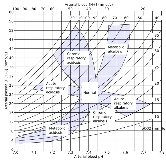
| Primary disorder | Expected compensation |
|---|---|
| Metabolic acidosis | Winter's formula: expected PaCO2 (kPa) is approximately 0.2 x bicarbonate + 1, give or take 0.3. If the measured CO2 is higher than expected, there is an additional respiratory acidosis - the patient is tiring. |
| Metabolic alkalosis | PaCO2 rises by about 0.1 kPa for each 1 mmol/L rise in bicarbonate, limited by the resulting hypoxaemia |
| Acute respiratory acidosis | Bicarbonate rises by about 1 mmol/L for each 1.33 kPa rise in PaCO2 |
| Chronic respiratory acidosis | Bicarbonate rises by about 4 mmol/L for each 1.33 kPa rise in PaCO2 - a much larger rise, which is what makes chronic retention recognisable |
| Acute respiratory alkalosis | Bicarbonate falls by about 2 mmol/L for each 1.33 kPa fall in PaCO2 |
| Chronic respiratory alkalosis | Bicarbonate falls by about 5 mmol/L for each 1.33 kPa fall in PaCO2 |
The four primary disorders and their causes
Respiratory acidosis - high CO2
Always a failure of alveolar ventilation. Work down the respiratory pump from the brain outwards:
- Reduced drive - opioids, benzodiazepines, alcohol, anaesthetics, brainstem stroke, raised intracranial pressure
- Neuromuscular - Guillain-Barre syndrome, myasthenia gravis, motor neurone disease, high cervical cord injury, muscular dystrophy
- Chest wall - kyphoscoliosis, flail chest, obesity hypoventilation, circumferential burns
- Airway and lung - COPD, severe asthma in the exhausted phase, upper airway obstruction, advanced bronchiectasis and cystic fibrosis
Respiratory alkalosis - low CO2
Always hyperventilation, and the important task is to identify why - it is far more often a sign of serious illness than of anxiety.
- Hypoxia driving ventilation - pulmonary embolism, pneumonia, pulmonary oedema, asthma, high altitude
- Sepsis - one of the earliest gas abnormalities in sepsis
- Pain and anxiety - a diagnosis of exclusion, never a first assumption in an unwell patient
- Salicylate poisoning - early respiratory alkalosis from direct respiratory centre stimulation, later joined by a metabolic acidosis
- CNS causes - stroke, meningitis, encephalitis, head injury
- Liver failure, pregnancy (progesterone-driven), and mechanical over-ventilation
Metabolic acidosis - low bicarbonate
Divide by the anion gap, which distinguishes acid being added to the system from bicarbonate being lost from it.
Anion gap = (sodium + potassium) - (chloride + bicarbonate), normally 10 to 18 mmol/L. Some laboratories omit potassium, giving a normal range of 8 to 16. In hypoalbuminaemia the gap is falsely low: add roughly 2.5 mmol/L for every 10 g/L the albumin falls below 40.
| Raised anion gap (acid added) | Normal anion gap (bicarbonate lost, hyperchloraemic) |
|---|---|
| Lactic acidosis - sepsis, shock, ischaemic bowel, metformin, seizures, severe hypoxia | Diarrhoea - the commonest cause |
| Ketoacidosis - diabetic, alcoholic, starvation | Renal tubular acidosis - types 1, 2 and 4 |
| Renal failure - retained urate, phosphate and sulphate | Addison's disease |
| Salicylate poisoning | Acetazolamide and other carbonic anhydrase inhibitors |
| Methanol and ethylene glycol - suspect with a raised osmolar gap | Ureteric diversion into bowel |
| Isoniazid, iron and paraldehyde | Large volumes of 0.9% sodium chloride - dilutional hyperchloraemic acidosis |
| Pancreatic or biliary fistula |
The mnemonics are MUDPILES for the raised gap (methanol, uraemia, DKA, propylene glycol, isoniazid, lactate, ethylene glycol, salicylates) and, for the normal gap, HARDUPS (hyperalimentation, acetazolamide, renal tubular acidosis, diarrhoea, ureteroenterostomy, pancreatic fistula, saline).
Metabolic alkalosis - high bicarbonate
- Vomiting or nasogastric aspiration - loss of gastric acid, with hypokalaemia and hypochloraemia. Classic in pyloric stenosis, where the gas shows a hypochloraemic, hypokalaemic metabolic alkalosis.
- Diuretics - loop and thiazide
- Hypokalaemia from any cause
- Mineralocorticoid excess - Conn syndrome, Cushing syndrome, ectopic ACTH, liquorice excess
- Bartter and Gitelman syndromes
- Excess alkali - antacids, milk-alkali syndrome, large citrate loads in massive transfusion
- Chronic respiratory acidosis being over-corrected - a raised bicarbonate persisting after the CO2 has been brought down by ventilation
Worked examples
| Gas | Interpretation |
|---|---|
| On air: pH 7.21, PaO2 7.4, PaCO2 9.1, HCO3 26, BE +1 | Acidaemia. High CO2 explains it, and the bicarbonate is normal. Acute respiratory acidosis with type 2 respiratory failure and no time for renal compensation - an acute deterioration. Think opioids, acute severe asthma in the exhausted phase, or an acute neuromuscular problem. |
| On 28%: pH 7.30, PaO2 7.8, PaCO2 8.8, HCO3 34, BE +8 | Acidaemia with a high CO2 and a high bicarbonate. The raised bicarbonate means retention has been present for days or longer. Acute-on-chronic type 2 respiratory failure - the classic infective exacerbation of COPD, and the patient who benefits from NIV. |
| On air: pH 7.38, PaO2 8.4, PaCO2 7.4, HCO3 33, BE +8 | pH normal, CO2 high, bicarbonate high. Fully compensated chronic respiratory acidosis - stable chronic type 2 failure. Nothing acute is happening; do not start NIV on this gas alone. |
| On air: pH 7.10, PaO2 13.5, PaCO2 2.6, HCO3 6, BE minus 22, lactate 1.2, glucose 31 | Severe acidaemia with a very low bicarbonate - metabolic acidosis. The low CO2 is appropriate respiratory compensation (Winter's predicts about 2.2 kPa). With hyperglycaemia this is diabetic ketoacidosis. Check ketones and the anion gap. |
| On air: pH 7.48, PaO2 8.9, PaCO2 3.4, HCO3 22, BE minus 1 | Alkalaemia with a low CO2 - respiratory alkalosis, uncompensated so it is acute. The hypoxaemia with a low CO2 and a clear chest is the classic gas of pulmonary embolism, though pneumonia and early sepsis look similar. |
| On air: pH 7.52, PaO2 11.8, PaCO2 6.4, HCO3 38, BE +12, K 2.9, Cl 88 | Alkalaemia with a high bicarbonate - metabolic alkalosis, with a modest compensatory rise in CO2. Hypokalaemic, hypochloraemic - consistent with prolonged vomiting or diuretic use. |
| On 15 L via reservoir mask: pH 7.33, PaO2 8.1, PaCO2 4.0, HCO3 15, BE minus 9, lactate 6.2 | Acidaemia with a low bicarbonate and a high lactate - metabolic (lactic) acidosis with respiratory compensation. Severe hypoxaemia despite maximal oxygen indicates a large shunt. This is septic shock or severe pneumonia; treat as sepsis and involve critical care. |
| On air: pH 7.40, PaO2 12.1, PaCO2 3.1, HCO3 14, BE minus 9 | pH normal but both the CO2 and the bicarbonate are markedly abnormal - a mixed disorder, here a metabolic acidosis with a coexisting respiratory alkalosis. Salicylate poisoning produces exactly this pattern. A normal pH is not the same as a normal gas. |
Common pitfalls
Beyond the numbers
Three additional values on the printout are worth reading deliberately rather than glancing at.
- Lactate - a marker of tissue hypoperfusion or impaired clearance. Above 2 mmol/L is abnormal, and above 4 mmol/L in suspected sepsis indicates high risk and triggers the sepsis pathway. Not all raised lactate is hypoperfusion: it also rises with salbutamol, metformin, seizures, liver failure and thiamine deficiency.
- Potassium - available within a minute, and in suspected hyperkalaemia this speed matters. Treat on the gas result while awaiting the laboratory sample if there are ECG changes.
- Carboxyhaemoglobin and methaemoglobin - reported by co-oximetry. Request them where there has been smoke exposure, a faulty boiler, or unexplained cyanosis with a normal PaO2.
References
- British Thoracic Society. Guideline for oxygen use in adults in healthcare and emergency settings. 2017. Available here
- British Thoracic Society / ICS. Guideline for the ventilatory management of acute hypercapnic respiratory failure in adults. 2016. Available here
- Joint British Diabetes Societies. The management of diabetic ketoacidosis in adults. Available here
- NICE NG51. Sepsis: recognition, diagnosis and early management. 2016, updated 2024. Available here
- Kraut JA, Madias NE. Serum anion gap: its uses and limitations in clinical medicine. Clinical Journal of the American Society of Nephrology. 2007. Available here
- Albert MS, Dell RB, Winters RW. Quantitative displacement of acid-base equilibrium in metabolic acidosis. Annals of Internal Medicine. 1967. Available here
- NICE Clinical Knowledge Summaries. Poisoning or overdose. Available here
- Huckfinne, public domain, via Wikimedia Commons. Available here
This article is written for revision and education. It is not clinical guidance and must not be used to make decisions about the care of a patient. Always check current NICE guidance and local protocols.