
Scleritis and Episcleritis
Key points
- Episcleritis: benign, self-limiting inflammation of the thin vascular layer over the sclera. Mild discomfort, normal vision, settles in 1-2 weeks without treatment.
- Scleritis: inflammation of the sclera itself. Severe boring pain that wakes the patient, deep violaceous redness, and a real risk of visual loss and globe perforation.
- The bedside test: phenylephrine 2.5% blanches episcleral vessels but not scleral vessels. Redness that blanches is episcleritis; redness that persists is scleritis.
- Look in daylight: the deep violet or bluish hue of scleritis is easiest to see in natural light and can be washed out by a bright examination lamp.
- Systemic association: up to half of patients with scleritis have an underlying systemic disease - most often rheumatoid arthritis or granulomatosis with polyangiitis.
- Episcleritis treatment: reassurance, lubricants, and oral NSAIDs if troublesome. Referral only if recurrent or diagnostically uncertain.
- Scleritis treatment: same-day ophthalmology. Oral NSAIDs for mild disease, then systemic corticosteroids and steroid-sparing immunosuppression.
- Worst variant: necrotising scleritis, and its painless form scleromalacia perforans in long-standing rheumatoid arthritis, which signals severe systemic vasculitis.
Introduction
Episcleritis and scleritis are frequently taught together because they look similar to the untrained eye and are separated by a small number of physical signs. They are otherwise entirely different conditions. Episcleritis is a nuisance that resolves on its own. Scleritis is a destructive inflammation that threatens the eye and often signals a systemic vasculitis that threatens the kidneys and lungs.1
The distinction is worth getting right in both directions. Treating scleritis as episcleritis delays immunosuppression in a patient who may be developing granulomatosis with polyangiitis. Treating episcleritis as scleritis leads to unnecessary systemic steroids and an alarming conversation about vasculitis in a patient whose eye would have settled by itself.
The anatomy explains the difference. The sclera is the dense, avascular collagenous shell of the eye, continuous with the corneal stroma anteriorly and the dural sheath of the optic nerve posteriorly. Overlying it is the episclera, a thin, loose, highly vascular layer between sclera and conjunctiva. Inflammation of a vascular layer is uncomfortable and self-limiting; inflammation of a poorly vascularised collagenous structure is intensely painful, slow to settle and destructive.
Aetiology and classification
Episcleritis
Around 70% of episcleritis is idiopathic, and it affects young to middle-aged adults with a slight female preponderance. Two forms are described:
- Simple episcleritis - diffuse or, more often, sectoral redness with mild discomfort. This is around 80% of cases and resolves within 1-2 weeks.
- Nodular episcleritis - a discrete, mobile, tender nodule within the episclera. It takes longer to settle, often several weeks, and is more likely to recur.
Where a cause is identifiable, it is most often an inflammatory bowel disease, rheumatoid arthritis, gout, rosacea, or a preceding infection. However, the diagnostic yield of investigating a first, isolated episode of episcleritis is very low, and a systemic workup is not indicated unless it is recurrent or accompanied by systemic symptoms.
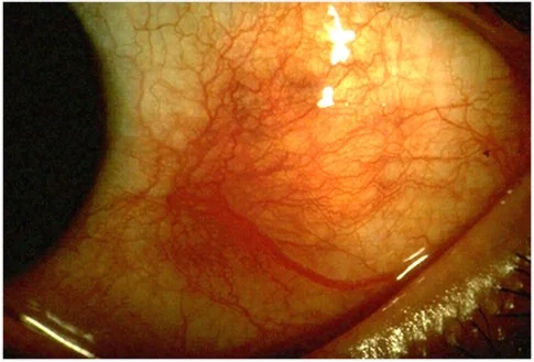
Scleritis
Scleritis is classified by the Watson and Hayreh system, which remains in use because the categories carry genuinely different prognoses.2
| Type | Proportion | Features and significance |
|---|---|---|
| Anterior diffuse | About 40-45% | Widespread anterior scleral oedema and redness; the commonest and most benign form |
| Anterior nodular | About 40-45% | A firm, immobile, deeply tender nodule that cannot be moved over the sclera; good prognosis |
| Anterior necrotising with inflammation | About 10-15% | Avascular patches, scleral thinning, intense pain; over half have an underlying systemic vasculitis and a substantial risk of visual loss |
| Anterior necrotising without inflammation (scleromalacia perforans) | Under 5% | Painless scleral thinning with a blue-grey appearance from underlying uvea, almost always in long-standing seropositive rheumatoid arthritis |
| Posterior scleritis | About 5-10% | Pain on eye movement, proptosis, reduced vision, exudative retinal detachment and choroidal folds; often missed because the eye may look white |
Systemic associations of scleritis
- Rheumatoid arthritis - the commonest association, particularly long-standing seropositive erosive disease
- Granulomatosis with polyangiitis - scleritis may be its presenting feature, and necrotising scleritis with peripheral ulcerative keratitis is highly suggestive
- Relapsing polychondritis, polyarteritis nodosa and other systemic vasculitides
- Systemic lupus erythematosus and other connective tissue diseases
- Inflammatory bowel disease, ankylosing spondylitis and other seronegative spondyloarthropathies
- Sarcoidosis
- Infection - herpes zoster, tuberculosis, syphilis, Lyme disease; and surgically induced necrotising scleritis after ocular surgery
- Gout and, rarely, drugs including bisphosphonates, which can cause an acute scleritis within days of a first infusion
Clinical features
The differences are best learned as a table, because the exam question is almost always a direct comparison.
| Feature | Episcleritis | Scleritis |
|---|---|---|
| Onset | Abrupt, over hours | Gradual, over days |
| Pain | Mild discomfort or gritty; often none | Severe, deep, boring; radiates to jaw, brow or temple; wakes the patient from sleep |
| Tenderness on palpation through the lid | Minimal | Marked and characteristic |
| Colour of redness | Bright salmon pink | Deep violaceous or bluish-red |
| Distribution | Usually sectoral, sometimes diffuse | Sectoral or diffuse; may be nodular |
| Vessels | Superficial, radially arranged, mobile over the sclera | Deep, criss-crossing, immobile |
| Phenylephrine 2.5% | Blanches | Does not blanch |
| Visual acuity | Normal | Often reduced |
| Photophobia and watering | Mild or absent | Common |
| Systemic disease | Uncommon (about 30%) | Common (about 50%) |
| Course | Self-limiting in 1-2 weeks | Weeks to months; recurrent; may be destructive |
Posterior scleritis
Posterior scleritis is the great mimic and the one most often missed, because the front of the eye can look entirely white. Suspect it in an adult with deep periocular pain, pain on eye movement, and reduced vision, particularly if there is proptosis or restricted movement. Signs on fundoscopy include choroidal folds, disc swelling, exudative retinal detachment and a subretinal mass that can be mistaken for a choroidal tumour. It is often misdiagnosed as orbital cellulitis, orbital myositis or optic neuritis, and B-scan ultrasonography settles the question.
Examination
- Visual acuity in each eye. Normal acuity favours episcleritis; reduced acuity mandates urgent referral.
- Look in natural daylight first if possible. Scleral violaceous discolouration is far easier to appreciate in daylight than under the bright white light of an examination lamp, which washes it out.
- Assess the depth of the injected vessels at the slit lamp. Episcleral vessels lie in a superficial radial plane and can be moved across the sclera with a cotton bud; scleral vessels are deeper, criss-crossing and immobile.
- Palpate through the closed lid with a cotton bud. Exquisite tenderness suggests scleritis; a nodule that moves freely is episcleral, one that does not is scleral.
- Instil phenylephrine 2.5% and re-examine after 10-15 minutes
- Examine the cornea with fluorescein for peripheral ulcerative keratitis and thinning
- Assess the anterior chamber for cells, which occur in scleritis but not episcleritis
- Dilated fundoscopy for disc swelling, choroidal folds and exudative detachment if posterior scleritis is suspected
- General examination for joints, skin, nasal crusting, respiratory signs and lymphadenopathy
Differential diagnosis
- Conjunctivitis - diffuse injection greatest in the fornices, discharge, grittiness, bilateral, no focal tenderness
- Anterior uveitis - photophobia, ciliary flush, small irregular pupil, cells and flare; may coexist with scleritis
- Pterygium or pinguecula with inflammation - a visible fleshy or yellowish lesion at the nasal limbus with surrounding injection
- Subconjunctival haemorrhage - a block of blood, painless
- Foreign body or trauma - focal injection with a history and fluorescein staining
- Orbital myositis and orbital cellulitis - pain on eye movement with proptosis; the main differential for posterior scleritis
- Optic neuritis - pain on eye movement with reduced acuity and a relative afferent pupillary defect, but no scleral injection
- Choroidal melanoma or metastasis - can mimic the subretinal mass of posterior scleritis on imaging
Investigations
A first isolated episode of simple episcleritis in an otherwise well patient needs no investigation at all. Scleritis, and recurrent or nodular episcleritis, warrant a systematic search for an underlying cause because up to half will have one.3
- FBC, CRP and ESR - a raised inflammatory response supports scleritis over episcleritis and helps monitor treatment
- Rheumatoid factor and anti-CCP antibodies - rheumatoid arthritis is the commonest association
- ANCA (both PR3 and MPO) - essential, because granulomatosis with polyangiitis may present with scleritis before any respiratory or renal disease
- ANA and extractable nuclear antigens - for lupus and connective tissue disease
- Urea, creatinine and urinalysis with microscopy - looking for the haematuria, proteinuria and red cell casts of a vasculitic nephritis. This is a cheap test that can change everything.
- Serum ACE and chest radiograph - for sarcoidosis, and for the cavitating lesions of granulomatosis with polyangiitis
- Serum urate - for gout
- Infection screen - syphilis serology, QuantiFERON or Mantoux for tuberculosis, and Lyme serology where the exposure history fits
- B-scan ultrasonography - for suspected posterior scleritis, showing scleral thickening and the T sign, where fluid in the sub-Tenon's space tracks around the optic nerve
- Orbital CT or MRI - if the differential includes orbital inflammation or a mass
Management
Episcleritis
- Explanation and reassurance - most cases settle within 1-2 weeks with nothing at all, and the eye is not at risk
- Cool compresses and preservative-free lubricants for symptomatic relief
- Topical or oral NSAIDs if symptoms are troublesome - for example oral ibuprofen or naproxen with gastric protection where indicated
- Avoid topical steroids in primary care. They shorten an episode that would settle anyway, and carry a real risk of rebound, steroid-induced ocular hypertension and masking of an alternative diagnosis.
- Refer routinely if episodes are frequent, if a nodule is not settling after several weeks, or if the diagnosis is in doubt
- Investigate for systemic disease only if episodes are recurrent or there are systemic features
Scleritis
Treatment escalates stepwise according to severity and response:
- Oral NSAIDs - for example flurbiprofen or indometacin at full anti-inflammatory dose, effective in most non-necrotising anterior scleritis
- Oral corticosteroids - prednisolone 0.5-1 mg/kg daily for disease not controlled by NSAIDs, tapered as inflammation settles, with bone and gastric protection5
- Steroid-sparing immunosuppression - methotrexate, mycophenolate mofetil, azathioprine or ciclosporin, started early where prolonged steroid would otherwise be needed
- Biologic therapy - rituximab, infliximab or other TNF inhibitors for refractory or necrotising disease, particularly in ANCA-associated vasculitis4
- Cyclophosphamide - reserved for severe necrotising scleritis with systemic vasculitis
- Surgery - scleral or corneal patch grafting for impending or actual perforation, always alongside systemic immunosuppression rather than instead of it
Treating the underlying systemic disease is the definitive treatment. In rheumatoid-associated scleritis and peripheral ulcerative keratitis, escalating systemic therapy improves both the eye and long-term survival, which is the clearest illustration of why this is not simply an eye problem.
Complications and prognosis
Episcleritis has essentially no complications. Individual episodes resolve fully, vision is unaffected, and although recurrence is common - up to two thirds of patients have further episodes - it remains benign. The main harm done is over-investigation and over-treatment.
Scleritis is quite different. Roughly 15-30% of patients lose vision, and complications include:
- Peripheral ulcerative keratitis with corneal thinning and possible perforation
- Scleral thinning and staphyloma, where the underlying uvea bulges through a thinned sclera and shows as a blue-grey patch
- Anterior uveitis, present in around a third and a marker of more severe disease
- Secondary glaucoma from trabecular inflammation or steroid response
- Cataract, from chronic inflammation and prolonged corticosteroid
- Cystoid macular oedema and exudative retinal detachment, particularly in posterior scleritis
- Globe perforation in necrotising disease - rare, but the reason necrotising scleritis is treated urgently and systemically
The prognosis depends far more on the underlying systemic disease than on the eye. Necrotising scleritis in association with rheumatoid arthritis or ANCA-associated vasculitis has historically carried a substantial mortality from the systemic disease itself, which modern immunosuppression has improved considerably. The practical point for finals is that scleritis is a reason to look hard at the kidneys, the lungs and the joints, not simply to prescribe an anti-inflammatory and arrange follow-up.
References
- Watson PG, Hayreh SS. Scleritis and episcleritis. British Journal of Ophthalmology. 1976. Available here
- Okhravi N, Odufuwa B, McCluskey P, Lightman S. Scleritis. Survey of Ophthalmology. 2005. Available here
- NICE Clinical Knowledge Summaries. Red eye. Available here
- Sainz de la Maza M, Molina N, Gonzalez-Gonzalez LA et al. Clinical characteristics of a large cohort of patients with scleritis and episcleritis. Ophthalmology. 2012. Available here
- BNF. Corticosteroids, general use and adverse effects. Available here
This article is written for revision and education. It is not clinical guidance and must not be used to make decisions about the care of a patient. Always check current NICE guidance and local protocols.