
Uveitis: Anterior, Intermediate and Posterior
Key points
- Definition: inflammation of the uveal tract - iris, ciliary body and choroid - classified anatomically as anterior, intermediate, posterior or panuveitis.
- Anterior uveitis: by far the commonest, accounting for around three quarters of cases. A unilateral painful red eye with photophobia, blurred vision and ciliary flush.
- The pupil: small (miotic) and often irregular from posterior synechiae, where the inflamed iris sticks to the anterior lens capsule.
- Slit lamp findings: cells and flare in the anterior chamber, keratic precipitates on the corneal endothelium, and in severe disease a hypopyon.
- The classic association: HLA-B27 - ankylosing spondylitis, reactive arthritis, psoriatic arthritis and inflammatory bowel disease. Ask about inflammatory back pain.
- Treatment: topical corticosteroid to suppress inflammation and cycloplegic to relieve ciliary spasm and break or prevent synechiae - both started by ophthalmology.
- When to investigate: not after a single unilateral episode in a well adult. Investigate bilateral, recurrent, granulomatous or posterior disease.
- Silent disease: uveitis in juvenile idiopathic arthritis is typically white and painless, which is why children are screened rather than waiting for symptoms.
Introduction
Uveitis is inflammation of the uveal tract, the pigmented vascular middle coat of the eye consisting of the iris and ciliary body anteriorly and the choroid posteriorly. In practice the term also covers inflammation of adjacent structures - the vitreous, retina and retinal vasculature - because they are almost never involved in isolation.
It matters out of proportion to its incidence. Uveitis accounts for roughly 10% of legal blindness in the working-age population of developed countries, and much of that loss is preventable with prompt treatment.1 It also has an unusually high yield as a clinical clue: a young man with a painful photophobic red eye and three months of early morning back stiffness has ankylosing spondylitis until proved otherwise, and the eye is what brought him in.
The uvea is heavily vascularised and pigmented, which explains both its susceptibility to immune-mediated and infective inflammation and its tendency to autoimmune targeting. The Standardization of Uveitis Nomenclature (SUN) working group classification, based on the primary anatomical site of inflammation, is the framework used clinically and is worth knowing.2
Classification
| Type | Primary site | Typical presentation | Approximate share |
|---|---|---|---|
| Anterior | Anterior chamber (iritis, iridocyclitis) | Painful red eye, photophobia, blurred vision, small irregular pupil | 60-80% |
| Intermediate | Vitreous (pars planitis) | Floaters and painless blurring; a white, quiet eye | 5-15% |
| Posterior | Retina or choroid (retinitis, choroiditis) | Painless visual loss, floaters, scotomas; a white eye | 10-20% |
| Panuveitis | All compartments | Any combination, usually with marked visual loss | 5-10% |
Uveitis is further described by onset (sudden or insidious), duration (limited, under three months, or persistent), course (acute, recurrent or chronic) and character (granulomatous, with large greasy mutton-fat keratic precipitates and iris nodules, or non-granulomatous, with fine keratic precipitates). Each of these narrows the differential considerably.
Aetiology
Around half of anterior uveitis is idiopathic. Of the remainder, the largest group by far is the HLA-B27-associated spondyloarthropathies.
Immune-mediated and systemic
- HLA-B27-associated - ankylosing spondylitis, reactive arthritis, psoriatic arthritis and inflammatory bowel disease. HLA-B27 is carried by around 8% of the UK population but by 50-60% of patients with acute anterior uveitis. It causes sudden, unilateral, recurrent, non-granulomatous, often alternating disease with a marked fibrinous reaction and sometimes hypopyon.
- Juvenile idiopathic arthritis - particularly the ANA-positive oligoarticular form in young girls. The uveitis is chronic, bilateral, painless and white, and can destroy vision before anyone notices.
- Sarcoidosis - granulomatous, often bilateral, with mutton-fat keratic precipitates, iris nodules and, in posterior disease, candle-wax drippings along retinal veins
- Behcet disease - a panuveitis with retinal vasculitis and a recurrent hypopyon that shifts with head position, in a patient with oral and genital ulceration
- Multiple sclerosis - classically intermediate uveitis
- Tubulointerstitial nephritis and uveitis (TINU) syndrome - a cause of bilateral uveitis in adolescents that is easily missed unless renal function is checked
- Vogt-Koyanagi-Harada disease and sympathetic ophthalmia - bilateral granulomatous panuveitis, the latter occurring weeks to years after penetrating injury or surgery to the other eye
Infective
- Herpes simplex and varicella zoster - unilateral, granulomatous, with raised intraocular pressure and sectoral iris atrophy, often with a history of keratitis or shingles
- Cytomegalovirus retinitis - a necrotising retinitis with the pizza-pie appearance of haemorrhage and exudate, in advanced HIV with a CD4 count below 50
- Toxoplasma gondii - the commonest cause of infectious posterior uveitis worldwide, giving a focal necrotising retinochoroiditis beside an old pigmented scar, described as a headlight in the fog because of overlying vitritis
- Tuberculosis - a great mimic, causing granulomatous anterior uveitis, choroidal granulomas or serpiginous-like choroiditis
- Syphilis - another great mimic, capable of producing almost any pattern; test for it in every atypical case
- Toxocariasis - in children, from soil contaminated by dog or cat faeces
- Endophthalmitis - post-operative or endogenous, presenting as a rapidly progressive panuveitis with hypopyon and profound visual loss
Other
- Trauma - traumatic iritis follows blunt injury by 24-72 hours and is a common emergency department diagnosis
- Drug-induced - rifabutin, cidofovir, bisphosphonates, sulphonamides and checkpoint inhibitors
- Lens-induced - phacoantigenic uveitis from a hypermature or ruptured lens
- Masquerade syndromes - intraocular lymphoma, leukaemia and retinoblastoma can all present as steroid-resistant uveitis, particularly in the very old and the very young
Clinical features
Anterior uveitis
- Pain - a deep, dull ache rather than a foreign body sensation, developing over hours to a day or two
- Photophobia - the most characteristic symptom, caused by movement of the inflamed iris and by ciliary spasm. It is often severe enough that the patient wears sunglasses indoors.
- Blurred vision - from cells and protein in the aqueous, corneal oedema, and later from cystoid macular oedema
- Redness - ciliary flush, densest at the limbus
- Watering without discharge - distinguishing it from conjunctivitis
- Consensual photophobia - shining light into the unaffected eye causes pain in the affected eye, because the consensual reflex constricts the inflamed iris. This is a specific and easily demonstrated sign.
- A small, sluggish, irregular pupil - miosis from iris sphincter spasm, irregularity from posterior synechiae
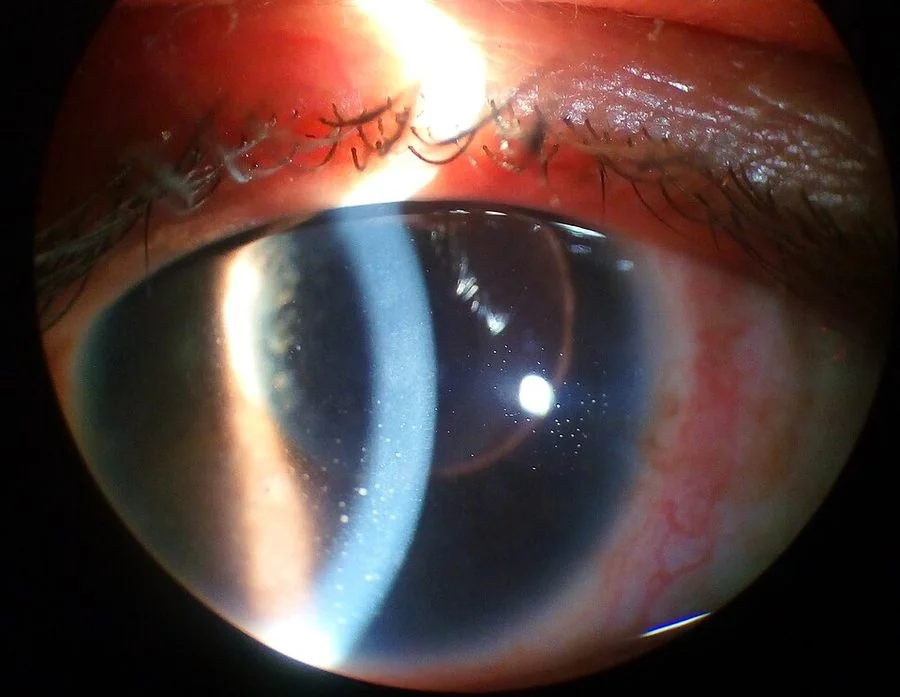
Slit lamp signs
- Cells - individual white cells drifting in the aqueous, seen in a narrow slit beam like dust in a sunbeam. Graded 0 to 4+ by the number in a 1 mm by 1 mm beam, and the principal measure of disease activity.
- Flare - a hazy Tyndall effect from protein leaking across a breached blood-aqueous barrier. Flare persists after cells settle and reflects chronic vascular damage rather than active inflammation, so it is not by itself a reason to escalate treatment.
- Keratic precipitates - clumps of cells on the corneal endothelium, classically in a triangular Arlt distribution inferiorly, where convection currents deposit them
- Posterior synechiae - adhesions between the pupil margin and the anterior lens capsule, distorting the pupil and, if extending through 360°, causing iris bombe and secondary angle closure
- Hypopyon - a layered collection of white cells inferiorly, seen in severe HLA-B27 disease, Behcet disease and endophthalmitis
- Iris nodules - Koeppe nodules at the pupil margin and Busacca nodules on the iris surface, both indicating granulomatous disease
- Intraocular pressure - usually low, because the inflamed ciliary body produces less aqueous; a raised pressure suggests herpetic uveitis, Posner-Schlossman syndrome or trabecular blockage
Intermediate and posterior uveitis
These present with floaters and painless blurring in a white eye, which is why they are missed. Intermediate uveitis shows vitreous cells, snowballs (aggregates in the inferior vitreous) and snowbanking over the pars plana. Posterior uveitis shows focal white retinal or choroidal lesions, vitritis obscuring the fundal view, retinal vasculitis with perivascular sheathing, and macular oedema. Any patient with new persistent floaters and reduced vision needs a dilated fundus examination.
Examination
- Visual acuity in each eye, with a pinhole
- Pattern of redness - looking specifically for ciliary flush
- Pupil size, shape and reactivity, and the consensual photophobia test
- Fluorescein staining to exclude a corneal dendrite or ulcer, since herpetic keratouveitis and microbial keratitis are the main mimics
- Slit lamp examination of the anterior chamber for cells, flare, keratic precipitates, synechiae and hypopyon
- Intraocular pressure
- Dilated fundus examination - mandatory, to grade vitreous cells and look for retinal or choroidal lesions and macular oedema
- Targeted systemic examination - spinal movement and sacroiliac tenderness, skin and nails for psoriasis, oral and genital ulceration, chest signs, lymphadenopathy, and joint examination
Differential diagnosis
| Condition | Key discriminating features |
|---|---|
| Acute anterior uveitis | Ciliary flush, small irregular pupil, cells and flare, normal or low intraocular pressure, consensual photophobia |
| Acute angle closure glaucoma | Fixed mid-dilated pupil, hazy oedematous cornea, stony hard globe, nausea and vomiting, pressure often above 40 mmHg |
| Microbial keratitis | Corneal white infiltrate with an epithelial defect that stains; contact lens wear |
| Herpetic keratouveitis | Uveitis with raised intraocular pressure, sectoral iris atrophy, reduced corneal sensation, previous dendrite |
| Scleritis | Deep boring pain that wakes the patient, violaceous non-blanching redness, focal tenderness |
| Conjunctivitis | Diffuse injection, discharge, normal vision, normal pupil |
| Endophthalmitis | Recent surgery or injection, rapid profound visual loss, hypopyon with dense vitritis |
| Intraocular lymphoma | Chronic steroid-resistant vitritis in an older adult; requires vitreous biopsy |
Investigations
A first episode of unilateral, non-granulomatous, acute anterior uveitis in a systemically well adult does not require investigation. The yield is low and the results are more likely to mislead than to help. Investigation is directed by the pattern.3
- Investigate if: the uveitis is bilateral, recurrent, granulomatous, intermediate or posterior; if it fails to respond to treatment; or if there are systemic symptoms
- FBC, CRP, ESR, U&Es and urinalysis - the last specifically for TINU syndrome, which is easy to miss
- HLA-B27 - most useful for prognosis and for prompting a rheumatology referral rather than for diagnosis, since it is common in the general population
- Chest radiograph and serum ACE - for sarcoidosis; add serum calcium
- Syphilis serology and QuantiFERON-TB Gold or Mantoux - performed in almost all cases of posterior or atypical uveitis, because both are treatable mimics and both are worsened by steroid
- Toxoplasma serology for focal retinochoroiditis, and HIV testing for any unexplained retinitis
- Sacroiliac joint imaging where inflammatory back pain is present
- Optical coherence tomography of the macula - to detect cystoid macular oedema, the commonest cause of visual loss
- Fluorescein angiography - for retinal vasculitis and ischaemia
- Anterior chamber tap or vitreous biopsy - for PCR in suspected viral retinitis, and for cytology in suspected lymphoma
Management
The aims are to suppress inflammation quickly, relieve pain, prevent structural damage from synechiae, and treat any underlying cause. Anterior uveitis is a same-day ophthalmology referral; the diagnosis needs a slit lamp and the treatment is not one a generalist should start.6
Topical corticosteroid
Prednisolone acetate 1% or dexamethasone 0.1% drops, given intensively - often hourly initially, including overnight in severe disease - and then tapered slowly over four to six weeks as cells settle.4 Tapering too quickly is the commonest cause of rebound. Steroid must be started only after the cornea has been examined with fluorescein and herpetic disease excluded.
Cycloplegia
Cyclopentolate 1% or atropine 1% serve two purposes. They paralyse the ciliary muscle, relieving the spasm that causes much of the pain and photophobia; and they dilate the pupil, breaking existing posterior synechiae and preventing new ones from forming across a constricted pupil. Warn the patient that vision will be blurred for near work and that they should not drive while dilated.
Escalation
- Periocular steroid injection - sub-Tenon's triamcinolone for severe anterior disease, macular oedema or poor adherence to drops
- Intravitreal steroid - a dexamethasone implant or fluocinolone implant for chronic posterior disease and macular oedema
- Systemic corticosteroid - oral prednisolone for bilateral, posterior or sight-threatening disease
- Steroid-sparing immunosuppression - methotrexate, mycophenolate, azathioprine or ciclosporin where steroid cannot be tapered below an acceptable dose
- Biologic therapy - adalimumab is licensed for non-infectious intermediate, posterior and panuveitis and is recommended by NICE for those inadequately controlled on corticosteroid5
- Treat the infection first in infective uveitis - aciclovir or valaciclovir for herpetic disease, and appropriate antimicrobial cover before any steroid in toxoplasmosis, tuberculosis and syphilis
Complications and prognosis
- Cystoid macular oedema - the commonest cause of visual loss in uveitis, and the reason OCT is performed routinely
- Posterior synechiae, iris bombe and secondary angle closure glaucoma
- Secondary open angle glaucoma - from inflammatory debris blocking the trabecular meshwork, from trabeculitis, or from steroid response
- Cataract - from both the inflammation and its treatment; posterior subcapsular cataract is characteristic of steroid use
- Band keratopathy - calcium deposition in a band across the interpalpebral cornea, particularly in chronic childhood uveitis
- Hypotony and phthisis bulbi - from chronic ciliary body damage in long-standing severe disease
- Retinal detachment, choroidal neovascularisation and optic atrophy in posterior disease
- Complications of treatment - steroid-induced hypertension, hyperglycaemia, osteoporosis and infection risk with systemic immunosuppression
Prognosis is generally good for acute anterior uveitis. A typical HLA-B27-associated episode settles within four to six weeks of treatment with full recovery of vision, although around half of patients have further episodes, sometimes in the other eye. The counselling point is therefore not that the eye is at risk this time, but that they should present quickly if it recurs, because early treatment prevents synechiae.
Chronic, bilateral, posterior and paediatric disease carries a much poorer prognosis. Juvenile idiopathic arthritis-associated uveitis deserves particular mention: because it is white and painless, children present late with band keratopathy, cataract and established glaucoma. This is why the British Society for Paediatric and Adolescent Rheumatology recommends formal ophthalmic screening on a defined schedule from the point of JIA diagnosis rather than waiting for symptoms - a screening programme that exists precisely because symptoms never come.
References
- Durrani OM, Meads CA, Murray PI. Uveitis: a potentially blinding disease. Ophthalmologica. 2004. Available here
- Jabs DA, Nussenblatt RB, Rosenbaum JT. Standardization of Uveitis Nomenclature (SUN) Working Group. American Journal of Ophthalmology. 2005. Available here
- Royal College of Ophthalmologists. Uveitis clinical guidance. Available here
- BNF. Corticosteroids - eye preparations. Available here
- NICE TA460. Adalimumab and dexamethasone for treating non-infectious uveitis. 2017. Available here
- NICE Clinical Knowledge Summaries. Red eye. Available here
This article is written for revision and education. It is not clinical guidance and must not be used to make decisions about the care of a patient. Always check current NICE guidance and local protocols.