
Retinal Detachment
Key points
- Definition: separation of the neurosensory retina from the underlying retinal pigment epithelium, depriving photoreceptors of their choroidal blood supply.
- Three types: rhegmatogenous (a retinal tear, by far the commonest), tractional (fibrovascular traction, as in proliferative diabetic retinopathy), and exudative (fluid from an underlying inflammatory, vascular or neoplastic process).
- Classic symptoms: flashes (photopsia), a sudden increase in floaters, and a curtain or shadow advancing across the visual field. Painless throughout.
- Signs: a relative afferent pupillary defect if extensive, reduced acuity if the macula is off, and a grey, elevated, mobile retina on dilated fundoscopy.
- The critical question: is the macula on or off? Macula-on detachment is a same-day surgical emergency; macula-off is repaired within a few days.
- Posterior vitreous detachment: the commonest cause of flashes and floaters, and usually benign - but around 10-15% with symptomatic PVD have a retinal tear, so all need urgent dilated examination.
- Treatment: laser or cryotherapy for a tear without detachment; vitrectomy, scleral buckle or pneumatic retinopexy for established detachment.
- Risk factors: myopia, previous cataract surgery, trauma, previous detachment in either eye, family history and lattice degeneration.
Introduction
Retinal detachment is separation of the neurosensory retina from the retinal pigment epithelium. Because the outer retina - including all the photoreceptors - is nourished by diffusion from the choroidal circulation across the pigment epithelium, separation starves the photoreceptors. They begin to degenerate within hours to days, and the loss becomes irreversible.
It affects around 1 in 10,000 people per year in the UK, rising sharply with myopia and after cataract surgery.1 Untreated, a rhegmatogenous detachment almost always progresses to involve the whole retina and blind the eye. Treated promptly, over 90% of eyes are successfully reattached anatomically.
The clinical urgency turns on a single anatomical question. Once the detachment reaches the macula, the cone photoreceptors responsible for central acuity are detached, and central vision does not fully recover even after successful surgery. Distinguishing macula-on from macula-off detachment therefore determines whether the patient goes to theatre today or later in the week, and it is the point on which exam questions turn.
Types and pathophysiology
Rhegmatogenous detachment
From the Greek rhegma, a break. This accounts for the great majority of detachments. A full-thickness break in the retina allows liquefied vitreous to pass through into the subretinal space, lifting the retina off the pigment epithelium.
The usual precipitant is posterior vitreous detachment. With age the vitreous gel liquefies (syneresis) and collapses forward, separating from the retina. In most people this is harmless and produces only floaters. But where the vitreous is abnormally adherent - at the vitreous base, at lattice degeneration, at retinal vessels or at old chorioretinal scars - the collapsing vitreous tugs on the retina and can tear it. Traction on the retina also mechanically stimulates photoreceptors, which the brain interprets as light: this is the origin of photopsia.
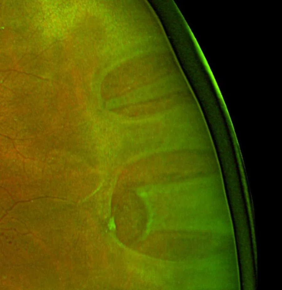
Tractional detachment
Fibrovascular or fibrous membranes on the retinal surface contract and physically pull the retina forward, without any retinal break. The commonest cause by far is proliferative diabetic retinopathy; others include retinopathy of prematurity, sickle cell retinopathy, penetrating trauma and proliferative vitreoretinopathy. The detached retina has a characteristically concave, taut, immobile appearance, in contrast to the convex and mobile retina of a rhegmatogenous detachment. Onset is typically insidious rather than sudden.
Exudative (serous) detachment
Fluid accumulates beneath the retina from a leaking choroidal or retinal source, with no break and no traction. The retina is smooth and convex, and the subretinal fluid characteristically shifts with posture - it pools inferiorly when the patient sits and moves posteriorly when they lie down, which is a distinctive and examinable sign. Causes include:
- Inflammatory - posterior scleritis, Vogt-Koyanagi-Harada disease, sympathetic ophthalmia
- Neoplastic - choroidal melanoma and choroidal metastases, which must always be excluded
- Vascular - severe hypertension, pre-eclampsia and eclampsia, and central serous chorioretinopathy
- Infective - toxoplasmosis and other posterior uveitides
- Iatrogenic - after extensive laser photocoagulation or intraocular surgery
Risk factors
| Risk factor | Mechanism and magnitude |
|---|---|
| Myopia | A longer eye means a thinner, stretched peripheral retina and earlier vitreous liquefaction. Risk rises roughly tenfold with myopia over -6 dioptres, and this is the commonest predisposing factor in younger patients. |
| Increasing age | Posterior vitreous detachment becomes near-universal, with peak incidence of detachment between 60 and 70 |
| Previous cataract surgery | Risk increases several-fold, and considerably more if the posterior capsule ruptured during surgery or a YAG capsulotomy has been performed |
| Ocular trauma | Blunt injury causes retinal dialysis at the vitreous base; penetrating injury causes direct tears. Accounts for most detachments in young men. |
| Detachment in the fellow eye | Around 10% risk in the other eye, since the predisposing anatomy is shared |
| Lattice degeneration | Areas of peripheral retinal thinning with abnormally strong overlying vitreous adhesion, present in about 8% of the population |
| Family history and hereditary vitreoretinopathies | Stickler syndrome, Marfan syndrome and Ehlers-Danlos syndrome all carry markedly increased risk |
| Previous retinal tear or detachment, retinopathy of prematurity, and diabetic retinopathy | Both direct predisposition and the tractional mechanism |
Clinical features
The classic triad is flashes, floaters and a field defect, and it is worth being able to explain the mechanism of each because that is what makes them memorable.
- Photopsia (flashes) - brief arcs or streaks of light, usually in the temporal peripheral field, worse in the dark and on eye movement. Caused by mechanical traction on the retina, which photoreceptors can only signal as light.
- Floaters - a sudden increase, often described as a shower of black spots, cobwebs, a swarm of flies or soot. A single large ring-shaped floater is the Weiss ring, the detached ring of glial tissue that surrounded the optic disc. A dense shower of small dark spots suggests red cells or pigment, meaning a tear has torn a vessel.
- A curtain, shadow or veil progressing across the field, corresponding to the detached retina. It appears in the field opposite to the detachment, since retinal images are inverted: a superior detachment gives an inferior field defect.
- Painless throughout - this is important, and distinguishes it from almost every other acute eye presentation
- Central visual loss once the macula detaches - typically a fall to 6/60 or worse
- Straight lines appearing bent as the detachment approaches the macula and distorts photoreceptor spacing
Signs
- Visual acuity - normal or near-normal if the macula is attached; markedly reduced if the macula is off. This is the fastest clue to which you are dealing with.
- Relative afferent pupillary defect - present in extensive detachment and a useful objective sign, but absent in small peripheral detachments
- Reduced intraocular pressure in the affected eye, typically by around 5 mmHg compared with the fellow eye
- Tobacco dust (Shafer's sign) - pigmented cells in the anterior vitreous, seen at the slit lamp, indicating a retinal break with high specificity
- Vitreous haemorrhage - obscuring the view and indicating a tear that has crossed a retinal vessel
- On dilated indirect ophthalmoscopy - a grey or opaque, elevated, folded retina that moves with eye movement, with retinal vessels running over its surface and appearing darker. A break may be visible as a red horseshoe or round hole.
- Visual field defect on confrontation, corresponding to the detached area
Differential diagnosis
- Posterior vitreous detachment alone - flashes and floaters with an intact retina on dilated examination; the commonest outcome, but only after the retina has been inspected
- Vitreous haemorrhage - sudden floaters and a red haze, most often from proliferative diabetic retinopathy, a retinal tear, or a retinal vein occlusion
- Retinoschisis - splitting within the retina rather than beneath it, giving a smooth, dome-shaped, immobile elevation with an absolute scotoma, usually bilateral, inferotemporal and asymptomatic
- Choroidal detachment - a smooth brown elevation, usually after surgery or with hypotony
- Migraine with aura - a scintillating zigzag scotoma expanding over 20-30 minutes, binocular and usually followed by headache, unlike the monocular flashes of traction
- Central retinal artery or vein occlusion - sudden painless visual loss but with a characteristic fundus and no flashes or curtain
- Ischaemic optic neuropathy - sudden loss with an altitudinal field defect and a swollen or pale disc
- Choroidal melanoma - can present with an exudative detachment; always considered when there is no break and no traction
Investigations
- Visual acuity in each eye - the practical proxy for whether the macula is on or off
- Pupil examination for a relative afferent pupillary defect
- Slit lamp examination of the anterior vitreous for pigment (tobacco dust) and red cells
- Dilated indirect ophthalmoscopy with scleral indentation - the definitive examination, which is the only reliable way to see the far peripheral retina where most breaks lie
- Optical coherence tomography - confirms whether subretinal fluid extends beneath the fovea, and is the objective way to answer the macula-on or macula-off question
- B-scan ultrasonography - essential when vitreous haemorrhage or dense cataract obscures the view, showing the detached retina as a mobile membrane tethered at the disc
- Visual field testing - documents the extent of the defect, though it should never delay treatment
- Investigations for the underlying cause in exudative detachment - blood pressure, inflammatory markers, autoimmune screen, and imaging for a choroidal mass
Management
Treating a break before detachment
A retinal tear without subretinal fluid is sealed by creating a controlled chorioretinal adhesion around it, which takes about ten days to become firm.
- Laser retinopexy - argon laser burns encircling the break, performed at the slit lamp under topical anaesthesia in a few minutes
- Cryotherapy - a freezing probe applied through the conjunctiva, used for very anterior breaks that laser cannot reach
- Success rates exceed 95% for preventing progression to detachment, which is why the urgency of assessing flashes and floaters is justified
Surgery for established detachment
| Procedure | Principle | Best suited to |
|---|---|---|
| Pars plana vitrectomy | Remove the vitreous and the traction on the break, drain subretinal fluid internally, apply laser around the break, and tamponade with gas or silicone oil | Most detachments in the UK, particularly pseudophakic eyes, posterior breaks, multiple breaks and eyes with vitreous haemorrhage |
| Scleral buckle | A silicone band or sponge sutured to the sclera indents the eye wall inwards to meet the detached retina and relieve traction on the break | Younger phakic patients with a single anterior break; avoids inducing cataract |
| Pneumatic retinopexy | An expanding gas bubble injected into the vitreous cavity tamponades the break, combined with laser or cryotherapy and strict posturing | Selected uncomplicated superior detachments with a single break |
| Combined vitrectomy and buckle | Both approaches together | Complex detachments, proliferative vitreoretinopathy and giant retinal tears |
Tractional and exudative detachment
Tractional detachment is repaired by vitrectomy with membrane peeling, and is operated on when the macula is threatened or involved; peripheral tractional detachment that is stable may simply be observed. Anti-VEGF injection beforehand reduces intraoperative bleeding. Exudative detachment is generally not treated surgically at all: the treatment is directed at the underlying cause - immunosuppression for posterior scleritis or Vogt-Koyanagi-Harada disease, blood pressure control in malignant hypertension, delivery in pre-eclampsia, and oncological treatment for a choroidal tumour.
Complications and prognosis
- Proliferative vitreoretinopathy - the commonest cause of surgical failure, in which retinal pigment epithelial cells escape into the vitreous and form contractile membranes that redetach the retina, typically at 4-8 weeks
- Recurrent detachment - requiring further surgery in around 10-15% of cases
- Cataract - almost universal after vitrectomy in a phakic eye, usually within 1-2 years
- Raised intraocular pressure - from gas expansion, silicone oil or steroid response
- Endophthalmitis - rare but devastating
- Refractive change and diplopia - a scleral buckle induces myopia and astigmatism and can cause strabismus by disturbing the extraocular muscles
- Epiretinal membrane and cystoid macular oedema - limiting the final acuity even after successful reattachment
- Detachment in the fellow eye - around 10%, which is why the other eye is examined and any lattice degeneration or break is considered for prophylactic treatment
Anatomical success - the retina reattached - is achieved in over 90% of eyes with a single operation, and in around 95-98% with repeat surgery. Functional success is a different matter, and depends almost entirely on the macula.2
If the macula was attached at the time of surgery, most patients retain excellent central vision. If the macula was detached, recovery is partial and the final acuity depends on how long it was off: repair within a week gives the best results, while detachment lasting more than a few weeks rarely recovers useful central acuity even with a perfect anatomical result. Many patients are left with persistent metamorphopsia, reduced contrast sensitivity or micropsia even when the Snellen acuity looks reasonable.
The practical lesson runs back to the beginning of the presentation. The interval that determines the outcome is not the one between diagnosis and surgery, which is usually short, but the one between the first flash or floater and the patient seeking help. Public and patient awareness - particularly among myopes, those who have had cataract surgery, and anyone who has had a detachment in the other eye - is where most of the preventable visual loss is to be found.
References
- Mitry D, Charteris DG, Fleck BW et al. The epidemiology of rhegmatogenous retinal detachment. British Journal of Ophthalmology. 2010. Available here
- Royal College of Ophthalmologists. Retinal detachment: clinical guidance. Available here
- NICE Clinical Knowledge Summaries. Retinal detachment. Available here
- Hollands H, Johnson D, Brox AC et al. Acute-onset floaters and flashes: is this patient at risk for retinal detachment? JAMA. 2009. Available here
This article is written for revision and education. It is not clinical guidance and must not be used to make decisions about the care of a patient. Always check current NICE guidance and local protocols.