
Central Retinal Artery Occlusion
Key points
- Definition: occlusion of the central retinal artery, causing sudden, painless, profound, unilateral visual loss. It is a stroke of the retina, not simply an eye problem.
- Presentation: vision typically drops to counting fingers or worse within seconds, with no pain and no warning.
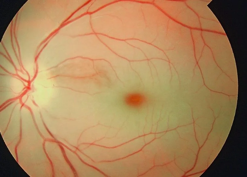
- The two signs: a relative afferent pupillary defect and, on fundoscopy, a pale oedematous retina with a cherry red spot at the fovea.
- Why the cherry red spot: the surrounding retina turns white with ischaemic oedema, while the thin fovea overlies choroidal circulation which is unaffected, so the underlying red choroid shows through.
- Two causes to separate immediately: embolic or thrombotic (usually carotid or cardiac) and arteritic (giant cell arteritis). The management diverges completely.
- Exclude GCA first: ask about jaw claudication, scalp tenderness, headache and polymyalgia; check ESR, CRP and platelets. If suspected, give high-dose corticosteroid immediately.
- Ocular treatment: of unproven benefit and rarely effective - retinal infarction is irreversible after around 90-240 minutes. Do not let it delay the systemic workup.
- The real priority: treat it as a TIA or stroke: same-day assessment, carotid imaging, ECG, and secondary prevention to protect the brain and the other eye.
Introduction
Central retinal artery occlusion (CRAO) causes sudden, painless, severe, unilateral visual loss. It is uncommon - around 1 to 2 per 100,000 people per year - but it is one of the presentations most often mismanaged, because attention goes to the eye when it should go to the patient.1
The single most important conceptual point is this: the central retinal artery is the first branch of the ophthalmic artery, which is the first branch of the internal carotid artery. A CRAO is therefore an anterior circulation stroke. Contemporary guidance from the American Heart Association and the Royal College of Ophthalmologists is explicit that it should be managed on a stroke or TIA pathway, not simply referred to an eye clinic.2
This matters because the risk after CRAO is not primarily to the eye - that damage is usually already done and irreversible - but to the brain. Around 15-25% of patients have a coexisting acute cerebral infarct on diffusion-weighted MRI, most of them clinically silent, and the risk of a subsequent stroke is highest in the first days. Sending a patient home from an eye clinic with a prescription for aspirin and an outpatient carotid Doppler in a fortnight is a recognised failure of care.
Aetiology
The causes divide into arteritic and non-arteritic, and the distinction determines everything about the immediate management.
Non-arteritic (around 95%)
- Carotid atherosclerosis with embolism - the commonest cause. Emboli originate from an ulcerated plaque at the carotid bifurcation. Cholesterol emboli (Hollenhorst plaques) appear as bright refractile yellow specks lodged at arteriolar bifurcations.
- Cardiac embolism - atrial fibrillation, mural thrombus after myocardial infarction, valvular disease, infective endocarditis (which may produce a septic embolus), atrial myxoma and a patent foramen ovale allowing paradoxical embolism
- Calcific emboli - from calcified aortic or mitral valves; these appear dull white and are more likely to cause permanent occlusion
- In situ thrombosis - on an atherosclerotic plaque at the lamina cribrosa, where the artery narrows
- Vasculitis and thrombophilia - antiphospholipid syndrome, systemic lupus erythematosus, hyperhomocysteinaemia, sickle cell disease and the oral contraceptive pill, particularly in younger patients
- Iatrogenic - retrobulbar injection, cosmetic facial filler injection (an increasingly recognised cause), and cardiac or carotid catheterisation
- Raised intraocular pressure exceeding retinal arterial perfusion pressure, as in acute angle closure or after orbital compression
Arteritic (around 5%)
Giant cell arteritis causes CRAO through inflammatory occlusion of the artery, and it must be actively excluded in every patient over 50. It is far more common as a cause of arteritic anterior ischaemic optic neuropathy than of CRAO, but the consequence of missing it is the same: untreated, the fellow eye is affected within days to weeks in a substantial proportion of cases, and that loss is entirely preventable with corticosteroid.3
Pathophysiology
The inner two thirds of the retina - the ganglion cell layer, inner nuclear layer and their supporting structures - are supplied by the central retinal artery, an end artery with essentially no collateral supply. The outer third, including the photoreceptors, is supplied by the choroidal circulation from the short posterior ciliary arteries.
When the central retinal artery occludes, the inner retina infarcts. Ischaemic cells swell, and because the inner retina is thick everywhere except at the fovea, the whole posterior pole becomes opaque and white. At the fovea the retina is thin, has no inner retinal layers, and lies directly over the intact choroidal circulation. The normal red-orange choroidal glow therefore shows through the surrounding white oedema, producing the cherry red spot.
The window for salvage is short. Experimental work in primates showed complete and irreversible inner retinal infarction after around 90-100 minutes of complete occlusion, with some retinal damage from as little as 97 minutes and total loss by 240 minutes.4 Most patients present far outside this window, which is the honest reason that ocular treatments so rarely work.
Clinical features
Symptoms
- Sudden visual loss - over seconds, described as a curtain coming down or the lights going out
- Painless - a defining feature. Pain suggests another diagnosis such as angle closure, giant cell arteritis with headache, or ocular ischaemic syndrome.
- Unilateral - bilateral simultaneous loss suggests giant cell arteritis, an embolic shower or a cortical cause
- Profound - typically counting fingers, hand movements or perception of light. Acuity better than 6/60 suggests either a branch occlusion or cilioretinal sparing.
- Preceding amaurosis fugax in around 10-15% - transient monocular visual loss lasting minutes and recovering fully, which is a warning that should have triggered urgent assessment
- No warning otherwise - patients are usually able to state the exact time of onset, which is diagnostically useful
Signs
- Relative afferent pupillary defect - present within seconds of the occlusion and always present in a complete CRAO. This is the objective sign that confirms an optic nerve or extensive retinal lesion rather than media opacity.
- Pale, opaque, oedematous retina at the posterior pole, giving a milky or ground-glass appearance
- Cherry red spot at the fovea
- Attenuated, thread-like retinal arterioles, sometimes with segmentation of the blood column into boxcarring - discrete segments of red cells separated by clear plasma
- A visible embolus in around 20% - a bright refractile Hollenhorst plaque at an arteriolar bifurcation, or a dull white calcific embolus
- Normal-looking optic disc initially; pallor and optic atrophy develop over 4-6 weeks
- Quiet, white eye - no redness, no inflammation, normal intraocular pressure
- Later - the retinal oedema clears over 4-6 weeks, the cherry red spot disappears, and the fundus can look almost normal apart from a pale disc and thin vessels. A patient presenting late may therefore have a near-normal fundus with no vision.
Differential diagnosis
| Condition | Distinguishing features |
|---|---|
| Central retinal artery occlusion | Profound loss over seconds, RAPD, pale retina with cherry red spot, attenuated arterioles |
| Central retinal vein occlusion | Loss over hours to days, usually less profound, blood and thunder fundus with widespread haemorrhages, dilated tortuous veins and disc swelling |
| Arteritic anterior ischaemic optic neuropathy | Giant cell arteritis with a pale, swollen disc, altitudinal field defect, RAPD, and systemic symptoms. Retina is not white. |
| Non-arteritic AION | Swollen hyperaemic disc with a small crowded 'disc at risk' in the fellow eye, altitudinal defect, vascular risk factors, no systemic inflammatory features |
| Retinal detachment | Flashes and floaters preceding a curtain, elevated retina on fundoscopy, low intraocular pressure |
| Vitreous haemorrhage | Floaters and a red haze, loss of red reflex, no view of the fundus |
| Occipital stroke | Homonymous field defect respecting the vertical midline, binocular, normal pupils and normal fundus |
| Branch retinal artery occlusion | Sectoral retinal whitening and a corresponding altitudinal or sectoral field defect, with central acuity often preserved |
| Ocular ischaemic syndrome | Chronic, with mid-peripheral haemorrhages, dilated non-tortuous veins, ocular pain, and severe carotid stenosis |
Investigations
Investigation runs in two parallel streams: excluding giant cell arteritis immediately, and identifying the embolic source urgently.
Immediate
- ESR, CRP and platelet count - the giant cell arteritis screen. A raised platelet count is a useful additional pointer. Normal inflammatory markers do not exclude GCA if the clinical picture is convincing.
- FBC, U&Es, glucose, HbA1c and lipid profile
- Blood pressure in both arms
- ECG - looking for atrial fibrillation, which is a treatable embolic source
- Blood cultures if there is fever or a murmur, to exclude infective endocarditis
Urgent, within 24 hours
- Carotid imaging - Doppler ultrasound or CT angiography, since symptomatic stenosis of 50-99% is an indication for endarterectomy and the benefit falls sharply with delay beyond two weeks
- Brain imaging - diffusion-weighted MRI is recommended, given the substantial rate of concurrent silent infarcts
- Echocardiography - transthoracic first, with transoesophageal or a bubble study where a cardiac source or patent foramen ovale is suspected
- Prolonged cardiac rhythm monitoring - 24-hour or longer, to detect paroxysmal atrial fibrillation
- Temporal artery ultrasound and biopsy where giant cell arteritis is suspected, arranged without delaying steroid
- Thrombophilia and vasculitis screen in patients under 50 or with no conventional risk factors - antiphospholipid antibodies, homocysteine, ANA and ANCA
Ocular
- Optical coherence tomography - shows thickening and hyper-reflectivity of the inner retinal layers acutely, and inner retinal atrophy later, which is useful when the patient presents late with a normal-looking fundus
- Fluorescein angiography - demonstrates delayed arterial filling; rarely needed for diagnosis
- Visual fields - to document the residual field
Management
Ocular treatments
A number of manoeuvres aim to dislodge an embolus distally or to lower intraocular pressure and improve perfusion. It is important to be honest about them: none has been shown in a randomised trial to improve visual outcome, and their theoretical window has usually passed by the time the patient arrives. They may be attempted within the first few hours but must never delay the systemic workup.
- Ocular massage - firm digital pressure on the globe for 10-15 seconds then release, repeated, aiming to fluctuate intraocular pressure and dislodge the embolus
- Lowering intraocular pressure - intravenous acetazolamide 500 mg, topical timolol, or anterior chamber paracentesis, to increase the arteriovenous perfusion gradient
- Rebreathing into a paper bag or carbogen inhalation - raising arterial CO2 to induce retinal vasodilatation
- Sublingual isosorbide dinitrate
- Intra-arterial thrombolysis - studied in the EAGLE trial, which was stopped early because it showed no benefit over conservative treatment and a higher adverse event rate; it is not standard care.5 Interest has more recently shifted to intravenous thrombolysis within 4.5 hours on stroke protocols, which is under active trial investigation.
Secondary prevention and follow-up
- Antiplatelet therapy - clopidogrel 75 mg daily, or aspirin with dipyridamole, following stroke secondary prevention guidance6
- Anticoagulation where atrial fibrillation or a cardiac source is identified
- High-intensity statin - atorvastatin 80 mg
- Blood pressure and diabetes optimisation, and smoking cessation
- Carotid endarterectomy for symptomatic stenosis of 50-99%, ideally within two weeks
- Corticosteroid and steroid-sparing therapy for giant cell arteritis, with tocilizumab as a steroid-sparing agent, and bone and gastric protection
- Ophthalmic follow-up at 4-6 weeks to check for rubeosis iridis, which develops in around 15-20% and can cause neovascular glaucoma; pan-retinal photocoagulation is given if it appears
- DVLA notification where the visual standard is no longer met, and low vision support
Prognosis
The visual prognosis is poor. Around two thirds of patients are left with acuity of counting fingers or worse, and meaningful spontaneous recovery occurs in under a fifth, usually where the occlusion was incomplete or a cilioretinal artery spared the fovea. Late presentation is the rule rather than the exception, and by the time most patients are seen the retina has infarcted.
The systemic prognosis is the part that can still be changed, and it is sobering. CRAO carries a subsequent stroke risk comparable to that after a hemispheric TIA, concentrated in the first days and weeks, and patients have significantly increased cardiovascular mortality over the following years. The 15-25% rate of concurrent silent cerebral infarction on MRI underlines that the retinal event is a marker of active, unstable arterial disease.
The message to carry into an exam and into practice is therefore consistent. The eye is usually beyond saving; the patient is not. A CRAO is an opportunity to identify a carotid stenosis, an unsuspected atrial fibrillation, or an untreated giant cell arteritis, and to prevent the stroke or the second blind eye that would otherwise follow. Judging the quality of the response by what was done for the retina is to judge it by the least modifiable part.
References
- Royal College of Ophthalmologists. Retinal artery occlusion: clinical guidance. Available here
- Mac Grory B, Schrag M, Biousse V et al. Management of central retinal artery occlusion: a scientific statement from the American Heart Association. Stroke. 2021. Available here
- NICE Clinical Knowledge Summaries. Giant cell arteritis. Available here
- Hayreh SS, Zimmerman MB, Kimura A, Sanon A. Central retinal artery occlusion: retinal survival time. Experimental Eye Research. 2004. Available here
- Schumacher M, Schmidt D, Jurklies B et al. Central retinal artery occlusion: local intra-arterial fibrinolysis versus conservative treatment (EAGLE). Ophthalmology. 2010. Available here
- NICE NG128. Stroke and transient ischaemic attack in over 16s: diagnosis and initial management. 2019, updated 2022. Available here
This article is written for revision and education. It is not clinical guidance and must not be used to make decisions about the care of a patient. Always check current NICE guidance and local protocols.