
Diabetic Retinopathy
Key points
- Definition: a microangiopathy of the retinal capillaries caused by chronic hyperglycaemia, leading to leakage, ischaemia and ultimately neovascularisation.
- Why it matters: the commonest cause of blindness in the UK working-age population, and it is asymptomatic until sight-threatening, which is why screening exists.
- Two separate processes: retinopathy (peripheral ischaemia leading to new vessels) and maculopathy (central leakage and oedema). They progress independently and are graded separately.
- NHS grading: R0-R3 for retinopathy and M0-M1 for maculopathy. R3 (proliferative) and M1 (maculopathy) are referable to hospital eye services.
- The key sign of ischaemia: cotton wool spots and venous beading indicate significant retinal non-perfusion and predict progression to new vessels.
- Proliferative disease: new vessels at the disc or elsewhere. Treated urgently with pan-retinal photocoagulation, which sacrifices peripheral retina to save central vision.
- Maculopathy: clinically significant macular oedema is treated with intravitreal anti-VEGF first line, with focal laser for some circinate patterns.
- Screening: annual digital retinal photography from age 12 for everyone with diabetes in the UK - one of the most successful screening programmes in the NHS.
Introduction
Diabetic retinopathy is the commonest cause of blindness in people of working age in the UK. Around a third of people with diabetes have some degree of retinopathy, and after 20 years of disease nearly everyone with type 1 diabetes and around 60% of those with type 2 will have retinopathy of some grade.1
Its defining clinical characteristic is that it is silent. Retinopathy can progress from a normal fundus to florid proliferative disease with new vessels ready to bleed, without the patient noticing anything at all, because the peripheral retina contributes little to conscious visual experience and the macula may be untouched. By the time vision changes, the damage is often advanced.
This is precisely why the NHS Diabetic Eye Screening Programme exists, and why it is one of the more successful screening programmes in the NHS. Since its introduction, diabetic retinopathy is no longer the leading cause of certifiable blindness in the working-age population in England and Wales, having been overtaken by inherited retinal disease - a genuine population-level success attributable to screening plus better glycaemic and blood pressure control.2
Pathophysiology
Chronic hyperglycaemia damages retinal capillaries through several converging mechanisms: non-enzymatic glycation producing advanced glycation end products, flux through the polyol pathway generating sorbitol and osmotic stress, activation of protein kinase C, and oxidative stress. The consequences at the capillary level are consistent.
- Pericyte loss - pericytes provide structural support and regulate capillary tone. Their selective loss weakens the vessel wall, producing microaneurysms, the earliest visible lesion.
- Basement membrane thickening and endothelial dysfunction - breaking down the inner blood-retinal barrier, so plasma leaks out. Fluid produces retinal oedema; lipoprotein that leaks and is left behind as fluid is reabsorbed produces hard exudates.
- Capillary occlusion - endothelial damage and increased leucocyte adhesion block capillaries, producing areas of retinal non-perfusion. Infarction of the nerve fibre layer at the edge of an ischaemic area, with stasis of axoplasmic flow, produces cotton wool spots.
- Ischaemia drives VEGF release - hypoxic retina secretes vascular endothelial growth factor, which is the central mediator of everything that follows
- Neovascularisation - VEGF stimulates new vessels to grow on the retinal surface and optic disc. These vessels are fragile, grow along the posterior hyaloid face, and bleed into the vitreous or contract to detach the retina.
Two processes therefore run in parallel and largely independently. Peripheral ischaemia drives the retinopathy sequence towards neovascularisation. Central leakage at the macula drives maculopathy and oedema. A patient can have severe maculopathy with minimal retinopathy or vice versa, which is why the two are graded separately.
Risk factors
- Duration of diabetes - the strongest predictor by a wide margin
- Glycaemic control - each 1% reduction in HbA1c reduces microvascular complications by around 35%. The DCCT and UKPDS both demonstrated this convincingly.3
- Hypertension - independently accelerates both retinopathy and maculopathy; UKPDS showed tight blood pressure control reduced progression by a third
- Dyslipidaemia - associated particularly with hard exudates and maculopathy; fenofibrate reduced progression in the FIELD and ACCORD-Eye studies
- Diabetic nephropathy - proteinuria is a strong marker of concurrent retinopathy, since the same microvascular process affects both organs
- Pregnancy - can cause rapid progression, which is why women with pre-existing diabetes are screened at booking and again at 28 weeks (and at 16-20 weeks if retinopathy is present)
- Puberty - accelerates progression in type 1 diabetes, which is why screening begins at age 12
- Rapid improvement in glycaemic control - paradoxically causes transient early worsening, particularly after starting insulin or a very tight regimen, or after bariatric surgery
- Smoking, anaemia and obstructive sleep apnoea
- Cataract surgery, which can accelerate maculopathy
Clinical features and grading
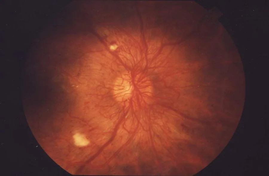
The signs are best learned in the order they appear, because that order is the pathophysiology.
| Sign | Appearance | What it means |
|---|---|---|
| Microaneurysms | Tiny discrete red dots | Capillary outpouchings from pericyte loss - the earliest visible lesion |
| Dot and blot haemorrhages | Round red lesions, deeper and larger than microaneurysms | Haemorrhage in the compact inner nuclear and outer plexiform layers |
| Flame haemorrhages | Streaky, feather-edged | Haemorrhage in the nerve fibre layer, tracking along the axons |
| Hard exudates | Sharply defined yellow deposits, often in a circinate ring | Lipoprotein left behind after oedema fluid is reabsorbed; a ring surrounds the leaking microaneurysm at its centre |
| Cotton wool spots | Fluffy pale grey-white patches with indistinct edges | Nerve fibre layer infarcts - a marker of ischaemia, not exudation |
| Venous beading and looping | Veins with a sausage-string calibre | Severe retinal ischaemia; strongly predicts progression to proliferative disease |
| Intraretinal microvascular abnormalities (IRMA) | Fine, irregular, tortuous intraretinal vessels near ischaemic areas | Shunt vessels bypassing occluded capillaries; a pre-proliferative sign |
| New vessels (NVD, NVE) | Fine, disorganised networks on the disc or elsewhere | Neovascularisation - proliferative disease, which threatens sight |
NHS screening grading
The UK screening programme uses a two-part grade: R for retinopathy and M for maculopathy. Knowing this scheme is expected, because it is the language used in every screening report a UK doctor will read.4
| Grade | Findings | Action |
|---|---|---|
| R0 | No retinopathy | Routine annual screening |
| R1 (background) | Microaneurysms, dot and blot haemorrhages, hard exudates, with no features of R2 or R3 | Routine annual screening |
| R2 (pre-proliferative) | Venous beading, venous loops or reduplication, multiple blot haemorrhages, IRMA | Refer to hospital eye service, seen within 13 weeks |
| R3A (active proliferative) | New vessels at the disc or elsewhere, pre-retinal or vitreous haemorrhage, pre-retinal fibrosis, tractional detachment | Urgent referral, seen within 2 weeks |
| R3S (stable treated proliferative) | Previously treated with pan-retinal photocoagulation and now stable | Annual digital surveillance |
| M0 | No maculopathy | Routine annual screening |
| M1 (maculopathy) | Exudate or microaneurysm/haemorrhage within defined distances of the fovea, or retinal thickening within 1 disc diameter of the fovea | Refer to hospital eye service, seen within 13 weeks |
Symptoms
- Usually none until sight-threatening disease is established - the single most important point
- Gradual central blurring and difficulty reading, from maculopathy
- Sudden onset of floaters, a shower of black spots, or a red haze - vitreous haemorrhage from new vessels
- Sudden profound visual loss if the vitreous haemorrhage is dense
- A curtain or shadow across the vision - tractional retinal detachment
- Fluctuating vision with changing blood glucose, from osmotic swelling of the lens rather than the retina
- A painful red eye with a very high pressure - neovascular glaucoma, a late and serious complication
Investigations
- Digital retinal photography with mydriasis - the screening modality, using two 45° fields per eye (macula-centred and disc-centred)
- Visual acuity at every screening episode and clinic visit
- Slit lamp biomicroscopy with a fundus lens - the clinic standard, giving a stereoscopic view that photography cannot, and the only reliable way to assess retinal thickening
- Optical coherence tomography - the definitive investigation for macular oedema, quantifying central retinal thickness and identifying intraretinal cysts and subretinal fluid. It also guides retreatment.
- Fluorescein angiography - maps areas of capillary non-perfusion and identifies leaking microaneurysms; used to plan focal laser and to assess unexplained visual loss
- B-scan ultrasonography - where vitreous haemorrhage obscures the fundus, to exclude tractional retinal detachment
- Systemic assessment - HbA1c, blood pressure, lipids, renal function and urine albumin:creatinine ratio. Retinopathy should always prompt a review of the whole microvascular picture.
Management
Risk factor modification
This is the foundation and applies at every grade. It slows progression more reliably than any ocular treatment and is the part most often skimmed over in answers.
- Glycaemic control - individualised HbA1c target, typically 48-58 mmol/mol, lowered gradually where the baseline is very high and retinopathy is present
- Blood pressure control - a target of 140/80 mmHg or below, or 130/80 mmHg with end-organ damage
- Lipid management - a statin for cardiovascular risk; fenofibrate has specific evidence for slowing retinopathy progression
- Smoking cessation
- Attend screening - non-attendance is the commonest reason for late presentation, and worth addressing directly
- Renal review, since nephropathy and retinopathy track together
Pan-retinal photocoagulation
The treatment for proliferative disease. Between 1,500 and 3,000 laser burns are applied to the peripheral retina, sparing the macula and the papillomacular bundle, usually over two or more sessions. Destroying ischaemic peripheral retina reduces total VEGF production, causing the new vessels to regress. The Diabetic Retinopathy Study showed it reduces the risk of severe visual loss by more than half.5
Diabetic macular oedema
- Intravitreal anti-VEGF - ranibizumab, aflibercept or faricimab, first line for centre-involving macular oedema, given as a loading course then according to OCT response. NICE recommends treatment where central retinal thickness is 400 micrometres or more.6
- Focal or grid laser photocoagulation - for non-centre-involving oedema and for circinate exudate rings with an identifiable leaking microaneurysm at the centre
- Intravitreal corticosteroid - a dexamethasone or fluocinolone implant, for eyes that respond poorly to anti-VEGF, particularly pseudophakic eyes. Steroid causes cataract and raised intraocular pressure, so it is generally reserved for eyes that have already had cataract surgery.
- Optimise systemic factors in parallel - poorly controlled blood pressure and fluid overload from renal disease both worsen macular oedema and limit the response to injections
Vitreoretinal surgery
- Pars plana vitrectomy - for non-clearing vitreous haemorrhage, tractional retinal detachment involving the macula, or combined tractional and rhegmatogenous detachment
- Anti-VEGF injection before surgery to reduce intraoperative bleeding from active new vessels
- Endolaser during vitrectomy to complete pan-retinal photocoagulation where the view previously prevented it
Complications and prognosis
- Vitreous haemorrhage - from bleeding new vessels; may clear spontaneously over weeks or require vitrectomy
- Tractional retinal detachment - fibrovascular tissue contracting on the retinal surface; requires surgery if the macula is threatened
- Neovascular glaucoma - a fibrovascular membrane grows across the iris and drainage angle, causing intractable secondary angle closure with severe pain and a poor visual prognosis
- Diabetic macular oedema - the commonest cause of visual impairment in diabetes, and the reason OCT is central to modern management
- Macular ischaemia - capillary dropout at the fovea, causing visual loss for which there is no treatment; the reason some patients do not improve despite an anatomically dry macula
- Cataract - earlier and faster than in people without diabetes
- Increased risk of other ocular disease - open angle glaucoma, and cranial nerve palsies affecting III, IV and VI
With screening, timely laser and injection therapy, and good systemic control, the outlook is far better than it was a generation ago. Pan-retinal photocoagulation reduces severe visual loss in proliferative disease by more than half, and anti-VEGF therapy has substantially improved outcomes in macular oedema. The overwhelming determinant of outcome, however, remains the stage at which the patient reaches treatment.
That makes attendance at screening the single most useful thing to reinforce in a diabetes review. Patients frequently see the annual photograph as bureaucratic, particularly when their vision is perfect - which of course is exactly the point at which it is most valuable. Explaining that the test is designed to find damage before it can be felt, and that treatment given at that stage works far better than treatment given later, is a better argument than simply reminding them that they are due.
References
- NICE Clinical Knowledge Summaries. Diabetes - type 2: retinopathy. Available here
- Public Health England. NHS Diabetic Eye Screening Programme: overview. Available here
- UK Prospective Diabetes Study (UKPDS) Group. Intensive blood-glucose control with sulphonylureas or insulin compared with conventional treatment. The Lancet. 1998. Available here
- NHS Diabetic Eye Screening Programme. Grading definitions for referrable disease. Available here
- Diabetic Retinopathy Study Research Group. Photocoagulation treatment of proliferative diabetic retinopathy. Ophthalmology. 1978. Available here
- NICE TA274. Ranibizumab for treating diabetic macular oedema. 2013. Available here
This article is written for revision and education. It is not clinical guidance and must not be used to make decisions about the care of a patient. Always check current NICE guidance and local protocols.