
Hypertensive Retinopathy
Key points
- Definition: retinal microvascular changes caused by systemic hypertension, ranging from asymptomatic arteriolar narrowing to bilateral disc swelling in a hypertensive emergency.
- Why examine the fundus: it is the only place in the body where arterioles can be seen directly, so it gives a free assessment of end-organ damage from hypertension.
- Grading: Keith-Wagener-Barker grades 1-4: 1 arteriolar narrowing, 2 AV nipping, 3 haemorrhages and cotton wool spots, 4 papilloedema.
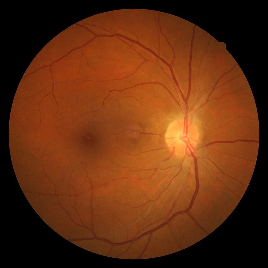
- Chronic signs: silver and copper wiring from arteriolosclerosis, and arteriovenous nipping where a thickened arteriole compresses the vein it crosses.
- Acute signs: flame haemorrhages, cotton wool spots, hard exudates and a macular star - a radial pattern of exudate around the fovea.
- The emergency: grade 4 (bilateral papilloedema) with blood pressure usually above 180/120 mmHg is malignant hypertension and needs same-day admission.
- Lowering pressure: in hypertensive emergency, reduce by no more than 25% in the first hour and to around 160/100 mmHg over 2-6 hours - too rapid a fall causes cerebral, retinal and renal infarction.
- Complications: retinopathy is an independent predictor of stroke, heart failure and cardiovascular death, over and above the blood pressure reading itself.
Introduction
Hypertensive retinopathy is the collective term for the retinal vascular changes produced by systemic hypertension. It is common - some degree of change is present in around 10% of non-diabetic adults, rising steeply with age and blood pressure - and it is nearly always asymptomatic.1
Its clinical value is largely as a window. The retina is the only site in the body where arterioles, venules and capillaries can be inspected directly and non-invasively. What is happening in the retinal circulation reflects what is happening in the cerebral, renal and coronary microcirculation, which is why the presence and severity of hypertensive retinopathy predicts stroke, cognitive decline, heart failure and cardiovascular mortality independently of the blood pressure reading itself.2
It matters in a second, much more acute way. Bilateral disc swelling in a patient with a very high blood pressure defines a hypertensive emergency - what used to be called malignant or accelerated hypertension - and mandates same-day admission. This is the one situation in which fundoscopy in a general medical setting genuinely changes management within the hour.
Pathophysiology
Retinal arterioles respond to raised systemic pressure in a predictable sequence, and understanding it makes the grading system intuitive rather than a list to be memorised.
- Vasoconstrictive phase - retinal arterioles autoregulate, constricting in response to raised pressure to protect the downstream capillary bed. This produces generalised and then focal arteriolar narrowing, and is fully reversible if pressure is controlled.
- Sclerotic phase - sustained hypertension causes intimal thickening, medial hyperplasia and hyaline degeneration in the arteriolar wall. The vessel becomes rigid and its wall becomes visible, giving copper wiring and eventually silver wiring. Where an arteriole crosses a venule and the two share a common adventitial sheath, the thickened arteriole compresses the vein: arteriovenous nipping.
- Exudative phase - once pressure exceeds the capacity of autoregulation, the blood-retinal barrier breaks down. Plasma and blood leak out, producing flame haemorrhages in the nerve fibre layer, hard exudates from residual lipoprotein, and cotton wool spots from nerve fibre layer infarction where arterioles occlude.
- Malignant phase - severe, rapid pressure elevation causes fibrinoid necrosis of arterioles, widespread ischaemia and optic disc swelling, reflecting failure of autoregulation in the optic nerve head circulation and often accompanying raised intracranial pressure.
Clinical features and grading
The Keith-Wagener-Barker classification dates from 1939 and remains the one used in UK exams and clinical letters, although modern schemes group the four grades into mild, moderate and malignant because grades 1 and 2 are not reliably distinguishable between observers.3
| Grade | Fundal findings | Interpretation |
|---|---|---|
| Grade 1 | Generalised arteriolar narrowing and tortuosity; mild copper wiring | Mild, chronic; no urgent action, treat the blood pressure |
| Grade 2 | Focal arteriolar narrowing and arteriovenous nipping; more marked copper or silver wiring | Moderate, chronic; a marker of established vascular disease |
| Grade 3 | The above plus flame haemorrhages, cotton wool spots and hard exudates, sometimes forming a macular star | Severe; assess urgently for end-organ damage |
| Grade 4 | All of the above plus bilateral optic disc swelling (papilloedema) | Hypertensive emergency - same-day admission |
Recognising the individual signs
- Arteriolar narrowing - judged by the arteriole-to-venule ratio, normally about 2:3. A ratio of 1:3 or less is abnormal. Focal narrowing, where a segment of arteriole is visibly constricted, is more specific than generalised narrowing.
- Copper wiring - the thickened arteriolar wall increases the light reflex, giving the vessel a burnished orange-brown appearance while the blood column is still visible
- Silver wiring - further wall thickening obscures the blood column entirely, so the vessel appears as a bright white line. This is a sign of severe chronic arteriolosclerosis.
- Arteriovenous nipping (Gunn's sign) - the vein appears to taper or vanish on either side of the crossing arteriole, and may be deflected at right angles (Salus's sign) or dilated distal to the crossing
- Flame haemorrhages - superficial, feather-edged, following the nerve fibre layer
- Cotton wool spots - pale, fluffy, indistinct; nerve fibre layer infarcts marking ischaemia
- Hard exudates - well-demarcated yellow deposits; when deposited radially around the fovea in Henle's layer they form a macular star
- Elschnig spots and Siegrist streaks - focal areas of choroidal infarction seen in severe hypertensive choroidopathy, particularly in young patients and in pre-eclampsia
Symptoms
- Usually none - grades 1 to 3 are almost always found incidentally
- Blurred vision - from macular oedema, exudate at the fovea or disc swelling
- Headache, nausea and vomiting - from raised intracranial pressure in a hypertensive emergency
- Visual field defects and transient visual obscurations - brief greyouts lasting seconds, associated with disc swelling
- Sudden visual loss - if the retinopathy has been complicated by a retinal vein or artery occlusion, or by ischaemic optic neuropathy
- Symptoms of another end organ - chest pain, breathlessness, focal neurology, oliguria or confusion, which are the ones that determine management
Differential diagnosis
Many of these signs are non-specific, and the same fundal appearances occur in other microvascular diseases. The distinction is usually made by the distribution of the lesions and by the clinical context.
| Condition | Distinguishing features |
|---|---|
| Diabetic retinopathy | Dot and blot haemorrhages predominate over flame haemorrhages, microaneurysms are prominent, exudates are often circinate, and changes are concentrated at the posterior pole. Many patients have both. |
| Central retinal vein occlusion | Unilateral, with haemorrhages in all four quadrants, dilated tortuous veins and a swollen disc - the blood and thunder fundus |
| Branch retinal vein occlusion | Sectoral haemorrhages confined to one venous territory, respecting the horizontal raphe |
| Radiation retinopathy | Identical appearance to diabetic retinopathy, months to years after orbital or head radiotherapy |
| Anaemia and leukaemia | Haemorrhages with pale centres (Roth spots), cotton wool spots, and often a striking degree of change with a normal blood pressure |
| HIV retinopathy | Cotton wool spots with few haemorrhages, in advanced immunosuppression |
| Papilloedema from raised intracranial pressure | Bilateral disc swelling without arteriolar narrowing or AV nipping; a normal blood pressure points away from hypertensive grade 4 |
| Ocular ischaemic syndrome | Unilateral, with mid-peripheral haemorrhages, dilated but not tortuous veins, and severe carotid stenosis |
Investigations
The retinopathy itself needs no ocular investigation beyond fundoscopy. The investigations are directed at the blood pressure, at other end-organ damage, and at a secondary cause if the presentation is atypical.
- Repeated blood pressure measurement in both arms, with ambulatory or home monitoring where the diagnosis of hypertension is being established4
- Urinalysis and urine albumin:creatinine ratio - proteinuria, haematuria and red cell casts point to hypertensive nephropathy or an underlying glomerulonephritis
- U&Es and eGFR - to assess renal end-organ damage, and as a baseline before starting an ACE inhibitor
- FBC and blood film - looking for the fragmented red cells of a microangiopathic haemolytic anaemia, which occurs in malignant hypertension
- HbA1c and lipid profile - for coexisting diabetes and for cardiovascular risk assessment
- ECG and echocardiography - for left ventricular hypertrophy, the cardiac counterpart of retinal end-organ damage
- Fundus photography - documents the appearance for comparison over time and is the standard record in most settings
- Secondary hypertension screen - renin and aldosterone, plasma or urinary metanephrines, cortisol and renal artery imaging, particularly in a young patient or where retinopathy is severe
- CT or MRI head - if there is headache, confusion or focal neurology, to distinguish hypertensive encephalopathy and posterior reversible encephalopathy syndrome from stroke or intracranial haemorrhage
Management
Chronic hypertensive retinopathy
There is no ocular treatment. Management is control of the blood pressure and of overall cardiovascular risk, following NICE guidance, and the retinal changes of grades 1 and 2 stabilise or partly regress once pressure is controlled.4
- Confirm hypertension with ambulatory or home monitoring rather than a single clinic reading
- Lifestyle - salt reduction, weight loss, alcohol moderation, exercise and smoking cessation
- Pharmacological treatment by the standard NICE algorithm: an ACE inhibitor or angiotensin receptor blocker for those under 55 and not of Black African or African-Caribbean family origin, a calcium channel blocker otherwise, then combination therapy with a thiazide-like diuretic
- Targets - clinic blood pressure below 140/90 mmHg under 80, and below 150/90 mmHg over 80
- Formal cardiovascular risk assessment with QRISK and a statin where indicated
- Recheck the fundus at subsequent reviews, since progression of retinopathy indicates inadequate control
Hypertensive emergency
Where hypertension has caused a retinal vein occlusion, macular oedema or an ischaemic optic neuropathy, those complications are managed on their own merits, but blood pressure control remains the intervention that alters the natural history and protects the fellow eye.
Complications and prognosis
- Retinal vein occlusion - hypertension is the commonest systemic association, and the fundal changes of retinopathy frequently coexist
- Retinal artery occlusion - through accelerated atherosclerosis and embolism
- Retinal arterial macroaneurysm - an outpouching of a major arteriole that can rupture and haemorrhage into the retina or vitreous
- Non-arteritic anterior ischaemic optic neuropathy - the small crowded disc plus vascular risk factors
- Hypertensive choroidopathy - Elschnig spots, Siegrist streaks and exudative retinal detachment, mainly in young patients with rapid pressure rises and in pre-eclampsia
- Macular oedema and exudate at the fovea, causing persistent central blurring
- Optic atrophy after prolonged grade 4 disc swelling
The ocular prognosis of mild and moderate retinopathy is good, and the signs stabilise or partially regress with treatment. Silver wiring and arteriovenous nipping, being structural, generally persist. Grade 3 and 4 changes resolve over weeks to months once pressure is controlled, though a macular star can take several months to clear and may leave residual visual blurring.
The systemic prognosis is what should dominate the conversation. Untreated malignant hypertension historically carried a one-year mortality above 80%, and even now hypertensive emergency carries substantial short-term risk. More broadly, moderate hypertensive retinopathy roughly doubles the risk of stroke and increases the risk of congestive heart failure and cardiovascular death, independently of the measured blood pressure.5 Finding retinopathy in a hypertensive patient is therefore a reason to intensify treatment and reassess overall risk, not simply an incidental note in the record.
References
- Wong TY, Mitchell P. Hypertensive retinopathy. New England Journal of Medicine. 2004. Available here
- Wong TY, Klein R, Couper DJ et al. Retinal microvascular abnormalities and incident stroke: the Atherosclerosis Risk in Communities Study. The Lancet. 2001. Available here
- Keith NM, Wagener HP, Barker NW. Some different types of essential hypertension: their course and prognosis. American Journal of the Medical Sciences. 1939. Available here
- NICE NG136. Hypertension in adults: diagnosis and management. 2019, updated 2023. Available here
- Duncan BB, Wong TY, Tyroler HA et al. Hypertensive retinopathy and incident coronary heart disease in high risk men. British Journal of Ophthalmology. 2002. Available here
This article is written for revision and education. It is not clinical guidance and must not be used to make decisions about the care of a patient. Always check current NICE guidance and local protocols.