
Glomerulonephritis: Recognising the Patterns and the Emergencies
Key points
- Glomerulonephritis: a group of conditions in which immune-mediated inflammation of the glomerulus causes haematuria, proteinuria, hypertension and a variable fall in GFR.
- Nephritic syndrome: haematuria (with red cell casts and dysmorphic red cells), proteinuria (usually sub-nephrotic), hypertension, oliguria and a rising creatinine.
- Commonest primary glomerulonephritis worldwide: IgA nephropathy - classically visible haematuria occurring within days of an upper respiratory tract infection in a young adult.
- The key timing distinction: IgA nephropathy occurs 1 to 2 days after a sore throat; post-streptococcal glomerulonephritis occurs 1 to 2 weeks after. This is one of the most heavily tested points in renal medicine.
- Rapidly progressive glomerulonephritis (RPGN): a nephrological emergency - renal function declining over days to weeks, with crescents on biopsy. Untreated it causes irreversible renal failure within weeks.
- Three immunological patterns of RPGN: anti-GBM disease (linear IgG), immune complex disease (granular; lupus, IgA, post-infectious) and pauci-immune ANCA-associated vasculitis (little or no staining).
- Diagnosis: urine dipstick and microscopy, quantified proteinuria, an immunological screen (ANCA, anti-GBM, ANA, complement, anti-PLA2R) and, in almost all adults, a renal biopsy.
- Management: blood pressure control and renin-angiotensin blockade for all, with immunosuppression - high-dose corticosteroids plus cyclophosphamide or rituximab, and plasma exchange in anti-GBM disease - for aggressive disease.
Introduction
Glomerulonephritis is not a single disease but a family of immune-mediated disorders of the glomerulus, unified by the finding of inflammation within the glomerular tuft and by a characteristic set of urinary abnormalities.1 Collectively these conditions are the third commonest cause of end-stage renal disease in the UK, after diabetes and hypertension.
They can be approached in several ways - by clinical syndrome, by histological pattern, or by immunological mechanism - and confusion usually arises from mixing these up. The most clinically useful starting point is the clinical syndrome, because it is available at the bedside from a dipstick and a blood pressure, and it determines urgency.
| Presentation | Features | Typical causes |
|---|---|---|
| Asymptomatic urinary abnormality | Incidental non-visible haematuria and/or proteinuria, normal function | IgA nephropathy, thin basement membrane disease, early disease of any type |
| Visible (macroscopic) haematuria | Brown or 'cola-coloured' urine, often recurrent and infection-related | IgA nephropathy, post-streptococcal GN, Alport syndrome |
| Nephrotic syndrome | Proteinuria over 3.5 g/day, hypoalbuminaemia, oedema | Minimal change disease, FSGS, membranous nephropathy - see the nephrotic syndrome article |
| Nephritic syndrome | Haematuria, hypertension, oliguria, proteinuria and a rising creatinine | Post-streptococcal GN, IgA nephropathy, lupus nephritis, membranoproliferative GN |
| Rapidly progressive glomerulonephritis | Renal function declining over days to weeks, with an active urinary sediment; crescents on biopsy | ANCA-associated vasculitis, anti-GBM disease, lupus nephritis, severe IgA nephropathy - a medical emergency |
| Chronic glomerulonephritis | Slowly progressive CKD with proteinuria, hypertension and shrunken kidneys | Any of the above, burnt out |
The individual glomerulonephritides
IgA nephropathy (Berger disease)
- The commonest primary glomerulonephritis worldwide, with a peak incidence in the second and third decades and a male predominance
- Pathogenesis - abnormally glycosylated IgA1 forms immune complexes that deposit in the mesangium, triggering complement activation and mesangial proliferation
- Classic presentation: visible haematuria occurring 1 to 2 days after an upper respiratory tract infection (so-called synpharyngitic haematuria), often recurrent over years. Many patients instead present with asymptomatic non-visible haematuria and proteinuria found incidentally.
- Histology - mesangial proliferation with IgA and C3 deposition in the mesangium on immunofluorescence, which is the diagnostic finding. Complement C3 in the blood is typically normal.
- Associations - coeliac disease, chronic liver disease (impaired IgA clearance), HIV, and IgA vasculitis (Henoch-Schonlein purpura), which is essentially the same process with systemic features
- Management - ACE inhibitor or ARB with blood pressure control for all with proteinuria; corticosteroids are used selectively where proteinuria persists above 1 g/day despite optimal supportive care and there is a risk of progression, though the benefit is debated because of infection risk. Newer agents (targeted-release budesonide, sparsentan, SGLT2 inhibitors) are changing practice.
- Prognosis - very variable. Around 20 to 30% reach end-stage renal disease over 20 years; isolated haematuria with normal function and no proteinuria has an excellent outlook
Post-streptococcal glomerulonephritis
- Follows infection with a nephritogenic group A beta-haemolytic Streptococcus - typically 1 to 2 weeks after pharyngitis or 3 to 6 weeks after impetigo or another skin infection
- Mainly affects children aged 3 to 12
- Presents with a full nephritic syndrome - cola-coloured urine, periorbital oedema, hypertension and oliguria
- Investigations - low complement C3 with a normal C4, raised ASOT or anti-DNase B titres, and a positive throat swab if still culturable
- Histology (rarely needed) - diffuse proliferative GN with subepithelial 'humps' of immune complex on electron microscopy and starry sky granular C3 deposition on immunofluorescence
- Management is supportive - salt and fluid restriction, diuretics and antihypertensives; antibiotics treat the infection but do not alter the renal course
- Prognosis is excellent in children, with over 95% making a full recovery; adults do less well
Anti-GBM disease (Goodpasture disease)
- Autoantibodies against the alpha-3 chain of type IV collagen in the glomerular and alveolar basement membranes
- Presents as a pulmonary-renal syndrome - rapidly progressive glomerulonephritis with pulmonary haemorrhage, causing haemoptysis, breathlessness and anaemia. A bimodal age distribution (young men, and older women).
- Smoking and hydrocarbon exposure markedly increase the risk of alveolar involvement
- Diagnosis - circulating anti-GBM antibodies and, on biopsy, linear IgG deposition along the glomerular basement membrane (the classic 'ribbon' pattern) with crescents
- Treatment is urgent: plasma exchange to remove circulating antibody, plus corticosteroids and cyclophosphamide to stop its production. Plasma exchange is central here in a way it is not for most other glomerulonephritides.
- Prognosis depends entirely on the creatinine at presentation - patients who are already dialysis-dependent rarely recover renal function, which is why same-day recognition matters
ANCA-associated vasculitis
- The commonest cause of rapidly progressive glomerulonephritis in older adults, and pauci-immune on immunofluorescence (little or no immunoglobulin deposition)
- Granulomatosis with polyangiitis - c-ANCA / anti-PR3; upper and lower respiratory tract involvement with epistaxis, nasal crusting, saddle-nose deformity, sinusitis, pulmonary nodules and cavitation, plus glomerulonephritis
- Microscopic polyangiitis - p-ANCA / anti-MPO; glomerulonephritis with pulmonary haemorrhage but without granulomas
- Eosinophilic granulomatosis with polyangiitis (Churg-Strauss) - p-ANCA, with asthma, eosinophilia and neuropathy; renal involvement is less common
- Systemic features are the clue - fever, weight loss, malaise, arthralgia, rash (purpura), mononeuritis multiplex, epistaxis and haemoptysis in a patient with an active urinary sediment5
- Treatment - corticosteroids with cyclophosphamide or rituximab for induction, then azathioprine, methotrexate or rituximab for maintenance; plasma exchange for severe renal disease or pulmonary haemorrhage
- See the vasculitis article for fuller detail
Lupus nephritis
- Occurs in around 40% of patients with SLE, and is a major determinant of prognosis
- Classified by the ISN/RPS system into six classes - class I and II (mesangial), class III (focal proliferative), class IV (diffuse proliferative - the commonest and most aggressive), class V (membranous - presents nephrotically) and class VI (advanced sclerosis). The class determines treatment, which is why biopsy is essential.
- Serology - positive ANA and anti-dsDNA, with low C3 AND low C4 (consumption of both classical and alternative pathways)
- Immunofluorescence shows a 'full house' - IgG, IgA, IgM, C3 and C1q all staining
- Treatment - mycophenolate or cyclophosphamide with corticosteroids for proliferative disease, increasingly combined with belimumab or voclosporin; hydroxychloroquine for all patients with lupus
Other patterns worth knowing
| Disease | Key features |
|---|---|
| Membranoproliferative (mesangiocapillary) GN | 'Tram-track' double contouring of the basement membrane; mixed nephrotic-nephritic picture; associated with hepatitis C, cryoglobulinaemia and endocarditis. C3 glomerulopathy is the complement-dysregulated subtype, with a persistently low C3 and normal C4. |
| Alport syndrome | X-linked (usually) mutation in type IV collagen, causing haematuria, progressive renal failure, bilateral sensorineural deafness and ocular abnormalities (lenticonus). Suspect in a young man with haematuria and hearing loss, and a family history of renal failure. |
| Thin basement membrane disease (benign familial haematuria) | Persistent isolated non-visible haematuria with normal renal function and no proteinuria, and a benign prognosis. An important 'do not over-investigate' diagnosis. |
| Amyloidosis | Nephrotic syndrome with apple-green birefringence on Congo red staining under polarised light |
| Diabetic nephropathy | The commonest glomerular disease overall - see its own article |
Rapidly progressive glomerulonephritis
Rapidly progressive glomerulonephritis is defined clinically - a decline in renal function over days to weeks accompanied by an active urinary sediment - and histologically by the presence of crescents in the glomeruli. A crescent is a proliferation of cells and fibrin filling Bowman's space, and it represents severe glomerular injury with rupture of the capillary wall.
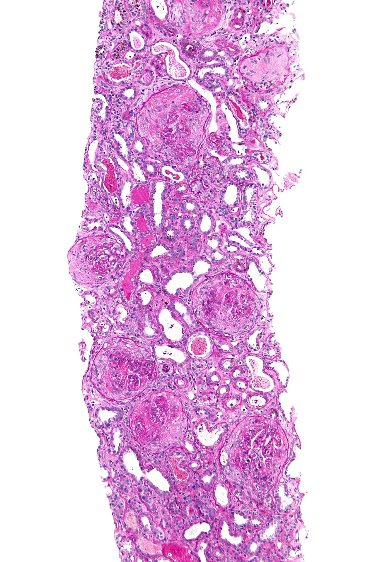
| Type | Immunofluorescence | Causes | Serology |
|---|---|---|---|
| Type I - anti-GBM | Linear IgG along the basement membrane | Anti-GBM (Goodpasture) disease | Anti-GBM antibodies |
| Type II - immune complex | Granular ('lumpy-bumpy') deposition | Lupus nephritis, IgA nephropathy / IgA vasculitis, post-infectious GN, membranoproliferative GN, cryoglobulinaemia | ANA and anti-dsDNA, low complement, raised IgA, ASOT |
| Type III - pauci-immune | Little or no staining | ANCA-associated vasculitis - granulomatosis with polyangiitis, microscopic polyangiitis, EGPA. The commonest type overall. | ANCA (anti-PR3 or anti-MPO) |
Investigations
Bedside and urine
- Urine dipstick - the essential first test; blood and protein together is the signature of glomerular disease
- Urine microscopy - dysmorphic red cells and red cell casts confirm a glomerular origin of bleeding; white cell casts suggest interstitial disease or infection
- Urine protein:creatinine ratio or ACR - to quantify proteinuria, which is both a diagnostic and prognostic marker
- Blood pressure, which is frequently raised and needs treating in its own right
- Urine culture - to exclude infection as a cause of haematuria
Blood tests and immunology
| Test | Significance |
|---|---|
| ANCA (anti-PR3, anti-MPO) | ANCA-associated vasculitis - pauci-immune RPGN |
| Anti-GBM antibodies | Anti-GBM (Goodpasture) disease |
| ANA and anti-dsDNA | Lupus nephritis |
| Complement C3 and C4 | Low C3 with normal C4 - post-streptococcal GN, C3 glomerulopathy. Low C3 AND low C4 - lupus nephritis, membranoproliferative GN, cryoglobulinaemia, endocarditis. Normal complement - IgA nephropathy, anti-GBM disease, ANCA vasculitis. |
| Anti-PLA2R antibodies | Primary membranous nephropathy |
| ASOT / anti-DNase B | Recent streptococcal infection - post-streptococcal GN |
| Immunoglobulins, serum free light chains, serum and urine electrophoresis | Myeloma and amyloidosis |
| Cryoglobulins | Cryoglobulinaemic vasculitis, often hepatitis C-related |
| Hepatitis B, hepatitis C and HIV serology | Secondary causes that change management entirely (and must be known before immunosuppression) |
| Blood cultures and echocardiography | Infective endocarditis can cause an immune complex glomerulonephritis and must not be immunosuppressed |
- Also send U&Es, full blood count (anaemia, eosinophilia in EGPA), CRP and ESR, LFTs, clotting, and a chest X-ray (looking for infiltrates, nodules or haemorrhage)
Renal biopsy
- Biopsy is the definitive investigation and is indicated in almost all adults with unexplained glomerular disease, because the histological diagnosis determines treatment and prognosis
- The specimen is examined by light microscopy (pattern and distribution of injury, crescents, sclerosis, interstitial fibrosis), immunofluorescence (the pattern of immunoglobulin and complement deposition - linear, granular or pauci-immune) and electron microscopy (foot process effacement, deposit location)
- Check clotting, platelets and blood pressure beforehand, and confirm two kidneys are present; the principal complication is bleeding
- Biopsy is often deferred in children with typical steroid-responsive nephrotic syndrome and in classic post-streptococcal GN following a predictable recovery
Management
Measures applying to all patients
- Blood pressure control - the most important intervention for long-term renal survival, with tighter targets where there is proteinuria3
- ACE inhibitor or ARB - reduces intraglomerular pressure and proteinuria beyond its antihypertensive effect, and is indicated in essentially all patients with persistent proteinuria
- Sodium restriction, diuretics for oedema, and fluid management
- Statin therapy and cardiovascular risk reduction
- Avoid nephrotoxins, particularly NSAIDs
- Treat the trigger where there is one - antibiotics for infection, antivirals for hepatitis B or C, withdrawal of a culprit drug, treatment of an underlying malignancy
- Monitor renal function and proteinuria to detect progression
Immunosuppression
Immunosuppression is reserved for histologically confirmed disease that is active and likely to progress, because the risks are substantial. It is emphatically not given for isolated haematuria with normal function.2
| Disease | Treatment |
|---|---|
| ANCA-associated vasculitis | Induction: high-dose corticosteroid plus cyclophosphamide or rituximab; plasma exchange for severe renal disease or pulmonary haemorrhage. Maintenance: rituximab, azathioprine or methotrexate for at least 2 years. Add co-trimoxazole for PCP prophylaxis. |
| Anti-GBM disease | Urgent plasma exchange plus corticosteroids and cyclophosphamide - the one setting where plasma exchange is unequivocally central |
| Lupus nephritis | Guided by ISN/RPS class: mycophenolate or cyclophosphamide with corticosteroids for proliferative (class III/IV) disease, often with belimumab or voclosporin; hydroxychloroquine for all |
| IgA nephropathy | Supportive therapy first for at least 3 to 6 months. Corticosteroids considered where proteinuria remains above 1 g/day with progressive disease, balanced against infection risk; targeted-release budesonide, sparsentan and SGLT2 inhibitors are newer options. |
| Post-streptococcal GN | Supportive only - immunosuppression is not indicated; the disease is self-limiting |
| Membranous nephropathy | Supportive for 6 months in low-risk disease; rituximab, the Ponticelli regimen or a calcineurin inhibitor for persistent or progressive disease |
| Minimal change disease and FSGS | Corticosteroids first line - see the nephrotic syndrome article |
Complications
- Acute kidney injury, sometimes requiring dialysis
- Progression to chronic kidney disease and end-stage renal disease
- Hypertension, often severe and requiring multiple agents, and accelerated cardiovascular risk
- Nephrotic complications where proteinuria is heavy - thromboembolism (including renal vein thrombosis), infection and hyperlipidaemia
- Pulmonary haemorrhage in anti-GBM disease and ANCA vasculitis - immediately life-threatening
- Hyperkalaemia, acidosis and fluid overload from renal failure
- Complications of immunosuppression - infection (including PCP and reactivated TB or hepatitis B), infertility from cyclophosphamide, malignancy, corticosteroid toxicity and bone marrow suppression
- Relapse - common in ANCA vasculitis and lupus nephritis, so long-term maintenance and monitoring are needed
- Recurrence in a transplanted kidney - particularly FSGS, IgA nephropathy and membranous nephropathy
Red flags
Prognosis
Outcomes range from complete spontaneous recovery to irreversible renal failure within weeks, and the histological diagnosis is the dominant determinant.
Post-streptococcal glomerulonephritis in children has an excellent prognosis, with over 95% recovering fully; the haematuria may persist for months and the low C3 normalises within 6 to 8 weeks, but permanent damage is unusual. Thin basement membrane disease is similarly benign and needs only reassurance and monitoring.
IgA nephropathy has a highly variable course. Patients with isolated haematuria, normal blood pressure and no proteinuria do very well, whereas roughly 20 to 30% of the overall cohort reach end-stage renal disease within 20 years. The predictors of progression are consistent across glomerular disease generally: the degree of proteinuria, the blood pressure, the creatinine at presentation, and the amount of interstitial fibrosis and tubular atrophy on biopsy.
Rapidly progressive glomerulonephritis was almost uniformly fatal before effective immunosuppression and remains the group in which prompt treatment makes the greatest difference. With modern induction regimens, remission is achieved in the large majority of ANCA-associated vasculitis, though relapse occurs in a substantial proportion and long-term maintenance is required. In anti-GBM disease the creatinine at presentation is decisive - patients who present before dialysis is needed often recover useful function, whereas those already dialysis-dependent seldom do, and that difference is frequently a matter of how quickly the diagnosis was considered.
Across all types, the treatment burden increasingly rivals the disease. Much of the morbidity in treated glomerulonephritis now arises from infection, corticosteroid toxicity, infertility and malignancy risk from cyclophosphamide - which is why the shift towards rituximab, targeted therapies and steroid-minimising regimens matters, and why immunosuppression is withheld in those whose disease is unlikely to progress.
References
- KDIGO 2021 Clinical Practice Guideline for the Management of Glomerular Diseases. Available here
- UK Kidney Association. Clinical practice guidelines - glomerular disease and vasculitis. Available here
- NICE NG203. Chronic kidney disease: assessment and management. 2021. Available here
- NICE Clinical Knowledge Summaries. Haematuria. Available here
- Jennette JC, Falk RJ. Small-vessel vasculitis. New England Journal of Medicine. Available here
- Nephron, CC BY-SA 3.0, via Wikimedia Commons. Available here
This article is written for revision and education. It is not clinical guidance and must not be used to make decisions about the care of a patient. Always check current NICE guidance and local protocols.