
Bladder Cancer
Key points
- Bladder cancer: the commonest urinary tract malignancy. Around 90% are urothelial (transitional cell) carcinomas; squamous cell carcinoma and adenocarcinoma are much rarer.
- Cardinal symptom: painless visible haematuria - this is bladder cancer until proven otherwise, and warrants urgent referral regardless of whether it settles.
- Biggest risk factors: smoking (responsible for around half of cases) and occupational exposure to aromatic amines in the rubber, dye, paint and textile industries.
- Schistosomiasis: chronic Schistosoma haematobium infection causes squamous cell carcinoma of the bladder - the key association in patients from endemic areas (notably Egypt and sub-Saharan Africa).
- Referral: urgent 2-week-wait for visible haematuria aged 45 or over, or non-visible haematuria with dysuria or a raised WCC aged 60 or over.1
- Diagnosis: cystoscopy is the definitive investigation, with CT urogram to image the upper tracts, since urothelial tumours can be multifocal.
- The critical division: non-muscle-invasive (NMIBC, ~75%) versus muscle-invasive (MIBC) disease. TURBT must include detrusor muscle in the specimen to make this distinction.
- Treatment: NMIBC: TURBT plus intravesical mitomycin C or BCG, with lifelong surveillance cystoscopy. MIBC: neoadjuvant chemotherapy then radical cystectomy or radical radiotherapy.
Introduction
Bladder cancer is the commonest malignancy of the urinary tract and around the tenth commonest cancer in the UK, with roughly 10,000 new cases annually. It is three to four times more common in men, and incidence rises sharply with age, with most cases diagnosed over 65.2
The urothelium lines the entire tract from the renal pelvis to the proximal urethra, and is exposed along its whole length to carcinogens excreted in urine. This produces the concept of a 'field change': urothelial tumours are frequently multifocal and recurrent, which is why the upper tracts must be imaged at diagnosis and why patients need lifelong cystoscopic surveillance rather than discharge after treatment.
| Type | Proportion | Associations |
|---|---|---|
| Urothelial (transitional cell) carcinoma | ~90% | Smoking, aromatic amines, cyclophosphamide, age |
| Squamous cell carcinoma | ~5% | Chronic inflammation - Schistosoma haematobium, long-term indwelling catheters, recurrent infection, bladder stones |
| Adenocarcinoma | ~1-2% | Urachal remnants (typically at the bladder dome), bladder exstrophy |
| Small cell carcinoma / sarcoma | Rare | Aggressive; small cell behaves like small cell lung cancer and is treated with chemotherapy |
Risk factors
- Smoking - the single largest risk factor, responsible for around 50% of cases and conferring a two- to four-fold increase in risk. Aromatic amines and polycyclic hydrocarbons are excreted in urine and concentrate in the bladder. Risk falls after cessation but never returns to baseline - and smoking cessation advice remains worthwhile even after diagnosis, as continued smoking increases recurrence
- Occupational exposure to aromatic amines - beta-naphthylamine and benzidine, historically used in the rubber, dye, paint, plastics, leather and textile industries. Also printers, hairdressers and painters. The latency is long (often 20-30 years), so take a lifetime occupational history
- Increasing age and male sex
- Schistosoma haematobium infection - causes chronic inflammation and squamous cell carcinoma; relevant in patients from Egypt, the Middle East and sub-Saharan Africa
- Chronic bladder inflammation - long-term indwelling catheters (a recognised risk in patients with spinal cord injury), recurrent urinary tract infection, and bladder calculi
- Cyclophosphamide - its metabolite acrolein causes haemorrhagic cystitis and later malignancy; mesna is given to reduce this
- Pelvic radiotherapy - for example previous prostate or cervical cancer treatment
- Family history and Lynch syndrome (particularly for upper tract urothelial tumours)
- Pioglitazone - a modest association reported
- Aristolochic acid exposure (some herbal preparations)
Clinical features
Other presentations
- Non-visible (microscopic) haematuria - often found incidentally on dipstick
- Storage lower urinary tract symptoms - frequency, urgency and dysuria; particularly characteristic of carcinoma in situ (CIS), which is a flat, high-grade lesion that may not be obvious cystoscopically but causes marked irritative symptoms and carries a high risk of progression
- Recurrent urinary tract infections or an infection that fails to clear
- Suprapubic pain or a palpable mass (late)
- Clot retention - acute urinary retention caused by blood clot, a urological emergency requiring irrigation
- Ureteric obstruction and hydronephrosis - if a tumour involves a ureteric orifice, causing loin pain or renal impairment
- Systemic features of advanced disease - weight loss, anorexia, bone pain, lymphoedema of the legs from pelvic nodal disease
Investigations
Referral criteria (NICE NG12)
- Urgent (2-week-wait) referral: aged 45 and over with unexplained visible haematuria (without UTI, or persisting after treatment of a UTI); or aged 60 and over with non-visible haematuria plus dysuria or a raised white cell count
- Consider non-urgent referral: aged 60 and over with recurrent or persistent unexplained urinary tract infection
Investigations
- Cystoscopy - the definitive diagnostic investigation. Flexible cystoscopy under local anaesthetic is used initially; rigid cystoscopy under general anaesthetic permits biopsy and resection
- CT urogram - images the entire upper urinary tract for synchronous urothelial tumours in the renal pelvis or ureter, and provides staging information. Ultrasound is an alternative in some settings but is less sensitive
- Urine dipstick and culture - to exclude infection, though a positive dipstick or a UTI does not remove the need for investigation in the presence of visible haematuria
- Urine cytology - may detect high-grade cells and CIS, but has poor sensitivity for low-grade tumours and cannot be used to exclude cancer
- U&Es and creatinine - renal function, relevant both to obstruction and to eligibility for cisplatin-based chemotherapy
- FBC - anaemia from chronic blood loss
- Transurethral resection of bladder tumour (TURBT) - both diagnostic and therapeutic; provides histology, grade and, crucially, depth of invasion
- Staging CT chest, abdomen and pelvis and, where indicated, bone scan or PET-CT for muscle-invasive disease
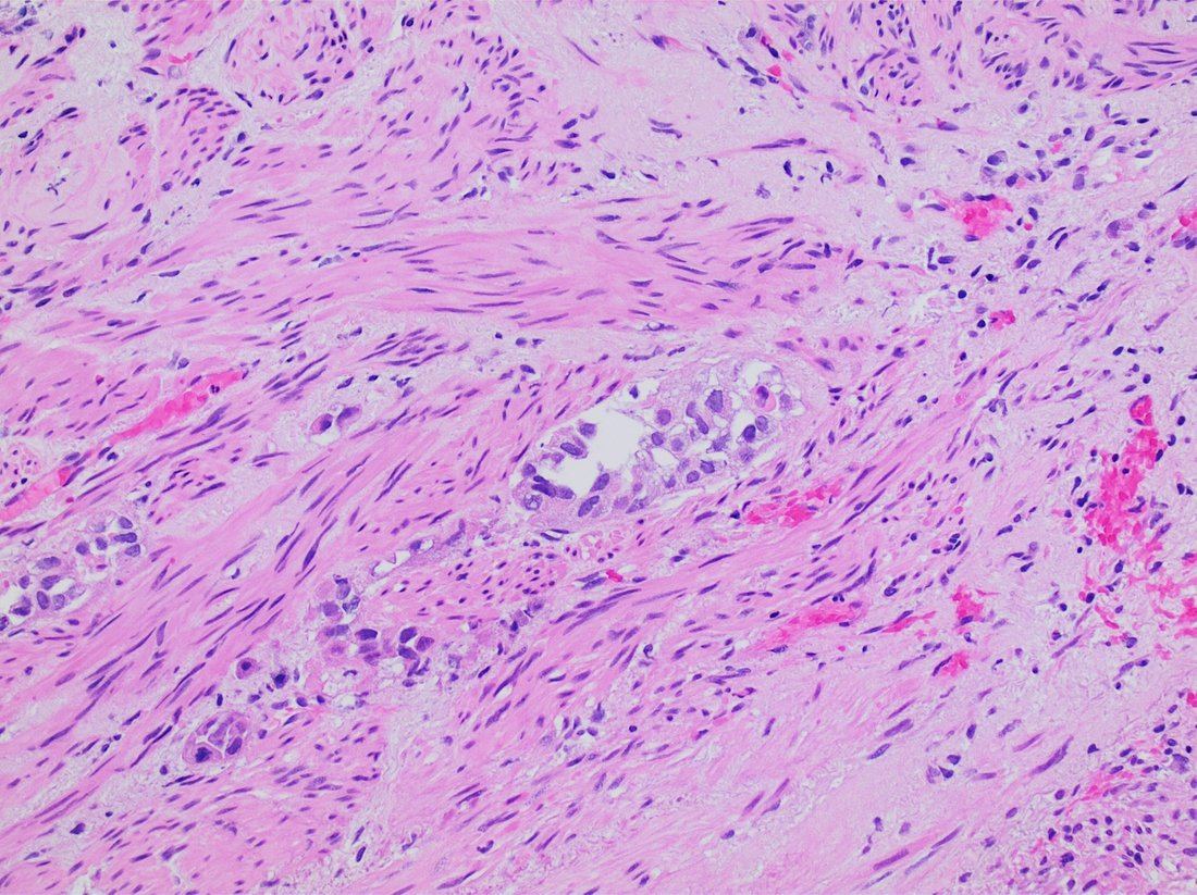
Staging
| Stage | Description | Category |
|---|---|---|
| Ta | Non-invasive papillary carcinoma, confined to the urothelium | Non-muscle-invasive (NMIBC) |
| Tis (CIS) | Flat, high-grade carcinoma in situ confined to the urothelium - looks like erythematous mucosa; high risk of progression | Non-muscle-invasive (NMIBC) |
| T1 | Invades subepithelial connective tissue (lamina propria) but not muscle | Non-muscle-invasive (NMIBC) |
| T2 | Invades detrusor muscle (T2a superficial half, T2b deep half) | Muscle-invasive (MIBC) |
| T3 | Invades perivesical fat | Muscle-invasive (MIBC) |
| T4 | Invades adjacent structures - prostate, uterus, vagina, pelvic or abdominal wall | Muscle-invasive (MIBC) |
Around 75% of bladder cancers are non-muscle-invasive at diagnosis. NMIBC is further stratified into low, intermediate and high risk based on grade, stage, size, number of tumours, recurrence and the presence of CIS - and this risk category determines whether intravesical chemotherapy, BCG, or in high-risk cases early cystectomy is offered.
Management
Non-muscle-invasive bladder cancer
- TURBT - complete resection of all visible tumour
- A single immediate post-operative dose of intravesical mitomycin C (within 24 hours) - reduces recurrence and is standard for low-risk disease
- Intravesical mitomycin C course - a course of instillations for intermediate-risk disease
- Intravesical BCG - for high-risk disease (high-grade T1, CIS). BCG is live attenuated Mycobacterium bovis and works by provoking a local immune response. Given as an induction course with maintenance
- Repeat TURBT - where muscle was absent from the specimen or the resection was incomplete, typically at 2-6 weeks
- Radical cystectomy - considered for high-risk disease that recurs or fails BCG
- Lifelong surveillance cystoscopy - the schedule depends on risk category; recurrence rates are high, which is why patients are never simply discharged
- Smoking cessation - reduces both recurrence and progression, and should be addressed at every visit
Muscle-invasive bladder cancer
- Neoadjuvant cisplatin-based combination chemotherapy before radical treatment - this provides a demonstrable survival benefit and is offered to patients fit enough with adequate renal function
- Radical cystectomy - removal of the bladder, with prostate and seminal vesicles in men or uterus, ovaries and part of the vagina in women, plus pelvic lymph node dissection. Requires urinary diversion: an ileal conduit (urostomy) or an orthotopic neobladder fashioned from bowel
- Radical radiotherapy with a radiosensitiser - an organ-preserving alternative for patients unfit for or declining surgery; outcomes are broadly comparable in selected patients
- Adjuvant chemotherapy in selected patients not given neoadjuvant treatment
- Immunotherapy - checkpoint inhibitors (e.g. atezolizumab, pembrolizumab) in advanced or metastatic disease, and increasingly in earlier settings
- Palliative care - radiotherapy for haematuria and pain, and symptom control in advanced disease
Patients undergoing cystectomy need thorough counselling about stoma care, body image, sexual dysfunction and, in men, infertility, along with specialist stoma nurse input. Long-term follow-up after diversion includes monitoring for B12 deficiency, metabolic acidosis and renal impairment.
Complications
- Recurrence - the defining feature of NMIBC, with recurrence in a substantial proportion despite complete resection
- Progression to muscle-invasive disease - the principal concern in high-risk NMIBC and CIS
- Clot retention - acute retention from blood clot, requiring three-way catheterisation and bladder irrigation
- Anaemia from chronic or acute haematuria
- Ureteric obstruction and hydronephrosis with renal impairment
- Metastatic disease - to pelvic lymph nodes, liver, lung and bone
- Complications of BCG - cystitis, and rarely BCG sepsis requiring antituberculous therapy
- Complications of cystectomy and urinary diversion - stoma problems, urinary leak, bowel obstruction, metabolic acidosis, B12 deficiency, recurrent infection, stones and renal impairment
- Sexual dysfunction and infertility after radical treatment
- Radiotherapy toxicity - radiation cystitis, proctitis and a contracted bladder
- Psychological impact - of stoma formation, body image change and the burden of lifelong surveillance
Red flags
Prognosis
Prognosis is dominated by whether the tumour has invaded muscle. Around 75% of patients present with non-muscle-invasive disease, which has an excellent survival - 5-year survival for Ta/T1 disease exceeds 90% - but a high burden of recurrence, affecting more than half of patients over time. This is a disease patients live with and are monitored for over decades, which explains both the lifelong surveillance and the considerable healthcare cost per patient.
Muscle-invasive disease is far more serious. Even with neoadjuvant chemotherapy and radical cystectomy, 5-year survival is in the region of 40-50%, and once there is nodal or distant metastatic disease it falls markedly. Neoadjuvant cisplatin-based chemotherapy improves absolute survival by around 5-8% and is therefore offered to all who are fit enough, though many patients are excluded by age, comorbidity or inadequate renal function.
Prognostic factors are stage, grade, the presence of carcinoma in situ, tumour size and multifocality, and response to BCG. Failure to respond to BCG in high-risk NMIBC is an adverse sign and generally prompts consideration of early cystectomy rather than further intravesical therapy. Two modifiable factors are worth emphasising because they are easily overlooked: continued smoking increases both recurrence and progression, so cessation support is genuinely disease-modifying rather than merely generic advice, and prompt investigation of haematuria is the main determinant of whether the disease is caught while still non-muscle-invasive.
References
- NICE NG12. Suspected cancer: recognition and referral. 2015, updated 2023. Available here
- NICE NG2. Bladder cancer: diagnosis and management. 2015, updated 2018. Available here
- Cancer Research UK. Bladder cancer statistics. Available here
- European Association of Urology. Guidelines on Non-muscle-invasive Bladder Cancer. Available here
- European Association of Urology. Guidelines on Muscle-invasive and Metastatic Bladder Cancer. Available here
- Advanced Bladder Cancer (ABC) Meta-analysis Collaboration. Neoadjuvant chemotherapy in invasive bladder cancer. European Urology. 2005. Available here
- Mikael Häggström, MD, CC0, via Wikimedia Commons. Available here
This article is written for revision and education. It is not clinical guidance and must not be used to make decisions about the care of a patient. Always check current NICE guidance and local protocols.