
Hydronephrosis
Key points
- Hydronephrosis: dilatation of the renal pelvis and calyces. It is a radiological finding, not a diagnosis - the task is always to find the cause.
- Hydroureter: dilatation of the ureter as well. Hydroureteronephrosis implies the obstruction is below the pelviureteric junction, which helps localise the lesion.
- The key rule: unilateral hydronephrosis implies obstruction at or above the ureter on that side; bilateral hydronephrosis implies obstruction at or below the bladder outlet, or bilateral ureteric pathology.
- Not always obstruction: dilatation can occur without obstruction - in pregnancy, vesicoureteric reflux, and a capacious extrarenal pelvis. MAG3 renography distinguishes obstructive from non-obstructive dilatation.
- Presentation: ranges from asymptomatic (found incidentally) to loin pain, and in bilateral or acute obstruction, anuria and acute kidney injury.
- First-line investigation: renal tract ultrasound, with CT to define the cause and level, and U&Es to assess function.
- Management: relieve the obstruction: catheter for bladder outlet obstruction, nephrostomy or ureteric stent for upper tract obstruction, then treat the underlying cause.
- The emergency: obstruction with infection or acute kidney injury - requires urgent decompression. Watch for post-obstructive diuresis afterwards.
Introduction
Hydronephrosis describes dilatation of the renal pelvis and calyces. It is important to be clear that this is a descriptive radiological finding rather than a diagnosis in itself - the clinical work lies in establishing whether there is true obstruction, where that obstruction is, what is causing it, and whether renal function is threatened.1
Sustained obstruction raises pressure within the collecting system, which is transmitted back to the nephron. This reduces glomerular filtration, causes progressive tubular atrophy and interstitial fibrosis, and ultimately produces cortical thinning and irreversible loss of function. The rate at which this happens is the reason obstruction is treated urgently: significant damage can begin within days to weeks, and a chronically obstructed kidney may never recover useful function.
Causes
Causes are conventionally divided by their relationship to the wall of the urinary tract, which is a reliable way to structure an answer.
| Category | Causes |
|---|---|
| Intraluminal (within the lumen) | Urinary calculi (the commonest acute cause), blood clot, sloughed renal papilla (papillary necrosis) |
| Intramural (in the wall) | Pelviureteric junction (PUJ) obstruction - the commonest congenital cause; ureteric stricture (post-instrumentation, TB, schistosomiasis); urothelial (transitional cell) carcinoma of the ureter; posterior urethral valves in male infants; neuromuscular dysfunction |
| Extramural (external compression) | Benign prostatic hyperplasia and prostate cancer (bilateral); pelvic malignancy - cervical, bladder, rectal, ovarian; retroperitoneal fibrosis; abdominal aortic aneurysm; lymphadenopathy; pregnancy; endometriosis; iatrogenic ureteric injury after pelvic surgery |
| Non-obstructive dilatation | Vesicoureteric reflux; pregnancy (physiological); a capacious extrarenal pelvis; high urine flow states such as diabetes insipidus; post-obstructive residual dilatation after the cause has been relieved |
Clinical features
Presentation depends entirely on how quickly the obstruction developed, whether it is unilateral or bilateral, and whether infection is present. Chronic, gradual obstruction is often silent until function is substantially lost.
Acute obstruction
- Severe colicky loin pain - rapid distension of the collecting system, typically from a stone
- Nausea and vomiting
- Haematuria
- Restlessness - the patient cannot lie still
Chronic obstruction
- Often completely asymptomatic, discovered incidentally on imaging performed for another reason
- Dull, persistent loin ache rather than colic, sometimes worse after drinking large volumes (classically in PUJ obstruction, 'Dietl's crisis')
- A palpable flank mass in gross hydronephrosis
- Recurrent urinary tract infections from stasis
- Insidious features of chronic kidney disease - fatigue, nausea, anaemia, hypertension
Features pointing to the underlying cause
- Lower urinary tract symptoms - hesitancy, poor stream, terminal dribbling, incomplete emptying, nocturia: suggests bladder outlet obstruction, usually prostatic in men
- Acute urinary retention with a painful, palpable bladder
- Anuria or marked oliguria - implies complete bilateral obstruction (or obstruction of a solitary kidney) and is a medical emergency
- Fever and rigors - infection above the obstruction; an emergency
- Weight loss, haematuria, bone pain or a pelvic mass - suggests malignancy
- Antenatal detection - hydronephrosis is one of the commonest findings on the routine 20-week anomaly scan, and prompts postnatal assessment for PUJ obstruction, vesicoureteric reflux or posterior urethral valves
Investigations
Imaging
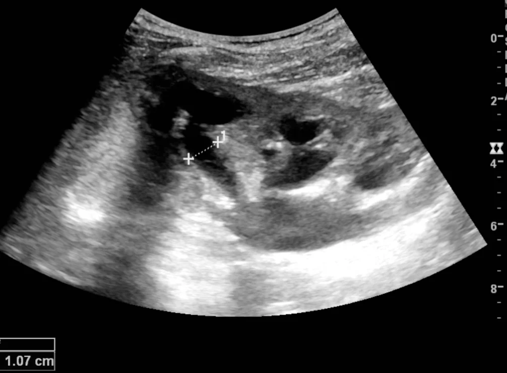
- Renal tract ultrasound - first line. It confirms hydronephrosis, grades its severity, assesses cortical thickness (thinning indicating chronicity and lost function), and shows bladder volume. It is quick, safe and radiation-free, making it the test of choice in pregnancy and children. Its weakness is that it cannot reliably identify the cause or the level of obstruction, and it may miss obstruction in a dehydrated patient or in retroperitoneal fibrosis, where the ureters are encased and cannot dilate
- Bladder scan (post-void residual) - simple, immediate, and essential: a large residual volume points to bladder outlet obstruction and means the first treatment is a catheter
- Non-contrast CT KUB - excellent for stones and for defining the level of obstruction
- CT with contrast / CT urogram - to characterise masses, retroperitoneal fibrosis and urothelial tumours
- MAG3 or DTPA renography (nuclear medicine) - the definitive test to distinguish obstructive from non-obstructive dilatation and to quantify differential function between the two kidneys. This determines whether a poorly functioning, chronically obstructed kidney is worth salvaging
- DMSA scan - assesses renal scarring and split function, particularly in children
- Micturating cystourethrogram (MCUG) - in infants and children to demonstrate vesicoureteric reflux or posterior urethral valves
- Antegrade or retrograde pyelography - defines the level of obstruction precisely, often performed at the time of stenting or nephrostomy
Laboratory
- U&Es and creatinine - to identify AKI and establish baseline function. Remember that these are often normal in unilateral obstruction
- FBC and CRP - evidence of infection
- Urine dipstick and culture - haematuria suggesting stones or tumour; nitrites and leucocytes suggesting infection
- Blood cultures if febrile
- Venous blood gas - for rapid potassium in AKI, and to assess acidosis
- PSA in men with suspected prostatic obstruction (interpreting with caution, as retention and catheterisation raise it)
- Calcium, urate and stone screen where calculi are the cause
- ESR, CRP and autoimmune screen if retroperitoneal fibrosis is suspected; IgG4 levels may be relevant
Management
Management has three components in sequence: decide whether decompression is urgent, relieve the obstruction at the appropriate level, and then treat the underlying cause.
Urgent decompression
Immediate relief of obstruction is required where there is infection, acute kidney injury, a solitary or transplanted kidney, bilateral obstruction, or intractable pain.
| Level of obstruction | Intervention |
|---|---|
| Bladder outlet (BPH, stricture, neurogenic bladder) - typically bilateral hydronephrosis with a large residual volume | Urethral catheter - simple, immediate and often completely effective. Suprapubic catheter if urethral catheterisation fails or is contraindicated |
| Upper tract / ureteric (stone, tumour, extrinsic compression) - typically unilateral | Percutaneous nephrostomy (radiologically placed, drains externally; preferred in sepsis) or retrograde ureteric stent (placed cystoscopically, internal). Choice depends on local practice, the patient's state and the cause |
| Infected obstructed system (pyonephrosis) | Emergency decompression plus IV antibiotics - nephrostomy is often favoured as it avoids instrumenting an infected ureter |
Treating the underlying cause
- Stones - ureteroscopy, lithotripsy or PCNL once any sepsis has settled
- Benign prostatic hyperplasia - alpha-blockers and 5-alpha reductase inhibitors, with TURP for those failing medical therapy or with high-pressure chronic retention
- Malignancy - oncological management, with stenting or nephrostomy for palliation of obstruction
- PUJ obstruction - pyeloplasty, where function is worth preserving
- Retroperitoneal fibrosis - corticosteroids, sometimes with other immunosuppression, plus stenting or ureterolysis
- Posterior urethral valves - endoscopic valve ablation in infancy
- Vesicoureteric reflux - antibiotic prophylaxis and surveillance in children, with surgery in selected cases
- Nephrectomy - considered for a chronically obstructed, non-functioning kidney that is causing pain, infection or hypertension
Complications
- Acute kidney injury - particularly with bilateral obstruction or a solitary kidney
- Chronic kidney disease and end-stage renal disease - from sustained obstruction causing tubular atrophy, interstitial fibrosis and cortical thinning
- Irreversible loss of the affected kidney - the risk rises steeply the longer obstruction persists
- Infection: pyelonephritis, pyonephrosis and urosepsis - stasis strongly predisposes to infection, and an infected obstructed system is life-threatening
- Stone formation - stasis promotes crystallisation, creating a self-perpetuating cycle
- Hypertension - from activation of the renin-angiotensin-aldosterone system in the obstructed kidney
- Post-obstructive diuresis with dehydration and electrolyte disturbance
- Renal tubular acidosis and hyperkalaemia - impaired distal tubular function in chronic obstruction
- Spontaneous rupture of the collecting system - rare, with acute severe obstruction
- Complications of intervention - stent discomfort, encrustation, migration and infection; bleeding after nephrostomy
Red flags
Prognosis
Prognosis is determined almost entirely by the duration and completeness of obstruction, and by whether infection supervenes. Acute obstruction relieved within a few days is generally followed by full recovery of renal function. Beyond a few weeks, recovery becomes progressively less complete, and obstruction persisting for months typically leaves permanent loss of function in that kidney.
Cortical thickness on ultrasound and differential function on MAG3 renography are the practical guides to salvageability. A kidney with preserved cortex and reasonable split function is worth decompressing and reconstructing; one with a thin cortex contributing very little function may be better managed conservatively or, if it is causing pain, infection or hypertension, by nephrectomy.
Unilateral obstruction with a healthy contralateral kidney rarely causes renal failure, because the normal kidney compensates - which is precisely why it can progress silently to complete loss of the affected side with an entirely normal creatinine throughout. Bilateral obstruction, by contrast, presents earlier with AKI but is frequently and dramatically reversible: relieving bladder outlet obstruction with a catheter can return a severely uraemic patient to near-normal function within days. In children, the long-term outlook depends on the underlying anomaly, with posterior urethral valves carrying the greatest risk of progressive chronic kidney disease despite early intervention.
References
- NICE Clinical Knowledge Summaries. Lower urinary tract symptoms in men. Available here
- NICE NG118. Renal and ureteric stones: assessment and management. 2019. Available here
- NICE NG148. Acute kidney injury: prevention, detection and management. 2019. Available here
- European Association of Urology. Guidelines on Paediatric Urology (antenatal hydronephrosis, posterior urethral valves). Available here
- Mevorach RA, Hulbert WC, Rabinowitz R. Hydronephrosis and obstructive uropathy. BMJ Best Practice. Available here
- Kristoffer Lindskov Hansen, Michael Bachmann Nielsen and Caroline Ewertsen, CC BY 4.0, via Wikimedia Commons. Available here
This article is written for revision and education. It is not clinical guidance and must not be used to make decisions about the care of a patient. Always check current NICE guidance and local protocols.