
Pyelonephritis
Key points
- Pyelonephritis: infection of the renal parenchyma and renal pelvis - an upper urinary tract infection and a systemic illness, in contrast to cystitis.
- Classic triad: fever (often with rigors), loin or flank pain, and nausea or vomiting, usually with renal angle tenderness and often preceded by lower urinary tract symptoms.
- Organism: Escherichia coli in around 70-80%, almost always reaching the kidney by ascending infection from the bladder rather than haematogenous spread.
- Investigations: urine dipstick and culture before antibiotics, bloods including FBC, CRP, U&Es and blood cultures if systemically unwell.
- Imaging: urgent renal tract ultrasound or CT if there is a suspicion of obstruction, stones or abscess, in all men, and in anyone failing to improve after 48-72 hours.
- Treatment: 7-10 days of an antibiotic that penetrates renal tissue - cefalexin, co-amoxiclav, trimethoprim or ciprofloxacin per sensitivities. Nitrofurantoin must not be used as it does not reach therapeutic tissue levels.
- The emergency: an obstructed, infected kidney (pyonephrosis) - antibiotics alone will fail; it requires urgent decompression with a nephrostomy or ureteric stent.
- Long-term risk: renal scarring, particularly with recurrent childhood infection and vesicoureteric reflux, leading to hypertension and chronic kidney disease.
Introduction
Pyelonephritis is infection of the renal parenchyma, calyces and renal pelvis. It is the principal form of upper urinary tract infection and, unlike cystitis, it is a systemic illness - patients are febrile, often vomiting, and a proportion are septic.1
The overwhelming majority arise by ascending infection: bacteria colonise the perineum, ascend the urethra into the bladder, and then travel up the ureter to the kidney. Haematogenous seeding is much less common and tends to occur with Staphylococcus aureus bacteraemia or in the immunosuppressed. Incidence is highest in young women, but the cases that cause the most trouble are those in men, pregnant women, diabetics and anyone with obstruction.
Aetiology and risk factors
The organisms are those of urinary tract infection generally: E. coli predominates, followed by Klebsiella, Proteus, Enterococcus and, in healthcare-associated infection, Pseudomonas. Proteus is again worth flagging because its urease production alkalinises urine and forms struvite stones, which then act as a persistent nidus for further infection.
Risk factors
- Female sex - a short urethra, as for lower UTI
- Structural abnormality or obstruction - stones, strictures, tumours, pelviureteric junction obstruction; the single most important group to identify because they change management
- Vesicoureteric reflux - allows bacteria to be propelled retrogradely to the kidney; the key mechanism in childhood pyelonephritis and subsequent scarring
- Pregnancy - progesterone-mediated ureteric dilatation and mechanical compression cause stasis; pyelonephritis complicates around 1-2% of pregnancies and is a leading cause of antenatal admission
- Diabetes mellitus - and the specific risk of emphysematous pyelonephritis
- Urinary catheterisation and instrumentation
- Immunosuppression, including renal transplant recipients
- Benign prostatic hyperplasia and incomplete bladder emptying in men
- Recent antibiotic use - predisposing to resistant organisms
- Previous UTI or pyelonephritis
Clinical features
The presentation is of an acutely unwell, febrile patient with loin pain. Lower urinary tract symptoms often precede it by a day or two, but their absence does not exclude the diagnosis.
- Fever, often high, with rigors and sweats - rigors in particular suggest bacteraemia
- Loin, flank or back pain, typically unilateral, and usually a constant ache rather than the colicky pain of a stone
- Nausea and vomiting - important practically, because it often prevents oral antibiotics and forces admission
- Renal angle (costovertebral) tenderness on examination - the classic sign, elicited by gentle percussion over the renal angle
- Preceding or concurrent lower urinary tract symptoms - dysuria, frequency, urgency
- Malaise, myalgia and anorexia
- Tachycardia, hypotension and confusion - suggest sepsis and demand immediate action
Differential diagnosis
| Condition | Distinguishing features |
|---|---|
| Renal or ureteric colic | Colicky loin-to-groin pain with restlessness, usually afebrile unless infection coexists; haematuria common. May coexist - and that combination is the emergency |
| Pyonephrosis (obstructed infected system) | Fever and loin pain with hydronephrosis on imaging; needs urgent drainage, not just antibiotics |
| Renal or perinephric abscess | Persisting fever beyond 72 hours of appropriate antibiotics; diagnosed on CT |
| Lower UTI (cystitis) | Bladder symptoms but systemically well, afebrile, no loin pain |
| Acute appendicitis / diverticulitis | Localised abdominal peritonism, shifting pain, altered bowel habit |
| Biliary sepsis (cholecystitis, cholangitis) | Right upper quadrant pain, jaundice, deranged LFTs, Charcot's triad |
| Basal pneumonia | Cough, pleuritic pain, hypoxia, focal chest signs and consolidation on chest X-ray |
| Ectopic pregnancy | Any woman of childbearing age with abdominal pain - do a pregnancy test |
| Renal infarction | Loin pain with markedly raised LDH, atrial fibrillation or a thrombotic risk; CT with contrast is diagnostic |
| Musculoskeletal back pain | No fever, no urinary symptoms, pain reproduced by movement or palpation |
Investigations
Bedside and laboratory
- Urine dipstick - usually positive for nitrites, leucocytes and blood; supportive but neither sensitive nor specific enough to rely on alone
- Midstream urine for microscopy, culture and sensitivity - should be sent in every case, before antibiotics. White cell casts on microscopy are specific for upper tract (renal) inflammation
- Blood cultures - if febrile or systemically unwell; positive in a significant minority and often the only way to identify the organism
- FBC - leucocytosis and neutrophilia; CRP raised
- U&Es and creatinine - to detect AKI, and to guide antibiotic dosing
- Lactate and venous blood gas - if sepsis is suspected
- Pregnancy test in any woman of childbearing age
- Blood glucose / HbA1c - undiagnosed or poorly controlled diabetes is a common contributor
Imaging
Most previously healthy women with a straightforward first episode who respond promptly do not need imaging. Imaging is directed at finding obstruction, stones, abscess or structural abnormality.2
- Renal tract ultrasound - the usual first-line test; readily available and detects hydronephrosis (indicating obstruction), stones and gross abscess. First line in pregnancy as it avoids radiation
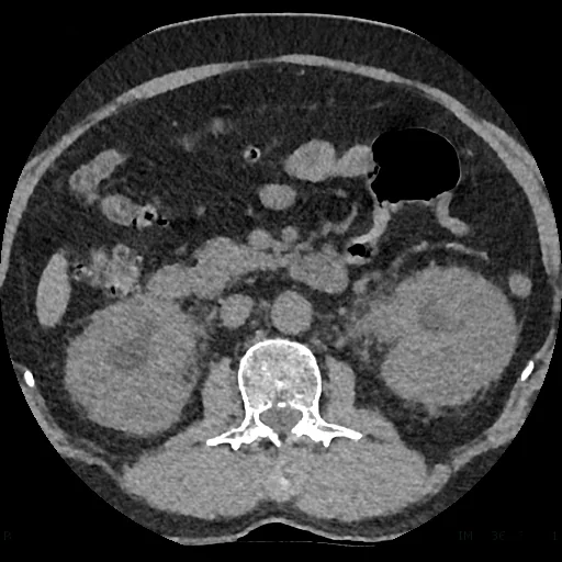
- CT of the kidneys, ureters and bladder (CT KUB), or contrast-enhanced CT - the most sensitive investigation; identifies stones, abscess, gas and the extent of parenchymal involvement. Preferred where the diagnosis is uncertain or complications are suspected
- Indications for urgent imaging: known or suspected stones, failure to improve after 48-72 hours, AKI, sepsis, a single functioning kidney, all men, recurrent episodes, diabetes (looking for emphysematous change), and immunosuppression
- DMSA scintigraphy - not used acutely, but detects renal scarring on follow-up, particularly in children
Management
Deciding on admission
Many patients can be treated at home with oral antibiotics, but a substantial proportion need admission. Admit if there is:
- Signs of sepsis or significant haemodynamic compromise
- Vomiting preventing oral antibiotics or maintenance of hydration
- Pregnancy - the threshold should be very low, and most are admitted
- Suspected obstruction, stones or abscess
- Acute kidney injury or significant pre-existing renal impairment
- Failure of oral therapy in the community
- Significant comorbidity, immunosuppression or frailty, or inability to cope at home
- Children, particularly infants under 3 months, who need urgent paediatric assessment
Antibiotics
Treatment is empirical initially and then tailored to culture sensitivities. NICE recommends 7 to 10 days of therapy for acute pyelonephritis.2
| Setting | Options |
|---|---|
| Oral (community or stepped down) | Cefalexin, co-amoxiclav (per sensitivities), trimethoprim (per sensitivities) or ciprofloxacin |
| Intravenous (severe or unable to tolerate oral) | Co-amoxiclav, cefuroxime, ceftriaxone, ciprofloxacin, or gentamicin - often with an aminoglycoside added in sepsis |
| Pregnancy | Cefalexin is the usual choice. Avoid ciprofloxacin and trimethoprim; avoid nitrofurantoin at term |
| Duration | 7-10 days total; IV therapy is usually stepped down to oral once the patient is afebrile and improving for 24-48 hours |
Supportive care and source control
- Intravenous fluids for dehydration and to maintain renal perfusion
- Analgesia and antipyretics - paracetamol; NSAIDs are best avoided where there is AKI or dehydration
- Antiemetics for vomiting
- Sepsis six within one hour if septic - blood cultures, lactate, urine output monitoring, oxygen, IV antibiotics, IV fluids
- Urgent urological decompression where there is obstruction - by percutaneous nephrostomy or retrograde ureteric stent. This is the definitive treatment of an obstructed infected system and must not be delayed while waiting to see whether antibiotics work
- Drainage of an abscess - percutaneous or, rarely, surgical, for collections that do not respond to antibiotics alone
- Remove or change an indwelling catheter where it is the likely source
Follow-up
- Repeat urine culture to confirm clearance where infection was complicated or in pregnancy
- Renal tract imaging in men, in recurrent episodes, in children, and where obstruction or stones are suspected
- Check renal function has recovered after any episode complicated by AKI
- Address underlying causes - stones, poor bladder emptying, glycaemic control, catheter care
- Blood pressure and urinalysis monitoring in those with evidence of scarring, because of the long-term risk of hypertension and CKD
Complications
- Sepsis and septic shock - the urinary tract is among the commonest sources of severe sepsis, particularly in older patients
- Acute kidney injury - from sepsis, dehydration, obstruction or direct parenchymal inflammation
- Renal abscess - suspect if fever persists beyond 72 hours despite appropriate antibiotics; needs CT and usually drainage
- Perinephric abscess - collection outside the renal capsule; often insidious and needs drainage
- Pyonephrosis - pus within an obstructed collecting system; a urological emergency requiring immediate decompression
- Emphysematous pyelonephritis - a necrotising, gas-forming infection occurring almost exclusively in poorly controlled diabetes, with gas visible in the renal parenchyma on CT; carries high mortality and requires aggressive resuscitation, antibiotics, drainage and sometimes nephrectomy
- Papillary necrosis - particularly in diabetes, sickle cell disease and analgesic nephropathy; sloughed papillae may themselves cause obstruction
- Xanthogranulomatous pyelonephritis - a rare chronic destructive granulomatous infection, classically associated with chronic obstruction and Proteus, producing a non-functioning kidney that may mimic renal cell carcinoma on imaging and usually requires nephrectomy
- Chronic pyelonephritis and renal scarring - leading to hypertension, proteinuria and chronic kidney disease
- In pregnancy: preterm labour, low birth weight, maternal sepsis and ARDS
- Recurrent infection
Red flags
Prognosis
Uncomplicated acute pyelonephritis in an otherwise healthy adult has a good prognosis. With appropriate antibiotics, fever typically settles within 48 to 72 hours and most patients make a complete recovery without lasting renal damage. Persisting fever beyond this window is the signal to look for a complication rather than simply to continue waiting.
Prognosis worsens considerably with obstruction, sepsis, diabetes, pregnancy, immunosuppression or advanced age. In these groups the determinant of outcome is usually how quickly source control is achieved - an obstructed infected system treated with antibiotics alone can progress to irreversible loss of the kidney and to death, whereas prompt decompression is often dramatically effective.
The long-term concern is renal scarring. A single episode in a healthy adult rarely causes significant permanent damage, but recurrent infection, particularly in childhood and in the presence of vesicoureteric reflux or obstruction, produces the fibrosis and calyceal blunting of chronic pyelonephritis. This is a recognised cause of hypertension, proteinuria and progressive chronic kidney disease in adult life, which is why childhood upper urinary tract infection is investigated and followed up as carefully as it is.
References
- NICE Clinical Knowledge Summaries. Pyelonephritis - acute. Available here
- NICE NG111. Pyelonephritis (acute): antimicrobial prescribing. 2018. Available here
- NICE NG51. Sepsis: recognition, diagnosis and early management. 2016, updated 2024. Available here
- NICE NG224. Urinary tract infection in under 16s: diagnosis and management. 2022. Available here
- Johnson JR, Russo TA. Acute pyelonephritis in adults. New England Journal of Medicine. 2018. Available here
- European Association of Urology. Guidelines on Urological Infections. Available here
- Cerevisae, CC BY-SA 4.0, via Wikimedia Commons. Available here
This article is written for revision and education. It is not clinical guidance and must not be used to make decisions about the care of a patient. Always check current NICE guidance and local protocols.