
Acute Angle Closure Glaucoma
Key points
- Definition: sudden obstruction of aqueous outflow when the peripheral iris occludes the trabecular meshwork, causing intraocular pressure to rise rapidly, often above 50 mmHg.
- Mechanism: pupil block - aqueous cannot pass from posterior to anterior chamber through a mid-dilated pupil, so it pushes the peripheral iris forward onto the drainage angle.
- Presentation: severe unilateral eye pain and headache, haloes around lights, blurred vision, and nausea and vomiting that can dominate the picture.
- Signs: a red eye with ciliary flush, a hazy oedematous cornea, a fixed mid-dilated oval pupil, and a stony hard globe on gentle palpation.
- The classic trigger: dim light - watching television or entering a dark cinema - which mid-dilates the pupil. Also pharmacological pupil dilatation and anticholinergic drugs.
- Immediate treatment: lie the patient flat, give acetazolamide 500 mg IV, topical pressure-lowering drops and pilocarpine, plus analgesia and an antiemetic.
- Definitive treatment: laser peripheral iridotomy to both eyes, creating a channel that bypasses the pupil and prevents recurrence.
- Never miss it: the vomiting and headache mean it is misdiagnosed as migraine, gastroenteritis or an intracranial event. Always examine the eye of a patient vomiting with a headache.
Introduction
Acute angle closure glaucoma is one of the few genuine emergencies in ophthalmology, in which irreversible optic nerve damage accumulates over hours. Intraocular pressure that normally sits between 10 and 21 mmHg can rise above 50 or even 70 mmHg within an hour, exceeding the perfusion pressure of the optic nerve head and infarcting axons.1
It is also one of the most misdiagnosed conditions in medicine. The prostaglandin-mediated systemic response to an acutely raised intraocular pressure produces nausea, vomiting, bradycardia and abdominal pain, and patients frequently present to acute medicine or surgery rather than to an eye department. Cases have been worked up as acute abdomen, migraine, meningitis and subarachnoid haemorrhage while the eye went unexamined.
Angle closure is uncommon in white European populations - around 1 in 1,000 people over 40 - but is considerably more common in East and South East Asian and Inuit populations, where it accounts for a much larger share of glaucoma blindness worldwide than open angle disease does.2 The disproportionate global burden reflects both anatomical differences and later presentation.
Pathophysiology
Aqueous humour is secreted by the ciliary epithelium into the posterior chamber, behind the iris. It flows forward through the pupil into the anterior chamber, then drains through the trabecular meshwork in the angle between iris and cornea, into Schlemm's canal and the episcleral veins. The entire circulation depends on the pupil being a patent channel and on the drainage angle being open.
Pupil block
In a predisposed eye - short, hypermetropic, with a shallow anterior chamber and a thick, anteriorly positioned lens - the iris lies in close apposition to the lens. When the pupil is mid-dilated, the area of iris-lens contact is maximal and the pressure needed to push aqueous through it is greatest. Aqueous accumulates behind the iris, and because the peripheral iris is the thinnest and most mobile part, it bows forward like a sail. This is iris bombe, and it apposes the peripheral iris to the trabecular meshwork, blocking drainage completely.
The result is a vicious cycle. Blocked drainage raises the pressure, and the rising pressure presses the iris more firmly against the meshwork. Pressure climbs steeply, corneal endothelial pumps are overwhelmed so the stroma imbibes fluid and the cornea becomes oedematous and hazy, the iris sphincter becomes ischaemic and paralysed at its mid-dilated position, and ganglion cell axons at the optic nerve head begin to die.
Other mechanisms
- Plateau iris - an anteriorly positioned ciliary body pushes the peripheral iris into the angle even when pupil block has been relieved by iridotomy; treated with laser iridoplasty
- Phacomorphic glaucoma - a large, intumescent, mature cataract pushes the iris forward
- Secondary angle closure - neovascular glaucoma from a fibrovascular membrane across the angle in proliferative diabetic retinopathy or after central retinal vein occlusion, and inflammatory angle closure from 360° posterior synechiae in uveitis
- Malignant (aqueous misdirection) glaucoma - aqueous is diverted posteriorly into the vitreous, pushing the whole lens-iris diaphragm forward; typically after intraocular surgery and made worse rather than better by pilocarpine
- Lens dislocation - in trauma, Marfan syndrome or homocystinuria
Risk factors
| Risk factor | Mechanism |
|---|---|
| Hypermetropia (long-sightedness) | A short axial length means a crowded anterior segment and a shallow anterior chamber. Ask whether the patient wears glasses for distance and reading. |
| Increasing age | The lens thickens and moves forward throughout life, progressively shallowing the anterior chamber |
| Female sex | Women have shallower anterior chambers on average and are affected around three times as often |
| East and South East Asian or Inuit ancestry | Anatomically shallower anterior chambers and thicker irises |
| Family history | First-degree relatives share the anatomical predisposition |
| Mature or intumescent cataract | Increases lens thickness and pushes the iris forward |
| Pharmacological pupil dilatation | Mydriatic drops for fundoscopy or retinal screening can precipitate an attack in a susceptible eye |
| Anticholinergic and sympathomimetic drugs | Any agent that mid-dilates the pupil - see below |
Clinical features
Symptoms
- Severe unilateral eye pain, often with an ipsilateral frontal headache, developing over minutes to hours
- Blurred or dramatically reduced vision, sometimes down to counting fingers or perception of light
- Haloes around lights - coloured rings caused by corneal epithelial oedema splitting white light. These may have occurred in transient episodes for weeks beforehand as intermittent subacute angle closure.
- Nausea and vomiting - present in a majority and frequently the presenting complaint
- Abdominal pain - occasionally severe enough to prompt a surgical referral
- Onset in dim light - the evening, a dark room, a cinema, or during emotional stress
- A history of previous self-limiting attacks that resolved on going to sleep, because sleep constricts the pupil and breaks the block
Signs
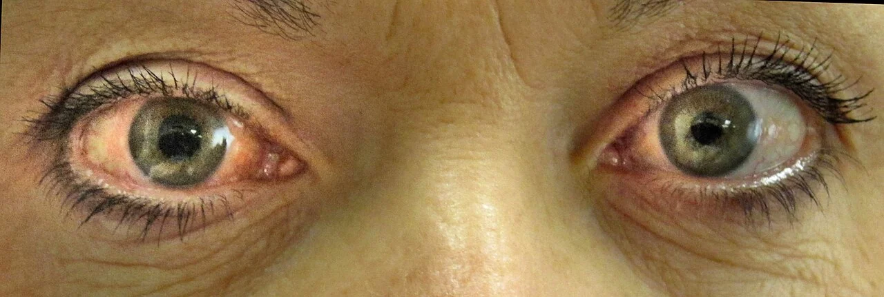
- Red eye with ciliary flush - injection densest at the limbus
- Hazy, oedematous cornea - giving a steamy appearance and obscuring the iris detail. This is why the fundus cannot be seen.
- Fixed, mid-dilated, often vertically oval pupil, unreactive to light because the sphincter is ischaemic
- Shallow anterior chamber - the iris appears to bow forward, and the peripheral chamber depth is less than a quarter of corneal thickness on oblique illumination
- Stony hard globe on gentle palpation through the closed upper lid, compared with the other eye. This is a crude test but it is dramatic and is a legitimate bedside finding.
- Markedly raised intraocular pressure - typically 50-70 mmHg
- Reduced visual acuity, often severely
- Systemic signs - bradycardia, sweating, and a distressed, unwell patient
Differential diagnosis
The differential runs in two directions, because the eye can be mistaken for other eye disease and the systemic symptoms can be mistaken for something else entirely.
- Anterior uveitis - small irregular pupil, clear cornea, normal or low pressure
- Microbial keratitis - corneal infiltrate staining with fluorescein, contact lens wear, normal pressure
- Scleritis - deep boring pain, violaceous non-blanching redness, normal pupil and pressure
- Acute conjunctivitis - normal vision, normal pupil, discharge
- Endophthalmitis - recent surgery or injection, hypopyon, vitritis
- Migraine with aura - headache with visual disturbance and vomiting, but a white, comfortable eye with a normal pupil
- Cluster headache - severe unilateral periorbital pain with a red watering eye, but with ptosis and miosis rather than a dilated pupil, and a normal cornea
- Subarachnoid haemorrhage or meningitis - headache and vomiting; the eye is normal and there are neurological signs
- Acute abdomen - where abdominal pain and vomiting dominate and nobody has looked at the eye
Investigations
The diagnosis is clinical and treatment must not wait for tests, but the following confirm it and guide subsequent management.
- Intraocular pressure by Goldmann applanation or a handheld tonometer - the confirmatory test, usually 50-70 mmHg. Corneal oedema can make applanation unreliable, so a normal-looking reading in a convincing clinical picture should not reassure you.
- Slit lamp examination - corneal oedema, a shallow anterior chamber, a fixed mid-dilated pupil, and often mild anterior chamber inflammation
- Gonioscopy - the definitive assessment of the drainage angle, performed once the cornea has cleared. It confirms closure and, critically, assesses the fellow eye, which shares the same anatomy.
- Visual acuity and visual fields, once the eye is quiet, to quantify any permanent damage
- Optic disc assessment - possible only after the cornea clears; look for glaucomatous cupping and disc pallor from ischaemia
- Anterior segment OCT or ultrasound biomicroscopy - used in specialist assessment of angle configuration and to distinguish plateau iris
- Refraction - documenting hypermetropia supports the diagnosis and identifies the fellow eye as at risk
Management
Emergency medical treatment
The aim is to lower the pressure by every available route simultaneously - reduce aqueous production, increase outflow, and osmotically draw fluid out of the eye.3
| Drug | Route and dose | Mechanism |
|---|---|---|
| Acetazolamide | 500 mg intravenously, then 250 mg orally four times daily | Carbonic anhydrase inhibitor; reduces aqueous production by the ciliary epithelium. The single most effective agent.6 |
| Pilocarpine 2-4% | Topically, every 5-15 minutes initially | Muscarinic agonist; constricts the pupil, pulling the peripheral iris out of the angle. Often ineffective until the pressure falls below about 40-50 mmHg because the ischaemic sphincter cannot respond. |
| Timolol 0.5% | One drop topically | Beta-blocker; reduces aqueous production. Avoid in asthma and bradycardia. |
| Apraclonidine or brimonidine | One drop topically | Alpha-2 agonist; reduces production and increases uveoscleral outflow |
| Dexamethasone 0.1% | Topically, four times daily | Reduces the associated inflammation and corneal oedema |
| Mannitol 20% | 1-2 g/kg intravenously over 30-60 minutes | Osmotic agent; draws water from the vitreous. Reserved for refractory cases; use cautiously in heart failure and renal impairment. |
Definitive treatment
- Laser peripheral iridotomy - a small hole made with a YAG laser in the peripheral iris, creating a direct channel between posterior and anterior chambers that permanently bypasses pupil block. Performed once the cornea has cleared enough to see through, usually within 24-48 hours.
- Prophylactic iridotomy to the fellow eye - essential, because the fellow eye shares the same anatomy and has around a 50% risk of an attack within five years if untreated. This is a very common exam point.
- Lens extraction - increasingly used as definitive treatment. The EAGLE trial found that clear lens extraction gave better pressure control, quality of life and cost-effectiveness than laser iridotomy in primary angle closure with raised pressure, and it is now a first-line option in many units.4
- Laser iridoplasty - for plateau iris configuration persisting after iridotomy
- Trabeculectomy or a glaucoma drainage device - if pressure remains uncontrolled and the angle is permanently damaged by peripheral anterior synechiae
- Long-term topical pressure-lowering therapy and glaucoma follow-up for those left with chronic angle closure glaucoma
Complications and prognosis
- Permanent optic nerve damage and visual field loss - proportional to the height and duration of the pressure rise
- Central retinal artery or vein occlusion - the intraocular pressure can exceed retinal arterial perfusion pressure
- Peripheral anterior synechiae - permanent adhesions between iris and trabecular meshwork, converting an acute episode into chronic angle closure glaucoma
- Glaukomflecken - small anterior subcapsular lens opacities from lens epithelial necrosis, which are pathognomonic evidence of a previous attack
- Iris atrophy with a permanently fixed, irregular, unreactive pupil
- Corneal endothelial cell loss and bullous keratopathy
- Cataract and, in severe cases, phthisis bulbi
- An attack in the fellow eye, which is entirely preventable
Prognosis depends almost entirely on the interval between onset and treatment. An attack broken within a few hours usually leaves vision intact. An attack lasting more than a day or two frequently leaves permanent field loss, and one lasting several days can leave the eye blind. The corollary is that recognition, not sophistication of treatment, is what determines the outcome.
The condition is also, unusually among causes of blindness, largely preventable at the level of the second eye. A patient who has had an attack in one eye and receives prophylactic iridotomy to the other has effectively been protected against the same catastrophe. Making sure that happens, and that any subacute warning symptoms - evening haloes, transient blurring, brow ache - are taken seriously, is where most of the achievable benefit lies.5
References
- NICE NG81. Glaucoma: diagnosis and management. 2017, updated 2022. Available here
- Quigley HA, Broman AT. The number of people with glaucoma worldwide in 2010 and 2020. British Journal of Ophthalmology. 2006. Available here
- Royal College of Ophthalmologists. Acute angle closure clinical guidance. Available here
- Azuara-Blanco A, Burr J, Ramsay C et al. Effectiveness of early lens extraction for the treatment of primary angle-closure glaucoma (EAGLE): a randomised controlled trial. The Lancet. 2016. Available here
- NICE Clinical Knowledge Summaries. Glaucoma. Available here
- BNF. Acetazolamide. Available here
This article is written for revision and education. It is not clinical guidance and must not be used to make decisions about the care of a patient. Always check current NICE guidance and local protocols.