
Subconjunctival Haemorrhage
Key points
- Definition: bleeding from a conjunctival or episcleral vessel into the space between conjunctiva and sclera, producing a flat, sharply demarcated block of blood.
- Presentation: painless, with normal vision, discovered by the patient in a mirror or pointed out by someone else. Usually entirely asymptomatic apart from appearance.
- The reassuring features: normal acuity, normal pupil, clear cornea, no discharge, a quiet eye and blood with a visible posterior border.
- Common precipitants: coughing, sneezing, vomiting, straining, heavy lifting, Valsalva manoeuvres, minor trauma and eye rubbing. Often no cause is found.
- Check three things: blood pressure, INR if on warfarin, and whether it is recurrent - recurrence warrants a clotting screen and platelet count.
- Treatment: none required. Reassurance, and lubricants if there is mild irritation. It resolves over 1-2 weeks, changing colour like a bruise.
- Red flags: trauma with no visible posterior border, reduced acuity, pain, a peaked pupil, or a 360° haemorrhage - all suggest globe rupture or orbital injury.
- In a child: consider non-accidental injury, particularly if bilateral, recurrent, or with other bruising and an inconsistent history.
Introduction
Subconjunctival haemorrhage is bleeding into the potential space between the conjunctiva and the underlying sclera. It looks alarming - a solid red block on the white of the eye, often extending across a whole quadrant - and patients frequently present frightened, having discovered it unexpectedly and assumed it means something serious.
In the overwhelming majority it means nothing at all. Vision is unaffected, the eye is not at risk, and no treatment is required. The clinical work is threefold: confirming that it really is a simple subconjunctival haemorrhage rather than a sign of something else; deciding whether it points to an underlying problem such as uncontrolled hypertension, over-anticoagulation or a bleeding disorder; and delivering reassurance that actually lands.
That last part deserves attention. The commonest reason a patient returns is not that the haemorrhage worsened but that they were not warned that the blood would spread and change colour over the following days before it cleared. A haemorrhage that starts bright red and turns green-yellow at the edges by day four looks, to a patient expecting steady improvement, like deterioration.
Anatomy and pathophysiology
The bulbar conjunctiva is a thin, transparent, mobile membrane loosely attached to the underlying episclera by fine connective tissue, and it is supplied by fragile conjunctival and anterior ciliary vessels. These vessels have relatively little supporting tissue around them, so a modest rise in venous pressure or a minor mechanical insult can rupture one.
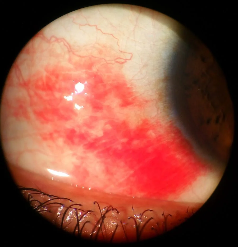
Because the conjunctiva is transparent and the sclera behind it is white, blood in this space shows through as a vivid, uniformly red patch. It looks flat and solid rather than made up of individual vessels, and no scleral white is visible through it - two features that distinguish it from conjunctival injection, where individual dilated vessels can be traced and the sclera shows between them.
The blood is confined by the loose attachments of the conjunctiva and does not enter the eye. This is why acuity, the pupil and the cornea are all normal, and why the haemorrhage does not need to be drained. It is reabsorbed by the conjunctival lymphatics and vasculature over one to two weeks, and the haemoglobin is broken down through the same biliverdin and bilirubin pathway as a skin bruise, which is why it goes through green and yellow phases before clearing.
Causes and risk factors
Spontaneous
- Idiopathic - no cause is found in a substantial proportion, particularly in older patients
- Valsalva manoeuvres - coughing, sneezing, vomiting, retching, straining at stool, heavy lifting, childbirth and strenuous exercise. A sudden rise in venous pressure ruptures a conjunctival vessel.
- Increasing age - conjunctival vessels become more fragile, and this is by far the largest risk factor
- Hypertension - present in a substantial proportion of patients with spontaneous haemorrhage, and the reason blood pressure is checked in every case
- Anticoagulants and antiplatelets - warfarin, direct oral anticoagulants, aspirin and clopidogrel. Warfarin in particular warrants an INR check.
- Diabetes mellitus
- Eye rubbing, contact lens wear and minor trauma from a fingernail
- Bleeding disorders - thrombocytopenia, haemophilia, von Willebrand disease, liver disease and vitamin K deficiency, particularly if the haemorrhage is recurrent or accompanied by bruising elsewhere
- Acute haemorrhagic conjunctivitis - enterovirus 70 and coxsackievirus A24, which cause epidemic outbreaks with multiple small subconjunctival haemorrhages and a follicular conjunctivitis
Traumatic
- Direct blunt or penetrating ocular trauma
- Foreign body, including a subtarsal one
- Ocular or orbital surgery, and after intravitreal injection - expected and harmless
- Orbital fracture - the haemorrhage may track forward from a fracture site
- Base of skull fracture - blood tracking forward beneath the conjunctiva, classically with no posterior border
- Non-accidental injury, in children and in vulnerable adults
Clinical features
Symptoms
- No symptoms at all in most cases - it is noticed in a mirror or pointed out by someone else
- Mild irritation, grittiness or a foreign body sensation if the haemorrhage is raised enough to disturb the tear film
- No pain - true pain suggests another diagnosis
- Normal vision - any visual disturbance means something else is going on
- No discharge or photophobia
- Anxiety about the appearance - which is usually the actual presenting complaint, and should be addressed as such
Signs
- A flat, sharply demarcated, uniformly red area of blood on the sclera, with no visible individual vessels and no white sclera visible through it
- Normal visual acuity
- Normal pupil size, shape and reactions
- A clear cornea, with no fluorescein staining
- A quiet anterior chamber with no hypopyon or hyphaema
- Full, painless eye movements and no proptosis
- A visible posterior border when the patient looks in the opposite direction
- Changing colour over days - from bright red to darker red, then green and yellow at the edges, before clearing
| Feature | Subconjunctival haemorrhage | Conjunctivitis | Uveitis or keratitis |
|---|---|---|---|
| Appearance of redness | Solid block of blood, no vessels visible | Diffuse dilated vessels, sclera visible between them | Ciliary flush, densest at the limbus |
| Vision | Normal | Normal | Reduced |
| Pain | None | Gritty | Significant, with photophobia |
| Discharge | None | Purulent, watery or mucoid | Watering, no discharge |
| Pupil | Normal | Normal | Small and irregular in uveitis |
| Cornea | Clear | Clear | Infiltrate or oedema |
Red flags
Investigations
Most cases need no investigation. A small number of targeted tests answer the questions that actually change management.
- Blood pressure - in every patient, since undiagnosed or poorly controlled hypertension is a recognised association and the haemorrhage may be the only prompt to check it1
- INR - in anyone taking warfarin, since a supratherapeutic INR is a common finding and needs correcting
- FBC including platelets, and a clotting screen - if the haemorrhage is recurrent, bilateral, spontaneous in a young person, or accompanied by bruising, epistaxis or bleeding elsewhere
- Liver function tests - if a coagulopathy from liver disease is suspected
- Fluorescein staining - to exclude a corneal abrasion or a Seidel-positive leak in any traumatic case
- Lid eversion and careful examination of the fornices - for a retained foreign body
- Orbital CT - if there has been significant trauma, if globe rupture or an intraocular foreign body is suspected, or if there is restricted eye movement. Not MRI first where a metallic foreign body is possible.
- Conjunctival swab - only if there is an accompanying follicular conjunctivitis suggesting acute haemorrhagic viral conjunctivitis in an outbreak setting
Management
There is no treatment that speeds resolution, and none is needed. The consultation is about confirming the diagnosis, addressing anything underlying, and explaining what will happen.
- Confirm it is benign - normal acuity, normal pupil, clear cornea, quiet eye, visible posterior border
- Reassure explicitly, explaining that the blood is on the outside of the eye, not inside it, that vision is not at risk, and that no treatment is needed
- Warn about the natural history - the haemorrhage may spread and appear to get bigger over the first 24-48 hours, and will then change colour through green and yellow before clearing, exactly like a bruise. Say this explicitly; it prevents most re-attendances.
- Give a timescale - typically 1-2 weeks for a small haemorrhage, up to 3 weeks for a large one
- Preservative-free artificial tears if there is mild irritation or a raised, drying area of conjunctiva
- Address the underlying cause - treat hypertension, correct a supratherapeutic INR according to guidance, treat a persistent cough, and advise against eye rubbing
- Do not stop anticoagulation for a subconjunctival haemorrhage alone. This is worth stating explicitly, because patients frequently stop their warfarin or DOAC themselves out of alarm, and the thrombotic risk of doing so vastly exceeds any risk from the eye.
- Safety-net - advise return if vision changes, if the eye becomes painful, or if the haemorrhage has not started to clear after three weeks
Prognosis
The prognosis of a simple subconjunctival haemorrhage is excellent. It resolves completely within one to three weeks depending on size, leaves no scar and no visual consequence, and requires no follow-up. Larger haemorrhages take longer and may temporarily look worse as blood spreads beneath the loosely attached conjunctiva before it is reabsorbed.
Recurrence in the same eye is common and usually reflects the same underlying tendency - age, fragile vessels, a persistent cough, anticoagulation or hypertension - rather than a new problem. Genuinely recurrent, multiple or bilateral spontaneous haemorrhages, particularly in a younger patient or one with bleeding elsewhere, are the group in whom a haematological cause should be sought.2
The clinical value of the topic, then, is not in the treatment - there is none - but in discrimination and communication. A structured examination confirms that the eye is safe in under a minute. A blood pressure reading and, where relevant, an INR catch the small number of cases pointing at something systemic. And a clear explanation of what the blood is going to do over the next fortnight is what determines whether the patient goes away reassured or comes back on day four convinced it is getting worse.3
References
- Mimura T, Usui T, Yamagami S et al. Recent causes of subconjunctival hemorrhage. Ophthalmologica. 2010. Available here
- Tarlan B, Kiratli H. Subconjunctival hemorrhage: risk factors and potential indicators. Clinical Ophthalmology. 2013. Available here
- NICE Clinical Knowledge Summaries. Red eye. Available here
- Royal College of Ophthalmologists. Ophthalmic trauma clinical guidance. Available here
This article is written for revision and education. It is not clinical guidance and must not be used to make decisions about the care of a patient. Always check current NICE guidance and local protocols.