
Periorbital and Orbital Cellulitis
Key points
- The dividing structure: the orbital septum, a fibrous sheet from the orbital rim to the tarsal plates. Infection anterior to it is periorbital (preseptal); posterior to it is orbital (postseptal).
- Periorbital cellulitis: lid swelling, redness and warmth with a normal eye - normal acuity, normal pupil, full painless eye movements, no proptosis.
- Orbital cellulitis: the same lid signs plus proptosis, painful or restricted eye movements, diplopia, reduced acuity or a relative afferent pupillary defect.
- The four discriminating signs: proptosis, ophthalmoplegia, pain on eye movement and visual impairment. Any one of them means orbital cellulitis until imaging says otherwise.
- Commonest source: ethmoid sinusitis in over 90% of orbital cases, spreading through the thin lamina papyracea.
- Imaging: contrast CT of orbits, sinuses and brain in every suspected orbital case, to identify a subperiosteal abscess and intracranial extension.
- Treatment: orbital cellulitis needs admission, IV broad-spectrum antibiotics and joint ENT and ophthalmology care, with surgical drainage for an abscess or failure to improve.
- Complications: orbital abscess, optic neuropathy, cavernous sinus thrombosis, meningitis, intracranial abscess and death. Mortality was around 17% before antibiotics.
Introduction
A child or adult presenting with a red, swollen, painful eyelid poses one of the cleanest diagnostic questions in acute medicine: is the infection in front of the orbital septum or behind it? The answer determines whether the patient goes home on oral antibiotics or is admitted for intravenous therapy, imaging and possible surgery.
The two conditions are not points on a continuum with the same treatment at different doses. Periorbital (preseptal) cellulitis is an infection of the eyelid and periorbital soft tissue that carries almost no risk to the eye and responds well to oral antibiotics. Orbital (postseptal) cellulitis is an infection within the closed bony orbit, and it threatens vision, threatens the brain, and killed around 17% of patients before antibiotics existed.1
Periorbital cellulitis is much the commoner of the two, and both are commoner in children than adults - the median age for orbital cellulitis is around 7 to 12 years. That matters clinically, because a frightened child with a swollen eye who will not open it is exactly the situation in which the necessary examination is hardest to perform and most often skipped.
Anatomy
The orbital septum is a thin fibrous membrane running from the periosteum of the orbital rim to the tarsal plates of the upper and lower lids. It is the anterior boundary of the orbit and forms a genuine anatomical barrier to the spread of infection, which is why it defines the whole classification.
Behind it lies a closed cone of bone containing the globe, the extraocular muscles, the optic nerve, orbital fat and the ophthalmic vessels. The orbit has essentially no capacity to expand. Infection or an abscess within it therefore raises orbital pressure, pushing the globe forward (proptosis), restricting the muscles (ophthalmoplegia and pain on movement), and compressing the optic nerve and its blood supply (visual loss). Those four consequences are precisely the four signs used to make the diagnosis, which is why they are worth deriving rather than memorising.
- Medially, the orbit is separated from the ethmoid air cells by the lamina papyracea - literally the paper-thin plate. It is perforated by vessels and is easily breached, which is why ethmoid sinusitis is the source in the great majority of cases.
- Superiorly lies the frontal sinus and the anterior cranial fossa; inferiorly the maxillary sinus; posteriorly the sphenoid sinus and the orbital apex
- The orbital veins are valveless and communicate freely with the facial veins anteriorly and the cavernous sinus posteriorly. This allows infection to spread in either direction and is the anatomical basis for cavernous sinus thrombosis - and for the historical concept of the danger triangle of the face.
- The superior orbital fissure and optic canal at the apex transmit cranial nerves III, IV, V1 and VI and the optic nerve, so posterior orbital infection can produce a rapidly progressive orbital apex syndrome
Aetiology
Sources of infection
- Sinusitis - the source in over 90% of orbital cellulitis, most often ethmoid, spreading directly through the lamina papyracea
- Local skin infection or trauma - insect bites, lacerations, impetigo, herpes zoster, and infected chalazia or styes; these are the usual cause of periorbital cellulitis
- Dacryocystitis and dacryoadenitis
- Dental infection, spreading via the maxillary sinus
- Post-surgical - after orbital, lacrimal, strabismus or sinus surgery, or after an orbital fracture repair
- Haematogenous spread - from a distant focus, and the mechanism in endogenous cases
- Retained orbital foreign body - always consider in penetrating trauma, particularly organic material
Organisms
- Staphylococcus aureus - including MRSA, particularly after trauma or skin infection
- Streptococcus pneumoniae and other streptococci, particularly Streptococcus anginosus group in sinus-derived disease
- Streptococcus pyogenes
- Anaerobes - Peptostreptococcus, Bacteroides and Fusobacterium, more common in older children and adults and in chronic sinus disease. Infections are frequently polymicrobial.
- Haemophilus influenzae type b - historically the classic paediatric cause, now uncommon in the UK because of Hib vaccination, but consider it in unimmunised children
- Fungi - Mucor and Aspergillus - invasive fungal sinusitis in diabetic ketoacidosis, haematological malignancy, neutropenia and immunosuppression. Rapidly fatal without urgent surgical debridement and systemic antifungals.
Clinical features
Both conditions present with a swollen, red, warm, tender eyelid, often with fever and malaise. The discriminating features all concern what the eye itself is doing.
| Feature | Periorbital (preseptal) | Orbital (postseptal) |
|---|---|---|
| Lid swelling, redness, warmth | Present, often marked | Present |
| Visual acuity | Normal | May be reduced |
| Pupil reactions | Normal | Relative afferent pupillary defect may be present |
| Eye movements | Full and painless | Restricted and painful; diplopia |
| Proptosis | Absent | Present - may need to be assessed from above or with an exophthalmometer |
| Chemosis | Absent or minimal | Often marked conjunctival oedema |
| Colour vision | Normal | Reduced - an early sign of optic nerve compression |
| Systemic upset | Mild; often afebrile | Fever, malaise, raised inflammatory markers |
| Management | Oral antibiotics as an outpatient in most cases | Admission, IV antibiotics, CT imaging, joint ENT and ophthalmology care |
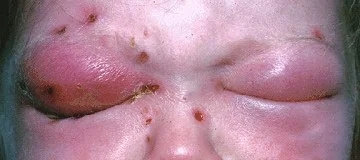
Features suggesting complications
- A subperiosteal abscess - progressive proptosis with globe displacement in a specific direction, usually laterally and downward from a medial ethmoid collection, with failure to improve after 24-48 hours of IV antibiotics
- Optic neuropathy - falling acuity, reduced colour vision, a relative afferent pupillary defect and a swollen disc; this is a surgical emergency
- Orbital compartment syndrome - a tense, proptosed globe with raised intraocular pressure, requiring immediate lateral canthotomy and cantholysis
- Cavernous sinus thrombosis - bilateral signs, cranial nerve palsies affecting III, IV, V1, V2 and VI, headache, reduced consciousness and a septic picture
- Meningitis or intracranial abscess - headache, neck stiffness, seizures, focal neurology and altered consciousness
Differential diagnosis
- Allergic angio-oedema - bilateral, non-tender, pale rather than erythematous, itchy, afebrile, often with a clear trigger
- Insect bite or contact dermatitis - itchy rather than painful, with a visible punctum or a distribution matching contact
- Thyroid eye disease - bilateral proptosis with lid retraction and lid lag, subacute rather than acute, with restricted movements but no fever or erythema
- Idiopathic orbital inflammatory disease (orbital pseudotumour) - painful proptosis with restricted movements, but with a subacute course, normal inflammatory markers and a dramatic response to corticosteroid
- Orbital myositis - painful diplopia with enlarged muscle bellies including the tendons on CT, unlike thyroid eye disease
- Rhabdomyosarcoma - the commonest primary orbital malignancy in children, presenting with rapidly progressive proptosis over weeks, which can be mistaken for infection
- Retinoblastoma, neuroblastoma metastasis and leukaemia - other paediatric causes of proptosis
- Ruptured dermoid cyst - sudden inflammatory reaction to leaked keratin, often at the superotemporal orbital rim
- Carotid-cavernous fistula - pulsatile proptosis with a bruit, chemosis and dilated corkscrew conjunctival vessels, usually after trauma
Investigations
Bedside and laboratory
- Visual acuity, colour vision, pupil reactions, eye movements and assessment for proptosis - the clinical examination is the primary investigation and must be documented
- Intraocular pressure - raised pressure suggests orbital compartment syndrome
- Observations - temperature, heart rate, respiratory rate and blood pressure, with a sepsis screen if unwell
- FBC, CRP and blood cultures - inflammatory markers guide response, though blood cultures are positive in a minority
- Swabs - from any discharging wound, conjunctiva or nasal cavity
- Blood glucose and ketones - specifically to identify diabetic ketoacidosis and the risk of mucormycosis
- Lumbar puncture - only if meningitis is suspected and after imaging has excluded raised intracranial pressure
Imaging
Management
Periorbital cellulitis
- Oral antibiotics - co-amoxiclav is the usual first choice in the UK, covering staphylococci, streptococci and anaerobes; clarithromycin with metronidazole in penicillin allergy2
- Review within 24-48 hours, with clear safety-netting about what to look for
- Admit for intravenous antibiotics if the patient is under one year old, systemically unwell, immunocompromised, unable to be examined adequately, or not improving on oral treatment
- Treat the source - a stye, dacryocystitis, sinusitis or a skin wound
- Warn explicitly to return immediately if the eye becomes painful to move, if vision changes, if double vision develops, or if the eye starts to protrude
Orbital cellulitis
- Admit under joint ophthalmology and ENT care, with paediatrics involved for children
- Intravenous broad-spectrum antibiotics - typically ceftriaxone with metronidazole, or co-amoxiclav, adjusted to local microbiology guidance and to culture results. Add vancomycin where MRSA is a concern.
- Neurological doses and CNS penetration should be used if intracranial extension is suspected
- Urgent contrast CT of orbits, sinuses and brain
- Four-hourly monitoring of visual acuity, colour vision, pupil reactions and proptosis - deterioration in any of these is the trigger for surgery, and this observation regimen is the single most important nursing instruction
- Nasal decongestants and topical steroid spray to promote sinus drainage, alongside treatment of the underlying sinusitis4,5
- Surgical drainage - endoscopic sinus surgery with drainage of any subperiosteal or orbital abscess, indicated for a large abscess, deteriorating vision, failure to improve after 24-48 hours, or intracranial complications
- Lateral canthotomy and cantholysis as an emergency bedside procedure if orbital compartment syndrome develops
- Switch to oral antibiotics once afebrile and clearly improving, completing a total course of 2-3 weeks
Complications and prognosis
- Subperiosteal and orbital abscess - the commonest complications, occurring in a substantial minority
- Optic neuropathy and permanent visual loss - from direct compression, from raised orbital pressure compromising perfusion, or from central retinal artery occlusion. Occurs in around 3-11% of orbital cellulitis.
- Exposure keratopathy and corneal ulceration from proptosis preventing lid closure
- Raised intraocular pressure and orbital compartment syndrome
- Cavernous sinus thrombosis - bilateral cranial neuropathies, headache and sepsis; treated with antibiotics and usually anticoagulation, and still carrying appreciable mortality
- Meningitis, subdural empyema, intracranial abscess and cerebral venous sinus thrombosis
- Osteomyelitis of the orbital walls or frontal bone (Pott's puffy tumour when the frontal bone is involved)
- Sepsis and death - now rare with prompt antibiotics, but the pre-antibiotic mortality of around 17% is a reminder of what is being prevented
With prompt recognition and treatment, the prognosis for both conditions is good. Periorbital cellulitis resolves fully on oral antibiotics in the great majority. Orbital cellulitis treated early with intravenous antibiotics and timely drainage usually resolves without lasting visual deficit, and most patients recover full acuity and eye movement.
The determinants of a poor outcome are consistent: delay in diagnosis, failure to image, failure to monitor visual function four-hourly, and failure to escalate when the patient is not improving. All four are process failures rather than knowledge failures, which is why the practical teaching points are so specific - open the eye, check the four signs, image if any are present or if you cannot examine, and set clear monitoring and escalation criteria.
The corresponding point for a discharge conversation is equally concrete. A patient sent home with periorbital cellulitis should leave knowing exactly which four things to watch for - pain on moving the eye, double vision, a change in vision, and the eye starting to bulge - because these are the symptoms that convert an outpatient problem into an emergency, and they are ones a patient can reliably report themselves.
References
- Chandler JR, Langenbrunner DJ, Stevens ER. The pathogenesis of orbital complications in acute sinusitis. Laryngoscope. 1970. Available here
- NICE Clinical Knowledge Summaries. Cellulitis - acute. Available here
- Garcia GH, Harris GJ. Criteria for nonsurgical management of subperiosteal abscess of the orbit. Ophthalmology. 2000. Available here
- NICE NG79. Sinusitis (acute): antimicrobial prescribing. 2017. Available here
- Royal College of Ophthalmologists. Clinical guidance and resources. Available here
This article is written for revision and education. It is not clinical guidance and must not be used to make decisions about the care of a patient. Always check current NICE guidance and local protocols.