
Metabolic Dysfunction-Associated Steatotic Liver Disease (MASLD)
Key points
- MASLD: hepatic steatosis affecting over 5% of hepatocytes plus at least one cardiometabolic risk factor, with no other cause of liver disease.
- New terminology: MASLD replaced 'non-alcoholic fatty liver disease' (NAFLD) in 2023, and MASH replaced NASH, to describe the condition positively rather than by exclusion.
- Prevalence: the commonest liver disease worldwide, affecting around 25-30% of adults, and rising in parallel with obesity and type 2 diabetes.
- Spectrum: simple steatosis → steatohepatitis (MASH, with inflammation and ballooning) → fibrosis → cirrhosis → hepatocellular carcinoma.
- Presentation: usually asymptomatic, found incidentally on ultrasound or through mildly raised ALT; ALT is often normal even with significant fibrosis.
- Key principle: fibrosis stage, not the degree of steatosis or inflammation, is what predicts liver-related and overall mortality.
- Assessment: risk-stratify with FIB-4 or the NAFLD fibrosis score, then transient elastography or the ELF test for those at intermediate or high risk.
- Management: weight loss of 7-10% is the cornerstone; control diabetes, lipids and blood pressure, and consider GLP-1 agonists or resmetirom in MASH.
Introduction and terminology
Metabolic dysfunction-associated steatotic liver disease (MASLD) is the accumulation of fat in the liver in people with cardiometabolic risk factors, in the absence of excess alcohol or another identifiable cause. It is the most common liver disease worldwide, affecting roughly a quarter to a third of adults, and its prevalence is rising in step with obesity and type 2 diabetes.1
Diagnostic criteria
MASLD requires hepatic steatosis (fat in more than 5% of hepatocytes on imaging or biopsy) plus at least one of five cardiometabolic criteria: raised BMI or waist circumference; raised fasting glucose, HbA1c, or type 2 diabetes; raised blood pressure or treatment for hypertension; raised triglycerides or lipid-lowering treatment; or low HDL cholesterol. Other causes of steatosis - significant alcohol intake, viral hepatitis, drugs, Wilson disease - must be excluded.
The disease spectrum
| Stage | Features | Significance |
|---|---|---|
| Steatosis (simple fatty liver) | Fat accumulation without significant inflammation | The large majority of patients; low risk of progression |
| MASH (steatohepatitis) | Steatosis plus lobular inflammation and hepatocyte ballooning | The progressive form; affects roughly 20% of those with MASLD |
| Fibrosis | Progressive collagen deposition, staged F0 to F4 | The key prognostic determinant; F2 or above is clinically significant |
| Cirrhosis (F4) | Irreversible architectural distortion | Risk of decompensation and hepatocellular carcinoma |
Risk factors and pathophysiology
Risk factors
- Obesity, particularly central adiposity - the strongest association
- Type 2 diabetes mellitus - present in a large proportion, and itself the strongest predictor of advanced fibrosis
- Insulin resistance and metabolic syndrome
- Dyslipidaemia, especially raised triglycerides and low HDL
- Hypertension
- Polycystic ovary syndrome, hypothyroidism, obstructive sleep apnoea and hypopituitarism
- Genetic factors, notably the PNPLA3 I148M variant, which increases susceptibility and is more prevalent in Hispanic populations
- Increasing age, and South Asian ethnicity at lower BMI thresholds
- Lean MASLD occurs in 10-20% of patients with a normal BMI, often with visceral adiposity or genetic predisposition, and should not be overlooked
Pathophysiology
The current model is one of multiple parallel hits. Insulin resistance in adipose tissue increases lipolysis, delivering excess free fatty acids to the liver, while hyperinsulinaemia drives hepatic de novo lipogenesis. Fat accumulates as triglyceride within hepatocytes.2
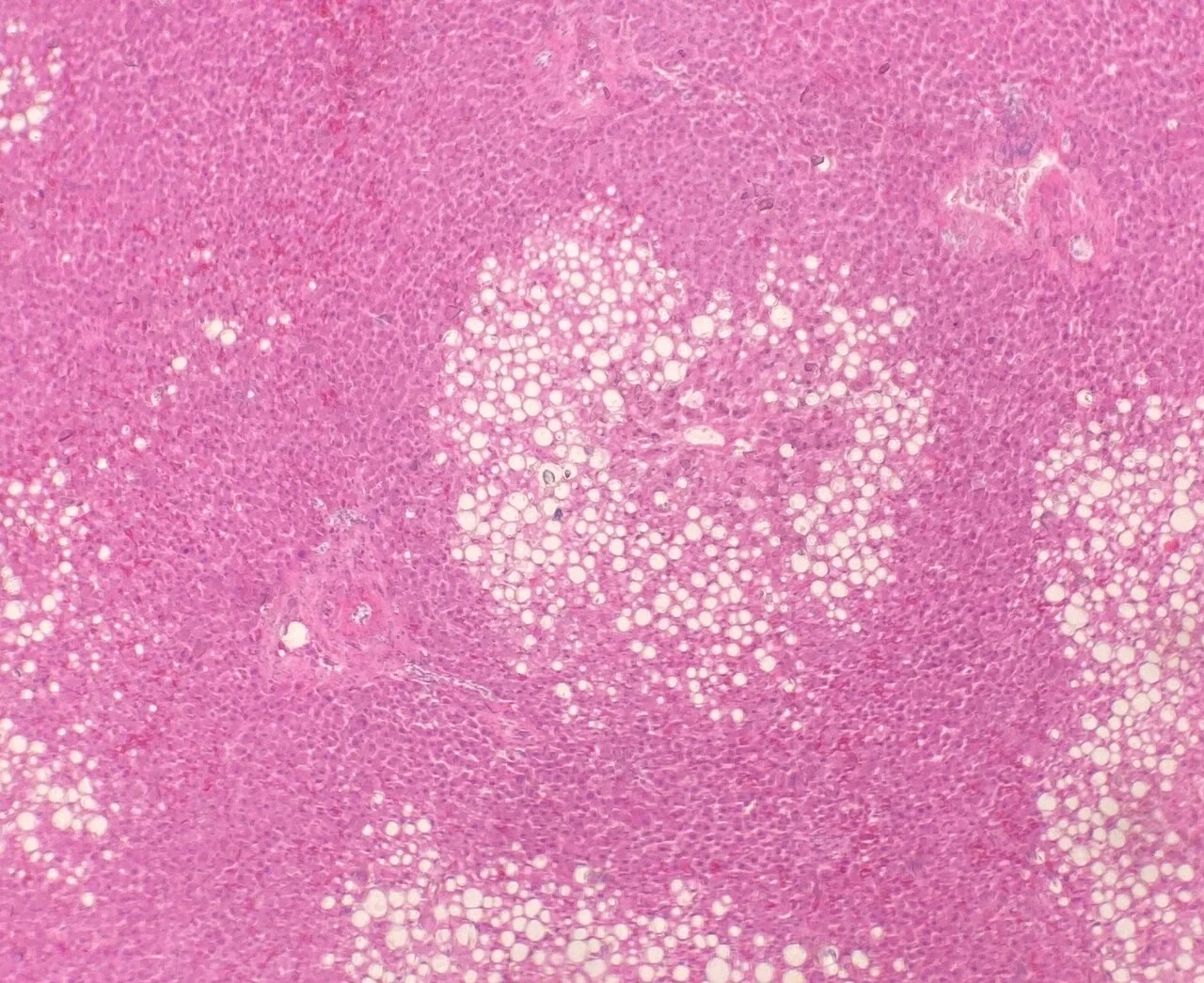
In susceptible individuals, this steatosis progresses to steatohepatitis through lipotoxicity from toxic lipid intermediates, mitochondrial dysfunction and oxidative stress, endoplasmic reticulum stress, and inflammatory cytokine release from dysfunctional adipose tissue and gut-derived endotoxin. Hepatocyte injury and apoptosis activate hepatic stellate cells, which deposit collagen and drive progressive fibrosis. Histology in MASH shows macrovesicular steatosis, lobular inflammation, hepatocyte ballooning and a characteristic pericellular "chicken wire" fibrosis pattern beginning in zone 3.
Clinical features
MASLD is usually asymptomatic and is most often discovered incidentally, either through mildly abnormal liver enzymes checked for another reason, or through a fatty liver reported on abdominal ultrasound performed for unrelated symptoms.3
- Fatigue and general malaise, which are common but non-specific
- Vague right upper quadrant discomfort or fullness
- Hepatomegaly, which may be smooth and non-tender
- Features of the metabolic syndrome: central obesity, acanthosis nigricans, hypertension
- Signs of chronic liver disease only once cirrhosis has developed - spider naevi, palmar erythema, jaundice, ascites and splenomegaly
- A proportion present for the first time with decompensated cirrhosis or hepatocellular carcinoma, having never been diagnosed with liver disease
Investigations
Initial assessment
- Liver function tests: ALT may be mildly raised, typically with an AST:ALT ratio below 1 (in contrast to alcohol-related disease, where it exceeds 2). A ratio that rises above 1 may signal advancing fibrosis
- Gamma-GT is often mildly raised
- Metabolic screen: HbA1c or fasting glucose, fasting lipid profile, BMI and waist circumference, blood pressure
- Full blood count: thrombocytopenia suggests portal hypertension and advanced disease
- Albumin and INR: to assess synthetic function
- Ferritin is often mildly raised as an acute phase reactant; check transferrin saturation before assuming haemochromatosis
- Non-invasive liver screen to exclude other causes: viral hepatitis serology, autoantibodies and immunoglobulins, caeruloplasmin in the young, alpha-1 antitrypsin, and coeliac serology
- Careful alcohol history with quantified units, to distinguish MASLD from MetALD and alcohol-related disease
Imaging
Abdominal ultrasound is first-line and shows a bright, echogenic ("fatty") liver with posterior beam attenuation, but it is insensitive to mild steatosis and cannot stage fibrosis. Controlled attenuation parameter (CAP) on FibroScan quantifies steatosis, and MRI proton density fat fraction is the most accurate but is largely a research tool.
Fibrosis assessment - the key step
Because fibrosis stage determines prognosis, non-invasive fibrosis assessment is central to management. A two-step approach is used:3
| Test | Components | Role |
|---|---|---|
| FIB-4 | Age, AST, ALT, platelet count | First-line triage in primary care. Below 1.3 makes advanced fibrosis unlikely; above 2.67 suggests high risk. Less reliable under 35 and over 65 |
| NAFLD fibrosis score (NFS) | Age, BMI, glucose, AST, ALT, platelets, albumin | Alternative first-line triage tool |
| Enhanced liver fibrosis (ELF) test | Hyaluronic acid, PIIINP, TIMP-1 | Recommended by NICE as a second-line test; a score of 9.8 or above indicates advanced fibrosis |
| Transient elastography (FibroScan) | Liver stiffness measurement in kPa | Widely used second-line test. Rising stiffness correlates with fibrosis stage; unreliable in acute hepatitis, cholestasis, heart failure or after eating |
| Liver biopsy | Histology | Remains the gold standard for distinguishing MASH from simple steatosis and staging fibrosis, but is invasive and reserved for diagnostic uncertainty or trial enrolment |
NICE recommends offering testing for advanced fibrosis with the ELF test to people with MASLD, repeating it every 3 years in adults, since fibrosis progresses slowly.3 People with type 2 diabetes warrant particular vigilance, having both the highest prevalence and the highest rate of progression.
Differential diagnosis
- Alcohol-related liver disease: distinguished by the alcohol history and an AST:ALT ratio above 2 - and remember the two commonly coexist as MetALD
- Chronic viral hepatitis B and C: hepatitis C genotype 3 in particular causes steatosis directly
- Drug-induced steatosis: amiodarone, methotrexate, tamoxifen, corticosteroids, sodium valproate, antiretrovirals
- Autoimmune hepatitis: raised IgG and positive autoantibodies
- Haemochromatosis: raised ferritin with a high transferrin saturation
- Wilson disease: consider in any patient under 40 with unexplained liver disease; check caeruloplasmin
- Alpha-1 antitrypsin deficiency
- Coeliac disease, hypothyroidism, and rarer causes such as lipodystrophy and abetalipoproteinaemia
- Acute fatty liver of pregnancy, a distinct obstetric emergency
Management
Weight loss - the cornerstone
Sustained weight loss is the most effective intervention, and the benefit is dose-dependent:1
- 3-5% weight loss improves steatosis
- 7-10% weight loss improves necroinflammation and resolves MASH in most patients
- 10% or more can regress fibrosis
Achieve this through a calorie-restricted diet - the Mediterranean diet has the best evidence - with reduced saturated fat, refined carbohydrate and particularly fructose, alongside at least 150 minutes of moderate exercise weekly. Both aerobic and resistance training reduce hepatic fat independently of weight loss. Bariatric surgery is highly effective for MASH with obesity and can resolve steatohepatitis and improve fibrosis.
Treating cardiometabolic risk
Because patients with MASLD are far more likely to die of cardiovascular disease than of liver disease, aggressive cardiovascular risk management is essential:
- Statins are safe and indicated in MASLD, including in compensated cirrhosis. They should not be withheld because of mildly raised transaminases - this is a common and harmful error
- Optimise glycaemic control. GLP-1 receptor agonists (semaglutide, liraglutide) and pioglitazone both improve steatohepatitis, and GLP-1 agonists additionally deliver weight loss and cardiovascular benefit. SGLT2 inhibitors also reduce hepatic fat
- Treat hypertension and dyslipidaemia to target
- Vitamin E may be considered in selected non-diabetic patients with biopsy-proven MASH, though concerns exist about long-term safety
- Resmetirom, a thyroid hormone receptor-beta agonist, is the first drug specifically licensed for MASH with moderate to advanced fibrosis in some jurisdictions
- Alcohol: advise minimising or avoiding alcohol entirely, as it acts synergistically with metabolic dysfunction
Monitoring and advanced disease
Patients with cirrhosis require six-monthly ultrasound surveillance for hepatocellular carcinoma, endoscopic screening for varices, vaccination, and assessment for transplantation if decompensated. MASLD is now among the fastest-growing indications for liver transplantation in the UK and the leading indication in some countries. Notably, hepatocellular carcinoma can arise in MASLD without cirrhosis, though surveillance in non-cirrhotic patients is not currently recommended.
Complications
- Progression to MASH, advanced fibrosis and cirrhosis
- Decompensated cirrhosis: ascites, variceal bleeding, hepatic encephalopathy, hepatorenal syndrome
- Hepatocellular carcinoma, which may occur even without established cirrhosis
- Cardiovascular disease - the leading cause of death in MASLD, exceeding liver-related mortality
- Type 2 diabetes, which MASLD both results from and independently predicts
- Chronic kidney disease
- Extrahepatic malignancy, particularly colorectal cancer
- Obstructive sleep apnoea and polycystic ovary syndrome as coexisting metabolic conditions
Red flags
Prognosis
The great majority of people with simple steatosis have a benign hepatic course and never progress. Roughly 20% have MASH, and of these a proportion progress through fibrosis stages at an average of about one stage per 7-14 years, with progression accelerated by type 2 diabetes, ongoing weight gain and continued alcohol intake.2
Fibrosis stage is the dominant prognostic factor - not the degree of steatosis or inflammation. Patients with F3-F4 fibrosis have substantially increased liver-related and all-cause mortality, whereas those with F0-F1 have near-normal liver-related outcomes.
The most important prognostic message, however, is that cardiovascular disease, not liver disease, is the leading cause of death in MASLD overall. This reframes management: the liver findings are best understood as a marker of systemic metabolic dysfunction, and treating that dysfunction benefits the patient far beyond the liver. Fibrosis is potentially reversible with sustained weight loss, so the outlook for engaged patients who achieve and maintain 10% weight loss is genuinely good.
References
- European Association for the Study of the Liver. EASL-EASD-EASO Clinical Practice Guidelines on the management of MASLD. J Hepatol. 2024. Available here
- Rinella ME et al. A multisociety Delphi consensus statement on new fatty liver disease nomenclature. Hepatology. 2023. Available here
- NICE NG49. Non-alcoholic fatty liver disease (NAFLD): assessment and management. 2016 (updated 2025). Available here
- Mikael Häggström, MD, CC0, via Wikimedia Commons. Available here
- British Society of Gastroenterology. Guidelines on the management of abnormal liver blood tests. Gut. 2018. Available here
- NICE Clinical Knowledge Summaries (CKS). Non-alcoholic fatty liver disease. 2024. Available here
- NHS. Non-alcoholic fatty liver disease (NAFLD). 2022. Available here
This article is written for revision and education. It is not clinical guidance and must not be used to make decisions about the care of a patient. Always check current NICE guidance and local protocols.