
Shock: Recognition and Management
Key points
- Shock: circulatory failure causing inadequate tissue oxygen delivery and cellular hypoxia. It is defined by inadequate perfusion, not by a blood pressure number.
- The four types: hypovolaemic, cardiogenic, obstructive and distributive. Distributive shock includes septic, anaphylactic and neurogenic.
- Early recognition: tachycardia, tachypnoea, prolonged capillary refill, oliguria and confusion appear before hypotension. Normal blood pressure never excludes shock.
- Lactate: a raised serum lactate is the key marker of anaerobic metabolism and tissue hypoperfusion, and clearance over time predicts outcome.
- Warm versus cold: warm peripheries with a bounding pulse and wide pulse pressure suggest distributive shock; cool, mottled peripheries suggest hypovolaemic, cardiogenic or obstructive shock.
- Treatment differs by type: fluid is the treatment for hypovolaemia and sepsis, but can be harmful in cardiogenic shock, where inotropes and treating the cause come first.
- Sepsis: give the Sepsis Six within one hour - oxygen, blood cultures, antibiotics, fluids, lactate and urine output measurement.
- Anaphylaxis: intramuscular adrenaline 500 micrograms into the anterolateral thigh, repeated every 5 minutes. Do not give it intravenously outside specialist settings.
Introduction
Shock is a state of circulatory failure in which oxygen delivery is inadequate to meet the metabolic demands of the tissues, causing cellular hypoxia, a switch to anaerobic metabolism, lactate accumulation and, if uncorrected, cell death and multi-organ failure.
The most important conceptual point is that shock is defined by inadequate tissue perfusion, not by hypotension. A previously hypertensive patient may be profoundly shocked at a systolic pressure of 110 mmHg; a young fit patient may compensate and maintain a normal blood pressure until they have lost 30-40% of their blood volume, and then deteriorate abruptly. Waiting for hypotension to diagnose shock means diagnosing it late.
The physiology
Oxygen delivery is the product of cardiac output and arterial oxygen content. Cardiac output is heart rate multiplied by stroke volume, and stroke volume depends on preload, contractility and afterload. Blood pressure in turn is cardiac output multiplied by systemic vascular resistance.
Every type of shock is a failure of one of these components, and identifying which one is failing dictates the treatment:
- Hypovolaemic shock - reduced preload from loss of circulating volume
- Cardiogenic shock - reduced contractility, so the pump fails
- Obstructive shock - mechanical obstruction to filling or ejection
- Distributive shock - profound vasodilatation reducing systemic vascular resistance, with maldistribution of flow
Classification
| Type | Mechanism | Causes | Peripheries | JVP | Cardiac output | SVR |
|---|---|---|---|---|---|---|
| Hypovolaemic | Loss of circulating volume | Haemorrhage, vomiting, diarrhoea, burns, third-space losses, diabetic ketoacidosis | Cold | Low | Low | High |
| Cardiogenic | Pump failure | Myocardial infarction, arrhythmia, acute valve failure, myocarditis, drug overdose (beta-blocker, calcium channel blocker) | Cold | High | Low | High |
| Obstructive | Mechanical obstruction to filling or outflow | Massive pulmonary embolism, cardiac tamponade, tension pneumothorax | Cold | High | Low | High |
| Distributive | Vasodilatation and maldistribution | Sepsis, anaphylaxis, neurogenic (spinal cord injury), adrenal crisis, liver failure | Warm (initially) | Low or normal | High (early) | Low |
Clinical features
Early signs - present before hypotension
- Tachycardia - usually the earliest sign, though absent in neurogenic shock and blunted in patients on beta-blockers or with a pacemaker
- Tachypnoea - a compensatory respiratory alkalosis, and one of the most sensitive and most frequently overlooked signs of deterioration
- Prolonged capillary refill time - over 2 seconds, tested centrally as well as peripherally
- Cool, mottled or clammy peripheries, with the mottling progressing proximally as shock worsens
- Reduced urine output - below 0.5 ml/kg/hour
- Anxiety, agitation, confusion or drowsiness - cerebral hypoperfusion, and a particularly important sign in older patients
- Narrowed pulse pressure - an early sign of vasoconstriction in hypovolaemia
- Postural hypotension - may be the only abnormality in early volume loss
Late signs
- Hypotension - systolic below 90 mmHg, or a mean arterial pressure below 65 mmHg, or a fall of more than 40 mmHg from the patient's usual pressure
- Anuria
- Reduced consciousness
- Bradycardia - a pre-terminal sign in the shocked patient
- Cyanosis and mottling extending to the trunk
Clues to the cause
- Fever, rigors, a source of infection, or a rash - sepsis. A non-blanching purpuric rash suggests meningococcal disease.
- Urticaria, angioedema, wheeze, stridor and a recent trigger - anaphylaxis
- Chest pain, ECG changes, raised JVP and a third heart sound - cardiogenic shock from myocardial infarction
- Distended neck veins with muffled heart sounds and pulsus paradoxus - cardiac tamponade
- Distended neck veins with tracheal deviation and absent breath sounds - tension pneumothorax
- Distended neck veins with hypoxia, pleuritic pain and a swollen leg - massive pulmonary embolism
- Haematemesis, melaena, abdominal pain, trauma or a pulsatile abdominal mass - haemorrhage
- Warm, dry, flushed skin with bradycardia after trauma - neurogenic shock from spinal cord injury
- Hyperpigmentation, hyponatraemia and hyperkalaemia, or recent steroid withdrawal - adrenal crisis
Investigations
Investigation runs in parallel with resuscitation, never before it.
Immediate bedside tests
- Arterial or venous blood gas with lactate - the single most useful early test. A raised lactate reflects anaerobic metabolism, and lactate clearance over the first hours predicts outcome better than any single value.
- Capillary blood glucose
- 12-lead ECG - ischaemia, arrhythmia, right heart strain in PE, electrical alternans in tamponade
- Point-of-care ultrasound - assesses inferior vena cava size and collapsibility, ventricular function, pericardial fluid, right heart strain, pneumothorax and free intra-abdominal fluid. In experienced hands it distinguishes the four types of shock within minutes.
- Urine output - insert a catheter and monitor hourly
Laboratory tests
- FBC - haemoglobin (which may be normal in acute haemorrhage before dilution), white cell count
- U&Es - renal function and electrolytes
- LFTs, amylase or lipase, and bone profile
- Coagulation screen and fibrinogen - and consider thromboelastography in major haemorrhage
- Group and save, and crossmatch - activate the major haemorrhage protocol early if bleeding
- CRP and blood cultures - two sets, before antibiotics where this does not delay them
- Troponin and BNP - if a cardiac cause is suspected
- Serum cortisol - before giving steroids if adrenal crisis is suspected
- Serum tryptase - in suspected anaphylaxis, taken as soon as possible after treatment and again at 1-2 hours and after 24 hours. It confirms the diagnosis retrospectively and should not delay adrenaline.
- Beta-hCG - in any woman of childbearing age with shock, to identify ruptured ectopic pregnancy
Imaging
- Chest X-ray - pneumothorax, pneumonia, pulmonary oedema, a widened mediastinum
- Echocardiogram - ventricular function, tamponade, valve failure, right heart strain
- CT - only in a patient stable enough to travel. CT aortogram for suspected dissection or ruptured aneurysm, CTPA for pulmonary embolism, and CT abdomen for an intra-abdominal source of sepsis or bleeding.
- FAST scan - in trauma, for free intraperitoneal fluid
General management
Assess and treat using an ABCDE approach, calling for senior and critical care help early. Reassess after every intervention.
- Airway - secure it if the conscious level is impaired
- Breathing - high-flow oxygen through a non-rebreathe mask initially, titrated once saturations allow. Support ventilation if needed, remembering that positive pressure ventilation reduces venous return and can worsen shock.
- Circulation - two large-bore cannulae (14 or 16 gauge) in the antecubital fossae, or intraosseous access if peripheral access fails. Take bloods on cannulation.
- Disability - conscious level, pupils and blood glucose
- Exposure - full examination for a source of bleeding, infection or rash, while avoiding hypothermia
- Monitoring - continuous ECG, oxygen saturations, blood pressure (invasive arterial monitoring in the sickest patients), urinary catheter and hourly urine output
- Escalate early - critical care outreach, the medical registrar, or the appropriate surgical team
- Treat the cause - this is what actually resolves shock; supportive measures only buy time
Vasopressors and inotropes
Used once fluid resuscitation is adequate, or earlier where fluid is contraindicated. These should be given through a central line where possible, in a critical care or resuscitation setting, and with invasive arterial monitoring.
| Drug | Action | Main use |
|---|---|---|
| Noradrenaline | Predominantly alpha-1 agonist - vasoconstriction | First-line vasopressor in septic and most distributive shock |
| Adrenaline | Alpha and beta agonist - vasoconstriction, inotropy and chronotropy | Anaphylaxis (intramuscular); cardiac arrest; second-line in refractory shock |
| Dobutamine | Beta-1 agonist - inotropy with some vasodilatation | Cardiogenic shock with low output; can lower blood pressure, so often combined with noradrenaline |
| Vasopressin | V1 receptor agonist - vasoconstriction independent of adrenergic receptors | Adjunct in septic shock refractory to noradrenaline |
| Dopamine | Dose-dependent dopaminergic, beta and alpha effects | Now largely superseded - associated with more arrhythmia than noradrenaline |
| Milrinone / levosimendan | Phosphodiesterase inhibition / calcium sensitisation | Specialist use in cardiogenic shock, particularly in patients on beta-blockers |
Management by type
Septic shock
Add noradrenaline to maintain a mean arterial pressure of at least 65 mmHg once fluid resuscitation is adequate. Consider hydrocortisone in shock refractory to fluids and vasopressors.
Anaphylaxis
Cardiogenic shock
- Identify and treat the cause urgently - primary PCI for myocardial infarction, cardioversion or pacing for arrhythmia, surgery for acute valve failure or ventricular septal rupture, and specific antidotes in drug overdose
- Fluid with great caution - a small 250 ml challenge may help if the patient is preload-depleted, but reassess immediately and stop if there is any worsening of oxygenation
- Inotropic support - dobutamine, often with noradrenaline to maintain perfusion pressure
- Diuretics and vasodilators once perfusion is restored, to relieve congestion
- Mechanical circulatory support - intra-aortic balloon pump, Impella or veno-arterial ECMO, as a bridge to recovery, revascularisation or transplantation
- Discuss with a tertiary cardiac centre early - transfer decisions are time-critical
Obstructive shock
The defining feature is that treatment is mechanical and immediate, and no amount of fluid or vasopressor will substitute for relieving the obstruction.
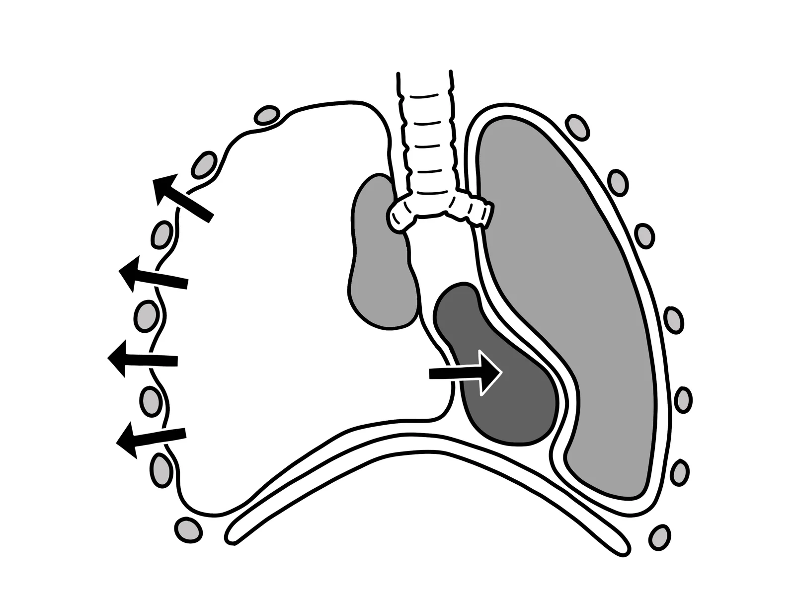
- Tension pneumothorax - immediate needle or finger thoracostomy followed by a chest drain. Do not wait for a chest X-ray.
- Cardiac tamponade - urgent echocardiographically guided pericardiocentesis, or surgery in trauma and aortic dissection. Give fluid as a holding measure and avoid diuretics and vasodilators.
- Massive pulmonary embolism - thrombolysis in the haemodynamically unstable patient, or surgical or catheter-directed embolectomy where thrombolysis is contraindicated
Hypovolaemic shock
- Stop the bleeding - direct pressure, tourniquet, pelvic binder, endoscopy, interventional radiology or surgery. This is the definitive treatment.
- Activate the major haemorrhage protocol early, and give blood products in a balanced ratio rather than crystalloid
- Tranexamic acid - within 3 hours in trauma and in gastrointestinal or obstetric haemorrhage where indicated
- Permissive hypotension in uncontrolled haemorrhage before surgical control, to avoid dislodging clot - a decision for senior clinicians
- Correct coagulopathy, acidosis and hypothermia - the lethal triad, each element worsening the others
- Non-haemorrhagic hypovolaemia - replace with balanced crystalloid and treat the underlying cause
Neurogenic and adrenal shock
Neurogenic shock follows spinal cord injury above T6 and causes hypotension with bradycardia and warm, dry skin. Treat with fluid, vasopressors (noradrenaline or phenylephrine) and atropine for bradycardia, alongside spinal immobilisation and neurosurgical referral. Adrenal crisis requires hydrocortisone 100 mg IV immediately, plus fluid and glucose - do not wait for a cortisol result before treating.
Complications
- Multi-organ dysfunction syndrome - the common final pathway, and the usual cause of death in patients who survive the initial insult
- Acute kidney injury - from renal hypoperfusion, often requiring renal replacement therapy
- Acute respiratory distress syndrome
- Hypoxic-ischaemic brain injury
- Ischaemic hepatitis and acute mesenteric ischaemia
- Disseminated intravascular coagulation - particularly in sepsis
- Stress ulceration and gastrointestinal bleeding
- Critical illness myopathy and neuropathy, with prolonged rehabilitation needs
- Post-intensive care syndrome - physical, cognitive and psychological sequelae persisting for months to years
- Complications of treatment - fluid overload, peripheral ischaemia and digital necrosis from vasopressors, and line-related infection
Red flags
Prognosis
Outcome depends on the cause, the speed of recognition and treatment, the degree of organ dysfunction reached, and the patient's physiological reserve.
Septic shock carries a mortality of the order of 30-40%, and every hour of delay in effective antibiotic administration is associated with a measurable increase in mortality. This time-dependence is the entire rationale for the Sepsis Six and for sepsis screening tools.
Cardiogenic shock complicating myocardial infarction remains the most lethal, with mortality that has improved with early revascularisation but still approaches 40-50%. Early primary PCI is the intervention with the clearest survival benefit.
Hypovolaemic shock from haemorrhage has an excellent prognosis if bleeding is controlled promptly, and a poor one once the lethal triad of coagulopathy, acidosis and hypothermia is established - which is why damage control resuscitation prioritises rapid surgical control and balanced blood product transfusion over restoring a normal blood pressure with crystalloid.
Anaphylaxis has a very low mortality when adrenaline is given promptly, and deaths are strongly associated with delayed adrenaline and with the patient having been sat or stood up. Both are avoidable.
Obstructive shock carries the most dramatic potential for reversal: relieving a tension pneumothorax or draining a tamponade can restore a peri-arrest patient to normal within minutes. Recognising these three diagnoses quickly - all of which present with hypotension and a raised JVP - is among the highest-yield skills in acute medicine.
Across all types, lactate clearance in the first 6 hours is one of the most useful prognostic markers, and serial reassessment rather than a single set of observations is the habit that most reliably improves outcomes.
References
- NICE NG51. Sepsis: recognition, diagnosis and early management. 2016, updated 2024. Available here
- Resuscitation Council UK. Emergency treatment of anaphylaxis: guidelines for healthcare providers. 2021. Available here
- Resuscitation Council UK. Adult advanced life support guidelines. Available here
- Evans L, Rhodes A, Alhazzani W et al. Surviving Sepsis Campaign: international guidelines for management of sepsis and septic shock 2021. Intensive Care Medicine. 2021. Available here
- NICE CG174. Intravenous fluid therapy in adults in hospital. 2013, updated 2017. Available here
- NICE NG39. Major trauma: assessment and initial management. 2016. Available here
- NICE NG185. Acute coronary syndromes. 2020, updated 2025. Available here
- NICE NG158. Venous thromboembolic diseases: diagnosis, management and thrombophilia testing. 2020, updated 2023. Available here
This article is written for revision and education. It is not clinical guidance and must not be used to make decisions about the care of a patient. Always check current NICE guidance and local protocols.