
Childhood Cancer: Recognition and Referral
Key points
- Scale: around 1,800 children aged 0-14 are diagnosed with cancer each year in the UK - roughly 1 in 500 children before their fifteenth birthday.
- The big three: leukaemia (about 30%, mostly ALL), CNS tumours (about 26%, the commonest solid tumours) and lymphoma (about 10%).
- The clinical task: recognition and referral, not treatment. Symptoms are common and non-specific, so the skill lies in identifying the pattern that does not fit a benign explanation.
- Leukaemia: pallor, fatigue, unexplained bruising or petechiae, bone pain and hepatosplenomegaly. NICE requires an FBC within 48 hours, and immediate referral for unexplained petechiae or hepatosplenomegaly.
- Brain tumour: morning headache with vomiting, new focal neurology, ataxia, visual change or an abnormal head posture. Newly abnormal cerebellar or central neurological function needs referral within 48 hours.
- Abdominal masses: a mass that crosses the midline suggests neuroblastoma; one that does not suggests a Wilms tumour. Either needs referral within 48 hours.
- Absent red reflex: leukocoria is retinoblastoma until proven otherwise and needs urgent ophthalmology referral - it is checked at every newborn and 6-week examination for this reason.
- Outcome: over 84% of children with cancer now survive 5 years, which makes recognising and managing the late effects of treatment part of the job.
Introduction
Childhood cancer is rare. A full-time GP might see one case every twenty years, and the symptoms - tiredness, headache, limp, a swollen gland, abdominal pain - are among the commonest reasons children are brought to a doctor at all. That combination of rarity and non-specific presentation is what makes early diagnosis difficult.
It nevertheless remains the second commonest cause of death in UK children aged 1-14, after injury.4 Around 1,800 children aged 0-14 are diagnosed each year, and a further large number of teenagers and young adults.
For finals and for practice, the expectation is not that you will treat these children - that happens in one of the specialist Principal Treatment Centres - but that you will recognise the presentations that warrant urgent referral, know the oncological emergencies, and understand the late effects that a growing population of survivors carries into adult life. NICE NG12 sets out the referral thresholds explicitly, and they are worth learning as written.1
The pattern of childhood cancer
| Cancer | Share of cases | Typical age | Notes |
|---|---|---|---|
| Leukaemia | About 30% | Peak 2-5 years | Around 80% acute lymphoblastic leukaemia, most of the rest acute myeloid leukaemia |
| CNS tumours | About 26% | All ages | The commonest solid tumours - astrocytoma, medulloblastoma, ependymoma, brainstem glioma |
| Lymphoma | About 10% | Hodgkin in adolescents; non-Hodgkin younger | Includes Burkitt lymphoma, which grows extremely rapidly |
| Neuroblastoma | About 6% | Under 5, median around 2 years | Arises from neural crest tissue in the adrenal medulla or sympathetic chain |
| Soft tissue sarcoma | About 6% | All ages | Mostly rhabdomyosarcoma |
| Wilms tumour (nephroblastoma) | About 5% | 1-5 years | Embryonal renal tumour |
| Bone tumours | About 4% | Adolescence | Osteosarcoma and Ewing sarcoma |
| Retinoblastoma | About 3% | Under 5 | Around 40% germline, from RB1 mutation |
| Germ cell tumours and hepatoblastoma | The remainder | Varies | Alpha-fetoprotein and beta-hCG are useful markers |
Predisposing conditions
Most childhood cancer is sporadic and no cause is identified. A recognisable predisposition is present in perhaps 10%, and knowing the classic associations is examinable.
- Down syndrome - a 10 to 20-fold increased risk of both ALL and AML, and transient abnormal myelopoiesis in the neonatal period
- Li-Fraumeni syndrome (TP53) - sarcoma, brain tumours, adrenocortical carcinoma
- Neurofibromatosis type 1 - optic pathway glioma, malignant peripheral nerve sheath tumour, juvenile myelomonocytic leukaemia
- Beckwith-Wiedemann syndrome - Wilms tumour and hepatoblastoma, warranting surveillance ultrasound
- WAGR and Denys-Drash syndromes (WT1) - Wilms tumour
- Hereditary retinoblastoma (RB1) - bilateral disease, and a lifelong excess risk of osteosarcoma
- Fanconi anaemia and ataxia telangiectasia - leukaemia and lymphoma, with marked radiosensitivity
- Immunosuppression - post-transplant lymphoproliferative disorder, and EBV-driven lymphoma
- Previous cancer treatment - ionising radiation and alkylating chemotherapy both cause second malignancies
Leukaemia
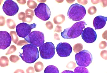
Symptoms arise from marrow failure - anaemia, neutropenia and thrombocytopenia - and from tissue infiltration by blasts.
- Pallor, fatigue and lethargy from anaemia
- Unexplained bruising, petechiae, purpura, epistaxis or gum bleeding from thrombocytopenia
- Persistent, recurrent or unusually severe infection, and unexplained fever, from neutropenia
- Bone and joint pain - a limp, refusal to weight bear, or night pain waking the child. Leukaemia is a well-recognised cause of an atraumatic limp and is frequently misdiagnosed as juvenile idiopathic arthritis or growing pains.
- Generalised lymphadenopathy and hepatosplenomegaly
- Testicular enlargement, and a mediastinal mass in T-cell disease
- Headache, vomiting or cranial nerve palsy from CNS involvement
CNS tumours
Brain tumours are the commonest solid tumour of childhood and have the longest average diagnostic interval of any childhood cancer, which is why the HeadSmart campaign was created to shorten it.5 Symptoms depend on site, age and rate of growth.
- Raised intracranial pressure - headache that is worse on waking or wakes the child from sleep, accompanied by vomiting, and often improving through the day
- Papilloedema, reduced visual acuity, double vision or a sixth nerve palsy as a false localising sign
- Cerebellar signs - ataxia, unsteadiness, nystagmus, intention tremor, deteriorating handwriting or coordination
- New focal neurology, seizures or persistent weakness
- Abnormal head position - head tilt or torticollis, which is a classic and easily missed posterior fossa sign
- Personality or behaviour change, declining school performance, or developmental regression
- Endocrine features from a suprasellar tumour - growth failure, diabetes insipidus with polyuria and polydipsia, precocious or delayed puberty, or weight change
- In infants - an abnormally increasing head circumference, a bulging fontanelle, the sunsetting sign, and irritability
Lymphoma and lymphadenopathy
Cervical lymphadenopathy is almost universal in young children and almost always reactive. The features that make a node worrying are its size, its consistency, its site and its behaviour over time.
- Persistent, painless, firm or rubbery nodes larger than 2 cm that do not resolve over 4-6 weeks or continue to enlarge
- Supraclavicular nodes at any size, which are almost never benign
- Matted or fixed nodes, or nodes with overlying skin change
- B symptoms - unexplained fever, drenching night sweats and weight loss of more than 10%
- A mediastinal mass - cough, breathlessness, orthopnoea, stridor, or superior vena cava obstruction with facial swelling and distended neck veins
- Hepatosplenomegaly
- Pruritus and alcohol-induced nodal pain - uncommon but classic in Hodgkin lymphoma
- NICE advises very urgent referral for unexplained lymphadenopathy or splenomegaly in a child, alongside an urgent full blood count1
Abdominal masses
| Neuroblastoma | Wilms tumour (nephroblastoma) | |
|---|---|---|
| Origin | Neural crest - adrenal medulla or sympathetic chain | Embryonal renal tissue |
| Typical age | Under 5, median around 2 years | 1-5 years |
| The mass | Irregular, firm, crosses the midline | Smooth, unilateral, does not cross the midline |
| The child | Often systemically unwell - weight loss, pallor, bone pain, fever | Often systemically well, with the mass found incidentally at bathing or nappy change |
| Characteristic features | Periorbital ecchymoses and proptosis from skull metastases; hypertension; Horner syndrome from a cervical primary; opsoclonus-myoclonus (dancing eye syndrome) | Haematuria in around 20%, hypertension, abdominal pain |
| Investigations | Raised urinary catecholamines (VMA and HVA), MIBG scan, biopsy, MYCN amplification status | Ultrasound then CT or MRI, and biopsy or primary nephrectomy per protocol |
| Associations | Sporadic; rarely familial (ALK, PHOX2B) | Beckwith-Wiedemann, WAGR and Denys-Drash syndromes, hemihypertrophy |
| Prognosis | Very variable - infants with localised disease may regress spontaneously; high-risk MYCN-amplified disease has around 50% survival | Good - overall survival around 90% |
Bone tumours, retinoblastoma and sarcoma
Bone tumours
- Persistent localised bone pain, worse at night and not related to activity, with or without a palpable swelling or a pathological fracture
- Osteosarcoma - typically the metaphysis of a long bone, most often the distal femur or proximal tibia, with a sunburst periosteal reaction and Codman triangle on radiograph
- Ewing sarcoma - typically the diaphysis of a long bone or the pelvis, with an onion-skin periosteal reaction. It often causes fever and raised inflammatory markers and is mistaken for osteomyelitis.
- NICE advises a radiograph within 48 hours for a child with unexplained bone swelling or persistent bone pain, and very urgent specialist referral if the radiograph suggests a bone sarcoma1
Retinoblastoma
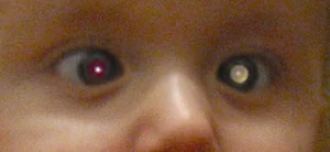
- Leukocoria - a white pupillary reflex, or an absent red reflex, which is why the red reflex is checked at the newborn examination and again at 6-8 weeks
- A new squint, particularly in a child under 2
- A red, painful or inflamed eye without an obvious cause, deteriorating vision, or proptosis
- Bilateral disease implies a germline RB1 mutation, with implications for siblings and for a lifelong risk of second cancers
- NICE advises urgent referral (within 2 weeks) to ophthalmology for a child with an absent red reflex1
Rhabdomyosarcoma
A painless, progressively enlarging mass that can arise almost anywhere. Site determines the presentation: head and neck disease causes proptosis, nasal obstruction or a cranial nerve palsy; genitourinary disease causes a bloody vaginal discharge, urinary retention or a scrotal mass; and limb or trunk disease presents simply as a lump. Any unexplained, enlarging soft tissue mass in a child needs urgent imaging and referral.
Oncological emergencies
- Tumour lysis syndrome - massive cell breakdown, spontaneously or after starting treatment, in bulky rapidly proliferating disease such as Burkitt lymphoma and T-cell ALL. Causes hyperkalaemia, hyperphosphataemia, hyperuricaemia, hypocalcaemia and acute kidney injury. Prevented with vigorous hydration and allopurinol or rasburicase, and managed with close biochemical monitoring and sometimes dialysis.
- Superior vena cava obstruction and mediastinal mass - facial and upper limb swelling, plethora, distended neck veins, orthopnoea and stridor. Do not lie the child flat and do not give a general anaesthetic without senior anaesthetic assessment, since the airway and venous return can be lost irretrievably on induction.
- Spinal cord compression - back pain, limb weakness, sensory level and sphincter disturbance. Needs urgent MRI of the whole spine and dexamethasone without waiting for imaging.
- Raised intracranial pressure and hydrocephalus - reduced consciousness, Cushing's triad, papilloedema. Needs urgent imaging and neurosurgical involvement.
- Hyperleucocytosis and leucostasis - a very high blast count causing sludging in the cerebral and pulmonary microcirculation, with confusion, hypoxia and haemorrhage
- Severe cytopenias - symptomatic anaemia or thrombocytopenic bleeding requiring irradiated blood products
Investigation and treatment principles
- FBC with a blood film, U&Es, LFTs, LDH, urate, calcium, phosphate, clotting and a group and save
- Imaging appropriate to the site - abdominal ultrasound as the first-line test for an abdominal mass, chest radiograph for a mediastinal mass, MRI for CNS and bone lesions
- Bone marrow aspirate and trephine, and a lumbar puncture for CNS staging in leukaemia and lymphoma
- Tissue biopsy for solid tumours, with molecular and cytogenetic characterisation that now determines risk stratification
- Tumour markers - urinary catecholamines for neuroblastoma, alpha-fetoprotein and beta-hCG for germ cell tumours and hepatoblastoma
- Treatment at a Principal Treatment Centre with shared care locally, almost always on a national or international trial protocol3
- Modalities - chemotherapy, surgery, radiotherapy, immunotherapy, haematopoietic stem cell transplantation and, increasingly, CAR-T cell therapy
- Supportive care - central venous access, antiemetics, nutritional support, transfusion, infection prophylaxis, and a multidisciplinary team including play specialists, psychologists, social workers and hospital teachers
Late effects and prognosis
Overall 5-year survival for childhood cancer in the UK now exceeds 84%, and for some diagnoses it is far higher - around 90% for acute lymphoblastic leukaemia and Wilms tumour, and over 95% for Hodgkin lymphoma. Outcomes remain poor for high-risk neuroblastoma at around 50%, and for diffuse intrinsic pontine glioma, where survival remains under 10%.4
The consequence of that success is a large and growing population of survivors - roughly 1 in 700 young adults in the UK - the majority of whom carry at least one long-term health consequence of their treatment. Long-term follow-up is a defined part of care, not an optional extra.
| System | Late effect | Usual cause |
|---|---|---|
| Endocrine | Growth hormone deficiency, hypothyroidism, precocious or delayed puberty, infertility, premature ovarian insufficiency | Cranial and gonadal radiotherapy; alkylating agents |
| Cardiac | Cardiomyopathy and heart failure, sometimes appearing decades later | Anthracyclines such as doxorubicin; mediastinal radiotherapy |
| Renal | Chronic kidney disease, tubulopathy, hypomagnesaemia | Cisplatin, ifosfamide, nephrectomy |
| Sensory | Sensorineural hearing loss, cataract | Platinum agents; cranial radiotherapy |
| Respiratory | Pulmonary fibrosis and restrictive lung disease | Bleomycin, busulfan, thoracic radiotherapy |
| Neurocognitive | Reduced processing speed, working memory and attention; educational underachievement | Cranial radiotherapy and intrathecal methotrexate, worst in young children |
| Musculoskeletal | Reduced bone density, avascular necrosis, growth asymmetry, scoliosis | Corticosteroids, radiotherapy to growth plates |
| Second cancers | Therapy-related leukaemia, sarcoma and breast cancer | Alkylating agents, topoisomerase inhibitors, radiotherapy |
| Psychosocial | Anxiety, depression, post-traumatic stress, difficulties with employment and insurance | The illness and its treatment |
Two practical points follow for a generalist. First, take an oncology history seriously in any young adult who presents with breathlessness, fatigue, subfertility or a new lump - previous anthracycline or radiotherapy exposure changes the differential entirely. Second, ask survivors whether they hold a treatment summary and long-term follow-up plan, since these documents are what allow risk-appropriate surveillance to continue once they have moved out of paediatric services.6
The message that should sit alongside all of this is the encouraging one. Most children with cancer are cured, most go on to lead ordinary adult lives, and the recognition step - the part that a student or foundation doctor is actually responsible for - is where the largest remaining gains in outcome lie.
References
- NICE NG12. Suspected cancer: recognition and referral. 2015, updated 2023. Available here
- NICE CG151. Neutropenic sepsis: prevention and management in people with cancer. 2012. Available here
- Children's Cancer and Leukaemia Group (CCLG). Available here
- Cancer Research UK. Children's cancers statistics. Available here
- HeadSmart. Be brain tumour aware. Available here
- NICE NG43. Transition from children's to adults' services for young people using health or social care services. 2016. Available here
This article is written for revision and education. It is not clinical guidance and must not be used to make decisions about the care of a patient. Always check current NICE guidance and local protocols.