
Cryptorchidism (Undescended Testis)
Key points
- Definition: failure of one or both testes to descend fully into the scrotum. It is the commonest congenital genitourinary abnormality in boys.
- How common: 3-5% of term male infants and up to 30% of preterm boys, falling to 1-2% by 3-6 months as spontaneous descent occurs.
- Spontaneous descent: happens in most affected boys within the first 3-6 months. Descent after 6 months of corrected age is rare, which is what sets the referral timing.
- Referral and surgery: refer by 3 months if still undescended; orchidopexy between 6 and 12 months, and by 18 months at the latest.
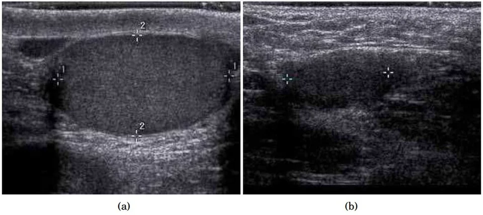
- Do not image: ultrasound, CT and MRI are not recommended for locating an impalpable testis - they miss intra-abdominal testes and do not change the plan, which is laparoscopy.
- Bilateral impalpable testes: a medical emergency. This may be a virilised 46,XX infant with congenital adrenal hyperplasia, so urgent endocrine and genetic assessment is required.
- Why it matters: germ cell loss begins from around 6 months, and there is a 2-8 fold increased risk of testicular germ cell tumour - in both testes, not only the affected one.
- Retractile testis: a normal variant caused by an active cremasteric reflex, but it needs annual review because a proportion later ascend.
Introduction
Cryptorchidism - literally hidden testis - describes a testis that has not completed its descent into the scrotum. It is the commonest congenital abnormality of the male genital tract, affecting 3-5% of boys born at term and a much higher proportion of those born preterm, because descent is one of the last events of fetal development.
Most of these testes descend spontaneously in the first few months of life, so the prevalence falls to around 1-2% by 3-6 months. Descent after that point is uncommon, which is why the referral and operating windows are defined as they are.1
The condition matters for three reasons: fertility, because germ cell development is impaired by the higher temperature outside the scrotum and the damage begins in the first year of life; malignancy, because an undescended testis carries a several-fold increased risk of germ cell tumour; and detectability, because a testis in the scrotum can be examined by the patient and a testis in the abdomen cannot. Orchidopexy addresses all three.
Embryology and classification
The testis develops from the gonadal ridge on the posterior abdominal wall and descends in two hormonally distinct phases. Knowing them explains both the associations and the terminology.
- Transabdominal phase (8-15 weeks) - driven by insulin-like 3 (INSL3) acting on its receptor RXFP2, which thickens the gubernaculum and anchors the testis at the internal inguinal ring while the abdomen grows away from it. Mullerian inhibiting substance contributes.
- Inguinoscrotal phase (25-35 weeks) - androgen-dependent, mediated by testosterone acting partly through the genitofemoral nerve and calcitonin gene-related peptide. The gubernaculum migrates to the scrotum and the testis follows it through the inguinal canal.
- The processus vaginalis, a peritoneal diverticulum that accompanies the testis, normally obliterates after descent. Persistent patency explains the strong association with inguinal hernia and hydrocele.
Types
| Type | Description | Management |
|---|---|---|
| True undescended | Arrested somewhere along the normal path of descent - intra-abdominal, within the inguinal canal, at the external ring, or high in the scrotum | Orchidopexy |
| Ectopic | Outside the normal path - most often the superficial inguinal pouch, and rarely femoral, perineal, prepenile or in the contralateral scrotum | Orchidopexy |
| Retractile | Can be brought fully into the scrotum and stays there temporarily; caused by a brisk cremasteric reflex in a normally descended testis | A normal variant, but needs annual review to puberty because a proportion subsequently ascend |
| Ascending (acquired) | A testis previously documented in the scrotum that rises out of it during childhood, usually because the spermatic cord fails to elongate with growth | Orchidopexy |
| Absent or vanishing | No testicular tissue, usually from prenatal torsion leaving a fibrous nubbin; around 4% of impalpable testes | Confirmed at laparoscopy; consider a prosthesis later |
Risk factors and associations
- Prematurity, low birth weight and being small for gestational age - the strongest risk factors, because the inguinoscrotal phase occurs in the third trimester
- Family history of cryptorchidism
- Maternal smoking, obesity and diabetes, and twin pregnancy
- Hypospadias - the combination of an undescended testis and hypospadias should always prompt assessment for a difference of sex development
- Inguinal hernia, present in a substantial proportion because of the patent processus vaginalis
- Syndromic associations - prune belly syndrome, Prader-Willi syndrome, Noonan syndrome, Klinefelter syndrome, and cerebral palsy
Clinical assessment
The testes are examined at the newborn examination and again at the 6-8 week check, precisely so that this diagnosis is made early.2 Examination technique determines the answer, and a poorly performed examination is the commonest reason a retractile testis is labelled undescended.
How to examine
- Warm room, warm hands, relaxed child. Cold and anxiety provoke the cremasteric reflex and pull a normal testis up.
- Examine supine first, then cross-legged or squatting in an older boy, which relaxes the cremaster.
- Sweep with the non-dominant hand from the anterior superior iliac spine along the inguinal canal towards the scrotum, milking the testis downwards, and catch it in the scrotum with the other hand.
- Decide whether it reaches the scrotum, and whether it stays there when released. A testis that reaches the base of the scrotum and remains without tension is retractile; one that springs straight back is undescended.
- Record the position, size and consistency, and compare with the other side. Compensatory hypertrophy of the contralateral testis suggests the impalpable testis is absent.
- Examine the penis and urethral meatus for hypospadias, and note scrotal development - a hypoplastic, poorly rugated hemiscrotum suggests the testis has never been there.
Differential diagnosis of the empty scrotum
An empty hemiscrotum is a finding, not a diagnosis, and working through the possibilities in order avoids both over-referral and missed pathology.
- Retractile testis - much the commonest explanation in a boy over 1 year. The testis can be brought to the base of the scrotum and stays there briefly; the scrotum is normally developed and rugated.
- True undescended testis - palpable in the inguinal canal or at the external ring, cannot be brought to the scrotum without tension, and springs back immediately when released
- Ectopic testis - palpable outside the line of descent, most often in the superficial inguinal pouch, and occasionally femoral or perineal
- Ascending testis - previously documented in the scrotum, now not; a specific reason to record the finding accurately at each check
- Absent or vanished testis - an underdeveloped, poorly rugated hemiscrotum with compensatory hypertrophy of the contralateral testis, usually the result of prenatal torsion
- Testicular torsion or atrophy after a missed torsion - acute pain in the former, an absent or small testis in the latter
- A difference of sex development - particularly with bilateral impalpable testes or an accompanying hypospadias
- A large hernia or hydrocele obscuring an otherwise normal testis, which becomes obvious once it is reduced or transilluminated
Investigations
Investigation is therefore reserved for specific situations rather than performed routinely.
- Karyotype or microarray, 17-hydroxyprogesterone, U&Es, testosterone, LH and FSH, and pelvic ultrasound - for bilateral impalpable testes or where a difference of sex development is suspected
- Anti-Mullerian hormone and inhibin B, or an hCG stimulation test, to establish whether functioning testicular tissue is present in bilateral impalpable testes
- Diagnostic laparoscopy - the gold standard for the impalpable testis, identifying an intra-abdominal testis, blind-ending vessels indicating a vanished testis, or vessels entering the internal ring
- Renal tract ultrasound only if there are other genitourinary anomalies, since isolated cryptorchidism is not associated with renal anomalies
Management
Timing
- Birth to 3 months - observe. Spontaneous descent occurs in the majority, so no action is needed beyond documenting the finding and arranging review.
- Still undescended at 3 months (corrected for gestation) - refer to paediatric surgery or urology
- Orchidopexy between 6 and 12 months of age, and by 18 months at the very latest. Operating in this window preserves germ cell development, since the histological damage becomes established during the second year of life.
- Retractile testis - no surgery, but annual review until puberty, because a proportion ascend and then require orchidopexy
- Ascending testis - orchidopexy at the point it is identified
- Post-pubertal or adult undescended testis - orchidectomy is usually recommended, since the testis contributes negligibly to fertility and carries an ongoing malignancy risk in a site that cannot be examined
Orchidopexy
- Palpable testis - an inguinal or scrotal approach, mobilising the testis and spermatic cord, ligating the patent processus vaginalis (which repairs the associated hernia), and fixing the testis in a subdartos pouch in the scrotum
- Impalpable testis - examination under anaesthetic first, since around a fifth become palpable once the child is relaxed, followed by diagnostic laparoscopy
- Intra-abdominal testis - a single-stage laparoscopic orchidopexy if the vessels are long enough, or a two-stage Fowler-Stephens procedure, in which the testicular vessels are divided at the first operation so that collateral supply from the vas deferens can develop before the testis is brought down at the second
- Atrophic nubbin or blind-ending vessels - excision, with the option of a prosthesis in adolescence
- Day-case surgery in most cases, with success rates above 90% for palpable testes and lower for intra-abdominal ones
Complications
Infertility
The scrotum keeps the testis 2-4°C below core temperature, which is required for normal spermatogenesis. In an undescended testis, transformation of gonocytes into adult dark spermatogonia fails and germ cell numbers fall progressively from around 6 months of age - the reason surgery is not deferred to school age as it once was.
- Unilateral undescended testis - paternity rates are close to those of the general population, with infertility in roughly 10%
- Bilateral undescended testes - infertility in a much larger proportion, quoted between about 35% and 65%
- Earlier orchidopexy is associated with better germ cell counts and better hormonal profiles, though the effect on eventual paternity is harder to demonstrate
Malignancy
- A 2 to 8-fold increased risk of testicular germ cell tumour, most often seminoma in a testis that has remained undescended
- The risk is increased in the contralateral, normally descended testis as well, which tells you that the cryptorchidism is a marker of an underlying testicular dysgenesis rather than simply a consequence of position
- Orchidopexy before puberty reduces but does not eliminate the risk, and its other benefit is that a scrotal testis can actually be examined
- Teach testicular self-examination from puberty, and have a low threshold for referral of a new testicular lump4
Other complications
- Testicular torsion - more likely in an undescended testis, and presenting atypically as groin or abdominal pain with an empty scrotum on that side. Always examine the scrotum in a boy with abdominal pain.6
- Inguinal hernia, from the patent processus vaginalis
- Trauma, where the testis lies superficially over the pubic bone
- Testicular atrophy after orchidopexy, particularly following a Fowler-Stephens procedure
- Psychological effects - body image and anxiety in adolescence, which are worth asking about rather than assuming
Prognosis
Orchidopexy is successful in more than 90% of boys with a palpable undescended testis, with lower rates for intra-abdominal testes where the blood supply is more precarious. Most boys who have had a unilateral orchidopexy in infancy go on to have normal fertility and normal testosterone production.
The residual risks are lifelong rather than resolved. The increased risk of germ cell tumour persists after surgery, peaking in the twenties and thirties, and applies to both testes. Boys who have had bilateral cryptorchidism should be counselled in adolescence about fertility and offered semen analysis if they wish it.
The practical message is about timing and about examination technique. Every boy has his testes checked at birth and at 6-8 weeks; anyone still lacking a scrotal testis at 3 months is referred; and surgery happens between 6 and 12 months. Getting that sequence right, and resisting the pull to order an ultrasound instead of referring, is essentially the whole of good management.3
References
- NICE Clinical Knowledge Summaries. Undescended testes. Available here
- GOV.UK. Newborn and infant physical examination (NIPE) screening programme handbook. Available here
- British Association of Paediatric Surgeons. Available here
- NICE NG12. Suspected cancer: recognition and referral. 2015, updated 2023. Available here
- European Association of Urology and European Society for Paediatric Urology. Guidelines on paediatric urology. Available here
- NICE Clinical Knowledge Summaries. Scrotal pain and swelling. Available here
This article is written for revision and education. It is not clinical guidance and must not be used to make decisions about the care of a patient. Always check current NICE guidance and local protocols.