
Testicular Torsion
Key points
- Testicular torsion: twisting of the spermatic cord, occluding the testicular blood supply and causing ischaemia and infarction. It is a surgical emergency.
- The time window: salvage rates are around 90-100% within 6 hours, falling to roughly 50% at 12 hours and under 10% after 24 hours. Time is testicle.
- Who gets it: bimodal - neonates, and above all adolescents and young men aged 12-25. It is the commonest cause of acute scrotal pain in this age group.
- The anatomy: the bell-clapper deformity - a high investment of tunica vaginalis leaves the testis hanging freely and able to rotate. It is usually bilateral, which is why both testes are fixed at surgery.
- Presentation: sudden, severe unilateral testicular pain, often waking the patient from sleep, with nausea and vomiting. Abdominal pain may be the only complaint in a boy.
- Signs: a high-riding testis lying horizontally, a swollen, exquisitely tender hemiscrotum, an absent cremasteric reflex, and negative Prehn's sign (elevation does not relieve pain).
- Investigation: this is a clinical diagnosis. Doppler ultrasound may be used only if it causes no delay - a normal scan does not exclude torsion.
- Management: nil by mouth, analgesia and immediate scrotal exploration. Viable testis: bilateral orchidopexy. Non-viable: orchidectomy with contralateral orchidopexy.
Introduction
Testicular torsion is twisting of the spermatic cord within or above the tunica vaginalis, obstructing venous drainage and then arterial inflow. The result is progressive congestion, ischaemia and ultimately infarction of the testis.1
It affects around 1 in 4,000 males under 25, with a bimodal age distribution: a neonatal peak (usually extravaginal torsion occurring perinatally) and a much larger adolescent peak between 12 and 25 years, coinciding with rapid testicular growth at puberty. It is the single most important diagnosis to exclude in any male presenting with acute scrotal pain.
Aetiology and anatomy
The bell-clapper deformity
Normally the tunica vaginalis covers only the anterior and lateral surfaces of the testis, leaving its posterior aspect anchored to the scrotal wall. In the bell-clapper deformity, the tunica vaginalis invests the testis completely and inserts high on the spermatic cord, leaving the testis suspended freely within the tunica like the clapper inside a bell, able to swing and rotate.
- This predisposes to intravaginal torsion, which is the type seen in adolescents and adults
- The deformity is present in around 12% of males and is bilateral in the majority
- This is precisely why the contralateral testis is fixed at the same operation - the anatomical predisposition affects both sides, and a patient who has torted one testis is at significant risk of torting the other
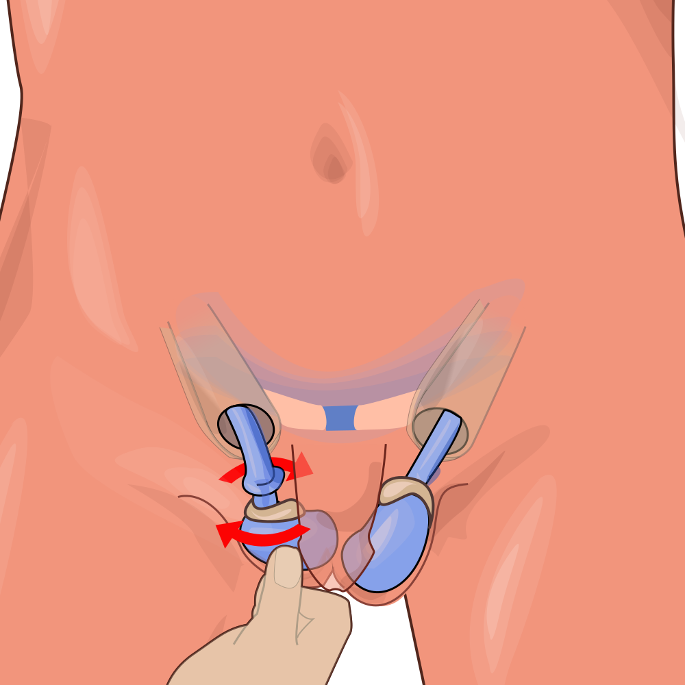
Types of torsion
- Intravaginal torsion - within the tunica vaginalis; the usual type in adolescents and adults, associated with the bell-clapper deformity
- Extravaginal torsion - the entire cord and tunica twist together, occurring in neonates (typically antenatally or perinatally, because the tunica has not yet adhered to the scrotal wall). Presents as a firm, painless, discoloured scrotal swelling in a newborn, and the testis is usually already non-viable
- Torsion of a testicular appendage (hydatid of Morgagni) - a common mimic in boys aged 7-12, causing more localised upper pole pain and sometimes the 'blue dot' sign; it is not dangerous, but is often indistinguishable clinically from true torsion, so exploration is frequently still required
Risk factors and triggers
- Age 12-25 and the pubertal growth spurt
- Bell-clapper deformity
- Undescended or maldescended testis (cryptorchidism) - a substantially increased risk
- Previous torsion or previous episodes of self-resolving severe scrotal pain (intermittent torsion)
- Family history of torsion
- Trauma - torsion may follow minor scrotal trauma, and trauma should never be assumed to be the sole explanation for pain
- Exercise or sudden movement, and cold weather (cremasteric contraction); pain frequently begins during sleep
- A testis with a horizontal lie on examination of the contralateral side
Clinical features
History
- Sudden onset, severe, unilateral testicular pain - typically reaching maximum intensity within minutes. Often wakes the patient from sleep or begins in the early morning
- Nausea and vomiting - present in a majority and a useful discriminator from epididymo-orchitis
- Referred lower abdominal or groin pain - crucially, a boy may complain only of abdominal pain. This is why the testes must be examined in every male presenting with abdominal pain; torsion is regularly missed because the scrotum was never examined
- Absence of urinary symptoms - dysuria, frequency and discharge point towards epididymo-orchitis instead
- Previous similar self-limiting episodes - suggests intermittent torsion and warrants elective fixation
- Establish the exact time of onset - this drives the urgency and informs the discussion about salvage
Examination
- Exquisitely tender, swollen, erythematous hemiscrotum - often too painful to examine properly
- High-riding testis - drawn upwards by the shortened, twisted cord
- Horizontal (transverse) lie of the testis, compared with the normal vertical orientation
- Absent cremasteric reflex - stroking the inner thigh normally elevates the ipsilateral testis; its absence is a sensitive sign, though not entirely specific and unreliable in very young boys
- Negative Prehn's sign - elevating the testis does NOT relieve the pain (in epididymo-orchitis, elevation typically eases it). Useful but not definitive
- Absent or reduced blood flow if Doppler is performed
- Examine the contralateral testis for a horizontal lie, which supports a bell-clapper anatomy
| Feature | Testicular torsion | Epididymo-orchitis | Torted appendage |
|---|---|---|---|
| Age | 12-25 (and neonates) | Any; often sexually active young men or older men with UTI | 7-12 years |
| Onset | Sudden, minutes; often wakes from sleep | Gradual, over days | Gradual, over 1-2 days |
| Pain severity | Severe | Moderate | Mild to moderate, localised to upper pole |
| Nausea/vomiting | Common | Uncommon | Uncommon |
| Fever/urinary symptoms | Absent | Common - dysuria, discharge, fever | Absent |
| Testicular lie | High, horizontal | Normal vertical | Normal |
| Cremasteric reflex | Absent | Present | Present |
| Prehn's sign | Negative (elevation does not help) | Positive (elevation relieves) | Variable |
| Other | - | Tender epididymis, possible urethral discharge | 'Blue dot' sign at upper pole |
Investigations
Management
Immediate management
- Contact urology or paediatric surgery immediately - this is a time-critical emergency
- Keep the patient nil by mouth and prepare for theatre
- Provide adequate analgesia - usually opioid; pain is severe and analgesia does not obscure the diagnosis
- Antiemetics and intravenous fluids as needed
- Obtain informed consent covering scrotal exploration, possible orchidectomy, and bilateral orchidopexy - the possibility of losing the testis must be discussed explicitly, along with fixation of the healthy side
- Manual detorsion may be attempted by an experienced clinician if surgery will be significantly delayed - classically rotating the testis outwards, like opening a book (medial to lateral). Success is signalled by prompt pain relief, but this is a temporising measure only and does not remove the need for surgical exploration and fixation
Surgical management
- Urgent scrotal exploration - the definitive management, through a scrotal (or midline raphe) incision
- The cord is untwisted and the testis wrapped in warm saline swabs and observed for return of perfusion
- If viable: bilateral orchidopexy - the affected testis is fixed within the scrotum with non-absorbable sutures, and the contralateral testis is fixed at the same operation because the bell-clapper deformity is usually bilateral
- If non-viable: orchidectomy of the infarcted testis with contralateral orchidopexy. Leaving necrotic tissue risks infection and may provoke anti-sperm antibodies
- A testicular prosthesis can be offered, usually at a later date, and should be discussed sensitively
- Torsion of an appendage, if found, is simply excised and carries no long-term consequence
Neonatal torsion
Neonatal (extravaginal) torsion presents with a firm, often painless, discoloured scrotal swelling noted at or shortly after birth. The affected testis is usually already infarcted and unsalvageable. Management remains debated, but urgent paediatric surgical assessment is required, and contralateral fixation is generally performed given the risk to the remaining testis.
Complications
- Testicular infarction and loss of the testis - the principal complication, and directly proportional to delay
- Subfertility and impaired spermatogenesis - may occur even with a salvaged testis, and even where the contralateral testis is normal, possibly through immune-mediated damage
- Anti-sperm antibody formation - from exposure of sperm antigens after breakdown of the blood-testis barrier
- Testicular atrophy - a salvaged testis may still atrophy over subsequent months, so follow-up is needed
- Hypogonadism - uncommon if the contralateral testis is healthy, but a concern after bilateral events
- Recurrent torsion - if fixation fails or was not performed
- Wound infection, haematoma and chronic scrotal pain after surgery
- Psychological effects - body image concerns and anxiety about fertility, particularly in adolescents; these deserve explicit attention
- Medicolegal consequences - delayed diagnosis of torsion is among the most frequent sources of successful negligence claims in emergency and primary care
Red flags
Prognosis
Prognosis is determined almost entirely by the time from symptom onset to detorsion. Testicular salvage rates are approximately 90-100% if surgery occurs within 6 hours, falling to around 50% by 12 hours and to less than 10% beyond 24 hours. This steep relationship is the reason for the aggressive approach to exploration, and it means that any avoidable delay - waiting for an ultrasound, a morning list, or a second opinion - translates directly into lost testes.
Where the testis is salvaged, the outlook is generally good, though a proportion undergo subsequent atrophy over the following months despite an apparently viable appearance at operation, and follow-up should include reassessment of testicular size. Recurrence after adequate bilateral orchidopexy is uncommon but not impossible if sutures fail.
Fertility after unilateral torsion is usually preserved. A single healthy testis is sufficient for normal testosterone production and fertility in most men, so a patient who has lost one testis can be given genuine reassurance on that point. That said, semen parameters are measurably abnormal in a proportion of men after torsion, even with a normal contralateral testis, and the mechanisms - ischaemia-reperfusion injury and possible autoimmune damage - are not fully understood. Because the patients are typically adolescents, the psychological impact and concerns about future fertility and body image are substantial and should be addressed directly rather than left unspoken, including offering a prosthesis where a testis has been removed.
References
- NICE Clinical Knowledge Summaries. Scrotal pain and swelling. Available here
- European Association of Urology. Guidelines on Paediatric Urology - acute scrotum. Available here
- British Association of Urological Surgeons (BAUS). Testicular torsion - patient and clinician information. Available here
- Sharp VJ, Kieran K, Arlen AM. Testicular torsion: diagnosis, evaluation, and management. American Family Physician. 2013. Available here
- Mellick LB, Sinex JE, Gibson RW, Mears K. A systematic review of testicle survival time after a torsion event. Pediatric Emergency Care. 2019. Available here
- Hariadhi, CC BY-SA 4.0, via Wikimedia Commons. Available here
This article is written for revision and education. It is not clinical guidance and must not be used to make decisions about the care of a patient. Always check current NICE guidance and local protocols.