
Stroke: Diagnosis and Management
Key points
- Stroke: a sudden-onset focal neurological deficit due to a vascular cause, lasting more than 24 hours or causing death. About 85% is ischaemic, 15% haemorrhagic.
- Recognition: FAST - Face, Arm, Speech, Time - identifies most anterior circulation strokes in the community.
- First investigation: urgent non-contrast CT head, primarily to exclude haemorrhage before any antithrombotic therapy.
- Thrombolysis: IV alteplase (or tenecteplase) within 4.5 hours of onset for ischaemic stroke, if no contraindication.
- Thrombectomy: mechanical clot retrieval for proximal large vessel occlusion, up to 24 hours in selected patients with salvageable tissue on perfusion imaging.
- Classification: the Bamford (Oxford Community Stroke Project) classification - TACS, PACS, LACS, POCS - predicts territory, prognosis and underlying pathology from the bedside.
- Secondary prevention: antiplatelet therapy (or anticoagulation if AF), high-intensity statin, blood pressure control and carotid endarterectomy where indicated.
- Prognosis: roughly a third of survivors are left with significant disability; early supported discharge and stroke unit care improve outcomes.
Introduction
Stroke is a clinical syndrome of rapidly developing focal (or global) disturbance of cerebral function, of presumed vascular origin, lasting more than 24 hours or leading to death.1 It is a medical emergency: the phrase 'time is brain' reflects the fact that an estimated 1.9 million neurons are lost every minute that a large vessel occlusion goes untreated.2
Around 85% of strokes are ischaemic, caused by occlusion of a cerebral artery, and 15% are haemorrhagic, caused by rupture of a blood vessel within the brain parenchyma. The distinction cannot be made clinically with any reliability and requires imaging - this is the single most important practical point in stroke medicine, because the two require opposite treatments.
Stroke is the fourth leading cause of death in the UK and the leading cause of adult disability. It is a very heavily examined topic because it combines anatomy, pharmacology, acute decision-making and public health in one condition.
Aetiology and classification
Ischaemic stroke
Ischaemic stroke follows occlusion of a cerebral artery, causing infarction of the territory it supplies. The commonest mechanisms are:
- Large artery atherosclerosis - a thrombus forms on a ruptured atherosclerotic plaque, typically at the carotid bifurcation or within a major intracranial vessel, or embolises distally from it
- Cardioembolism - a thrombus forms in the heart, most often the left atrial appendage in atrial fibrillation, and embolises to the brain. Also follows myocardial infarction, valvular disease and infective endocarditis
- Small vessel disease (lacunar infarction) - occlusion of a single deep perforating arteriole, related to chronic hypertension and diabetes, producing small subcortical infarcts
- Cryptogenic - no cause identified despite full work-up, accounting for a substantial minority
- Less common - arterial dissection (consider in younger patients, especially after neck trauma or manipulation), vasculitis, hypercoagulable states, venous sinus thrombosis with venous infarction
Haemorrhagic stroke
Primary intracerebral haemorrhage results from rupture of a blood vessel within the brain parenchyma, most often a small perforating artery weakened by chronic hypertension (hypertensive arteriopathy) or, in older patients with a lobar bleed, cerebral amyloid angiopathy. Subarachnoid haemorrhage, subdural haemorrhage and extradural haemorrhage are distinct entities covered separately.
Anatomical classification - the Bamford criteria
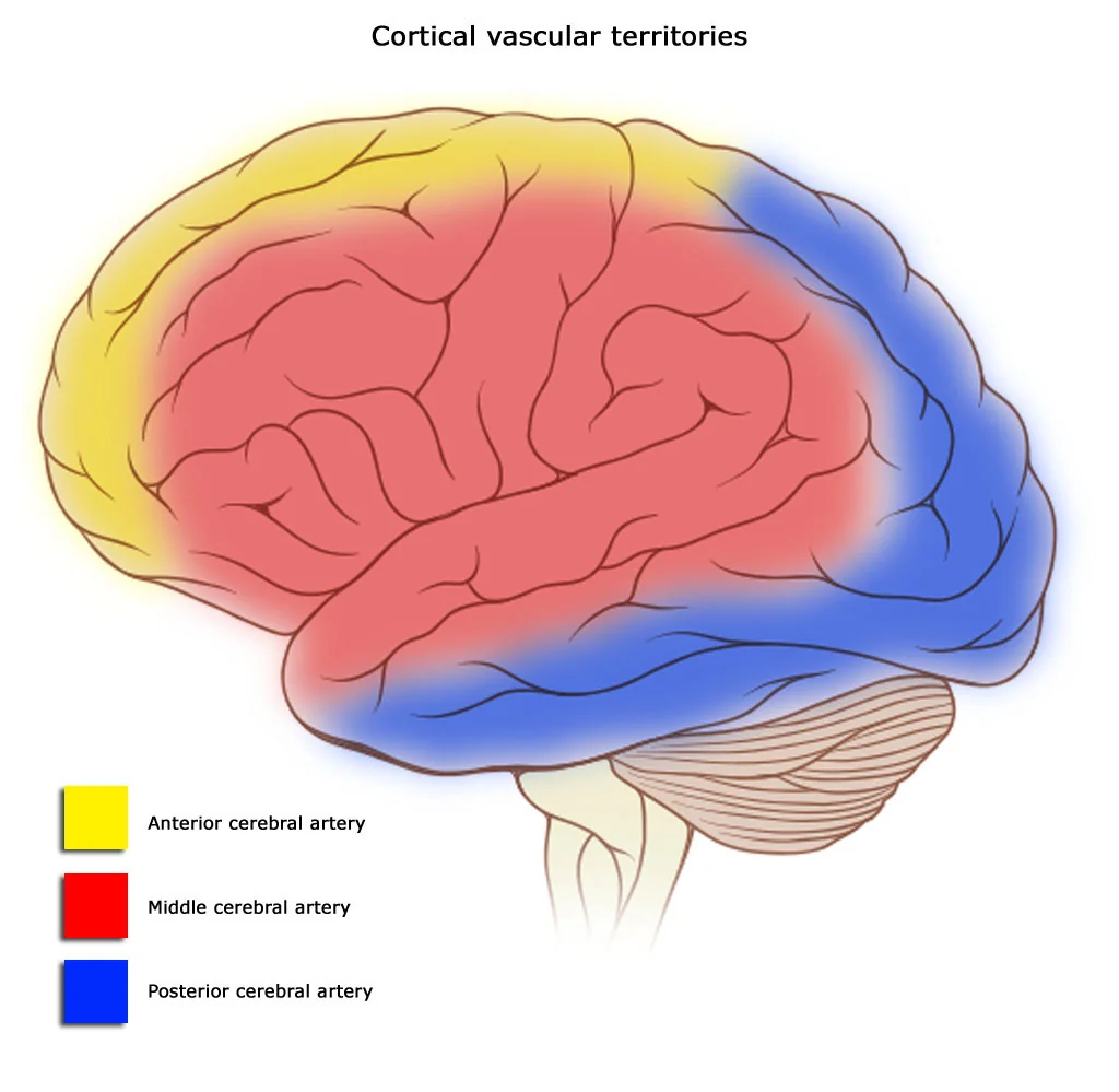
The Oxford Community Stroke Project (Bamford) classification predicts the arterial territory, and therefore prognosis and likely mechanism, from four clinical features assessed at the bedside: unilateral weakness (and/or sensory deficit) of the face, arm and leg; homonymous hemianopia; higher cerebral dysfunction (dysphasia, neglect, visuospatial disturbance); and brainstem or cerebellar signs.3
| Type | Criteria | Territory | Approx. proportion |
|---|---|---|---|
| Total anterior circulation stroke (TACS) | All three of: motor/sensory deficit, homonymous hemianopia, higher dysfunction | Large MCA +/- ACA territory | ~15% |
| Partial anterior circulation stroke (PACS) | Two of the three TACS criteria, or higher dysfunction alone | Smaller/branch MCA or ACA territory | ~35% |
| Lacunar stroke (LACS) | Pure motor, pure sensory, sensorimotor, or ataxic hemiparesis - no cortical signs | Small perforating vessels (basal ganglia, internal capsule, pons) | ~25% |
| Posterior circulation stroke (POCS) | Isolated hemianopia, brainstem signs, or cerebellar signs | Vertebrobasilar, PCA or cerebellar arteries | ~25% |
Risk factors
- Hypertension - the single most important modifiable risk factor for both ischaemic and haemorrhagic stroke
- Atrial fibrillation and other cardioembolic sources
- Smoking
- Diabetes mellitus
- Dyslipidaemia
- Previous TIA or stroke
- Carotid artery stenosis
- Increasing age
- Excess alcohol intake
- Combined oral contraceptive pill and pregnancy (increase venous and, less commonly, arterial thrombotic risk)
- Sickle cell disease, especially in children
- Cerebral amyloid angiopathy and anticoagulant use (haemorrhagic stroke specifically)
Clinical features
The onset is characteristically sudden, with a deficit that is maximal at (or very shortly after) onset - this contrasts with the evolving course of a mass lesion or the fluctuating course of a metabolic disturbance. Symptoms depend entirely on the territory affected.
- MCA territory: contralateral face and arm weakness (leg relatively spared), contralateral sensory loss, homonymous hemianopia, dysphasia if the dominant hemisphere is involved, neglect and visuospatial disturbance if the non-dominant hemisphere is involved
- ACA territory: contralateral leg weakness (face and arm relatively spared), personality change, urinary incontinence
- PCA territory: homonymous hemianopia (often with macular sparing), visual agnosia, memory disturbance
- Vertebrobasilar/brainstem: diplopia, vertigo, dysarthria, dysphagia, ataxia, crossed sensory/motor signs (ipsilateral face, contralateral body), reduced consciousness - a large basilar occlusion can cause locked-in syndrome
- Cerebellar: ataxia, nystagmus, intention tremor, dysmetria, vomiting
Community recognition - FAST
The FAST test (Face - Arm - Speech - Time) is a validated screening tool for paramedics and the public. It has good sensitivity for anterior circulation stroke but is less sensitive for posterior circulation strokes, which may present with isolated vertigo, ataxia or visual disturbance without limb weakness - a common source of missed or delayed diagnosis.
Clinical examination
- General: airway and conscious level (GCS), pulse (irregularly irregular suggests AF), blood pressure, blood glucose (hypoglycaemia is a critical stroke mimic), temperature
- Cranial nerves: visual fields to confrontation, pupillary response, eye movements and gaze deviation (the eyes may 'look towards the lesion' in a hemispheric stroke or away from it in a seizure), facial symmetry (forehead sparing suggests an upper motor neurone lesion), swallow assessment before any oral intake
- Limbs: tone, power (formally graded), reflexes, plantar response, coordination, sensation to light touch and pinprick
- Cardiovascular: listen for an irregular pulse, murmurs (endocarditis, valvular disease) and carotid bruits
- Fundoscopy: looking for papilloedema or hypertensive retinopathy
Differential diagnosis
Around 1 in 4 patients presenting with a stroke-like syndrome have a stroke mimic. Getting this wrong in either direction has consequences: missing a mimic exposes the patient to thrombolysis they do not need, while dismissing a genuine stroke as a mimic delays treatment.
- Hypoglycaemia - always check capillary glucose immediately; can produce a focal deficit indistinguishable from stroke
- Todd's paresis - transient post-ictal weakness following a focal seizure, which may not have been witnessed
- Migraine with aura - especially hemiplegic migraine, typically with a preceding visual aura and headache, spreading over minutes rather than seconds
- Functional (non-organic) neurological disorder
- Space-occupying lesion - tumour or abscess, usually a more gradual onset but can present acutely with haemorrhage into the lesion
- Sepsis with decompensation of an old deficit - a previous stroke can 're-present' during a systemic illness
- Bell's palsy - forehead involvement distinguishes a lower motor neurone facial palsy from a stroke
- Peripheral vestibular disorders - for isolated vertigo, distinguishing from a posterior circulation stroke (see the HINTS examination in Vestibular disorders)
Investigations
Immediate
- Capillary blood glucose - to exclude hypoglycaemia as a mimic before anything else
- Non-contrast CT head - the critical first investigation. Its purpose is to exclude haemorrhage, since ischaemic stroke and haemorrhage are managed in opposite directions. Early ischaemic change may be subtle or absent in the first few hours - loss of grey-white differentiation, sulcal effacement, or a hyperdense artery sign - but a normal early CT does not exclude ischaemic stroke
- CT angiography - performed alongside the non-contrast CT if thrombectomy is being considered, to identify a large vessel occlusion
- CT perfusion - identifies the ischaemic penumbra (hypoperfused but salvageable tissue) versus the infarct core, used to select patients for thrombectomy beyond the standard time window
- ECG - looking for atrial fibrillation as a cardioembolic source
- Bloods: FBC, U&Es, clotting, glucose, lipid profile
Subsequent
- MRI brain with diffusion-weighted imaging - far more sensitive than CT for acute infarction, especially small or posterior circulation strokes, but not usually needed to make the hyperacute treatment decision
- Carotid Doppler ultrasound - for anterior circulation ischaemic stroke or TIA, to identify carotid stenosis amenable to endarterectomy
- Echocardiography - if a cardioembolic source is suspected, particularly in younger patients or those with a structural cardiac history
- 24-hour (or prolonged) ECG monitoring - to detect paroxysmal atrial fibrillation when the initial ECG is normal but cardioembolism is suspected
- Vasculitis and thrombophilia screen - in younger patients with no conventional risk factors
Management
All patients with suspected acute stroke should be admitted directly to a hyperacute stroke unit7, bypassing the standard emergency pathway where a local 'stroke call' system exists, since specialist unit care itself reduces death and disability independent of any specific therapy.4
Hyperacute ischaemic stroke
Intravenous thrombolysis (alteplase, or increasingly tenecteplase) is offered to patients presenting within 4.5 hours of clearly known symptom onset (or last known well), provided haemorrhage has been excluded on CT and there is no contraindication.5 Selected patients with an unknown onset time (for example, wake-up strokes) may still be eligible if MRI shows a mismatch pattern suggesting recent onset.
| Category | Examples |
|---|---|
| Haemorrhage risk | Any evidence of haemorrhage on CT; recent major surgery or trauma; active bleeding; known bleeding diathesis |
| Anticoagulation | Therapeutic anticoagulation with a raised INR or recent DOAC dose (reversal agents may allow thrombolysis in specific cases) |
| Blood pressure | Persistently >185/110 mmHg despite treatment |
| Recent events | Ischaemic stroke or head injury within the preceding 3 months; history of intracranial haemorrhage |
| Glucose | Severe hypo- or hyperglycaemia mimicking or complicating the presentation |
Mechanical thrombectomy is offered for confirmed proximal large vessel occlusion (typically internal carotid or proximal MCA) in the anterior circulation, usually within 6 hours of onset, and up to 24 hours in carefully selected patients where perfusion imaging shows a large volume of salvageable tissue relative to the infarct core.6 Thrombectomy and thrombolysis are complementary, not alternatives - eligible patients should receive both.
Once haemorrhage and thrombolysis eligibility have been addressed, aspirin 300 mg is given (orally or rectally if dysphagic) as soon as haemorrhage is excluded, continued for two weeks and then switched to long-term secondary prevention. Blood pressure is not routinely lowered in the acute phase of ischaemic stroke, since this can worsen perfusion to the ischaemic penumbra - it is only actively treated if very high (generally >220/120 mmHg) or if thrombolysis is planned, when it must be brought below 185/110 mmHg first.
Supportive care
- Swallow screening before any oral intake - aspiration pneumonia is a leading cause of death after stroke
- VTE prophylaxis - intermittent pneumatic compression rather than pharmacological prophylaxis in the acute phase of haemorrhagic stroke, and used cautiously in ischaemic stroke
- Early mobilisation and multidisciplinary input - physiotherapy, occupational therapy and speech and language therapy from day one
- Glucose and temperature control - both hyperglycaemia and pyrexia worsen outcomes and should be actively managed
- Nutrition - nasogastric feeding if the swallow is unsafe and unlikely to recover quickly
Haemorrhagic stroke
Management centres on reversing any anticoagulation immediately, controlling blood pressure (target systolic around 140 mmHg if presenting early and systolic is 150-220 mmHg), and neurosurgical referral for large or cerebellar haematomas causing mass effect or hydrocephalus. Antiplatelets and anticoagulants are avoided; VTE prophylaxis with intermittent pneumatic compression is used instead of pharmacological agents in the acute phase.
Secondary prevention
- Antiplatelet therapy - clopidogrel 75 mg once daily is first-line long-term after aspirin has been given for the first two weeks
- Anticoagulation - for ischaemic stroke associated with atrial fibrillation, a DOAC is started once haemorrhagic transformation has been excluded and the infarct size assessed as safe to anticoagulate (often after 2 weeks for large infarcts, earlier for small ones - 'the 1-3-6-12 day rule' by increasing severity)
- High-intensity statin - atorvastatin 20-80 mg, regardless of baseline cholesterol, for ischaemic stroke
- Blood pressure control - once past the acute phase, treated to standard targets
- Carotid endarterectomy - for symptomatic carotid stenosis of 50-99% (NASCET criteria), ideally within 2 weeks of a non-disabling stroke or TIA in that territory
- Smoking cessation, glycaemic control, and lifestyle modification
- Driving - patients must not drive for at least 1 month after a stroke or TIA8, and Group 2 licence holders face a much longer disqualification; the DVLA must be informed for Group 2 licences and for Group 1 if there is residual deficit
Complications
- Aspiration pneumonia from an unsafe swallow
- Haemorrhagic transformation of an ischaemic infarct
- Cerebral oedema and raised intracranial pressure, particularly with large MCA territory ('malignant MCA syndrome') or cerebellar infarcts, which may require decompressive hemicraniectomy
- Seizures, both acute symptomatic and later post-stroke epilepsy
- Venous thromboembolism from immobility
- Urinary tract infection and pressure sores from reduced mobility and continence
- Post-stroke depression and cognitive impairment/vascular dementia
- Contractures, spasticity and shoulder pain from hemiplegia
Red flags
Prognosis
Roughly a third of patients who survive a stroke make a good functional recovery, a third are left with moderate disability, and a third are left severely dependent or die within the following year - though this varies enormously with stroke type and severity, and TACS carries a substantially worse prognosis than LACS.
Early admission to a specialist stroke unit, timely thrombolysis and thrombectomy where eligible, and structured secondary prevention all improve outcomes. Recovery continues for many months after the event, and rehabilitation input should not be curtailed simply because early gains have plateaued.
References
- NICE NG128. Stroke and transient ischaemic attack in over 16s: diagnosis and initial management. 2019, updated 2022. Available here
- Saver JL. Time is brain - quantified. Stroke. 2006. Available here
- Bamford J, Sandercock P, Dennis M et al. Classification and natural history of clinically identifiable subtypes of cerebral infarction. The Lancet. 1991. Available here
- Stroke Unit Trialists' Collaboration. Organised inpatient (stroke unit) care for stroke. Cochrane Database of Systematic Reviews. 2013. Available here
- Emberson J, Lees KR, Lyden P et al. Effect of treatment delay, age, and stroke severity on the effects of intravenous thrombolysis (alteplase) - a meta-analysis. The Lancet. 2014. Available here
- Goyal M, Menon BK, van Zwam WH et al. Endovascular thrombectomy after large-vessel ischaemic stroke - a meta-analysis (HERMES). The Lancet. 2016. Available here
- Royal College of Physicians. National clinical guideline for stroke for the UK and Ireland. Available here
- DVLA. Assessing fitness to drive: a guide for medical professionals. Available here
This article is written for revision and education. It is not clinical guidance and must not be used to make decisions about the care of a patient. Always check current NICE guidance and local protocols.