
Extradural Haemorrhage: Diagnosis and Management
Key points
- Extradural haemorrhage (EDH): arterial bleeding into the space between the skull and the dura mater, most often from a torn middle meningeal artery.
- Mechanism: a temporal or temporoparietal skull fracture lacerates the middle meningeal artery running in a groove on the inner skull surface.
- Classic presentation: head injury with brief loss of consciousness, a lucid interval of hours, then rapid deterioration as the haematoma expands.
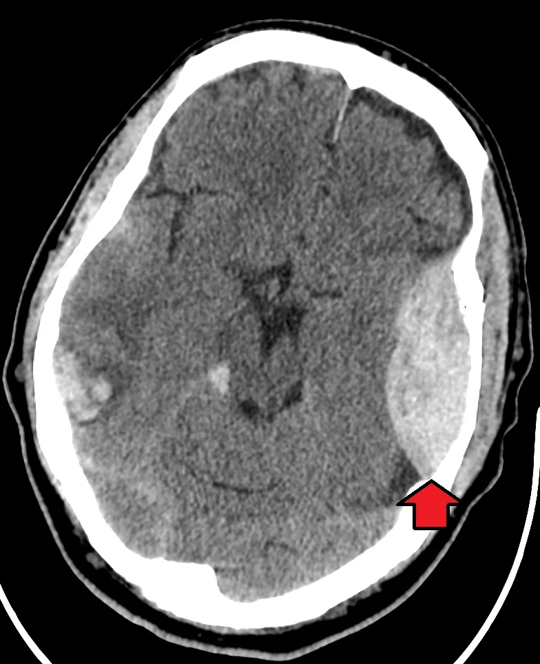
- CT appearance: a biconvex (lentiform, lemon-shaped) hyperdense collection that does not cross suture lines, because it is limited by dural attachment.
- This is a neurosurgical emergency: rapid arterial bleeding can cause fatal herniation within hours if not evacuated.
- Management: urgent craniotomy and clot evacuation for a significant or symptomatic haematoma; a small, asymptomatic collection may be observed closely.
- Classic sign: a fixed, dilated ('blown') pupil ipsilateral to the haematoma from uncal herniation compressing the third cranial nerve.
- Prognosis: excellent if evacuated before herniation occurs; the interval between deterioration and surgery is the single greatest determinant of outcome.
Introduction
Extradural (epidural) haemorrhage (EDH) is bleeding into the potential space between the inner table of the skull and the dura mater. It is typically arterial, most often from laceration of the middle meningeal artery by an overlying skull fracture, which makes it capable of expanding fast enough to cause death within hours if not recognised.1
It is one of the most time-critical diagnoses in neurosurgery and a favourite exam topic because the classic story - a young patient, a blow to the temple, a period of apparent normality, then rapid collapse - is dramatic, testable, and genuinely reflects how the condition can present.
EDH is less common than subdural haemorrhage overall but occurs in a younger population, since it depends on a skull fracture rather than the brain atrophy that predisposes older adults to subdural bleeding. The dura is normally adherent to the inner surface of the skull, so blood must strip it away to accumulate - which is why an extradural haematoma expands as a tense, lens-shaped collection under arterial pressure, and why the firm dural attachments at the suture lines halt its spread. This single anatomical fact explains both the biconvex shape and the failure to cross sutures.
It also explains the tempo. Because the bleeding is arterial and confined by a resistant dural attachment, pressure rises steeply once the compensatory reserve described by the Monro-Kellie doctrine is exhausted - which is exactly what produces the characteristic pattern of a well patient who then deteriorates precipitously rather than gradually.
Extradural haematomas account for a relatively small proportion of significant head injuries but occur disproportionately in young adults, with a peak in the second and third decades, and are strongly male-predominant. Because these patients typically have healthy brains and no significant comorbidity, they stand to gain more from timely intervention than almost any other group in neurosurgery - and correspondingly, they are the group in whom a missed diagnosis represents the greatest loss of potential years of healthy life.
Aetiology
The great majority of extradural haematomas follow trauma to the temporal or temporoparietal region, where the skull is thin and the middle meningeal artery runs in a bony groove on its inner surface, making it vulnerable to laceration by an overlying fracture.
The pterion - where the frontal, parietal, temporal and sphenoid bones meet - is the classic site, because the skull is at its thinnest there and the anterior division of the middle meningeal artery runs directly beneath it. A blow to the side of the head over this point, such as in assault or a sporting injury, is the archetypal mechanism, and knowing this anatomy explains why a seemingly modest impact to the temple can be far more dangerous than a heavier blow elsewhere.
- Middle meningeal artery laceration - by far the commonest source, associated with a temporal bone fracture
- Venous EDH - less common, from a torn dural venous sinus or diploic vein, tends to accumulate more slowly
- Skull fracture is present in the large majority of cases, particularly in adults - its absence should prompt reconsideration of the diagnosis, though children's more pliable skulls occasionally allow EDH without a visible fracture
Risk factors
- Head trauma with a temporal or temporoparietal skull fracture
- Young adult males - the group at highest risk of the relevant trauma mechanisms (assault, sport, road traffic collisions)
- Coagulopathy or anticoagulant use, which worsens the rate and volume of bleeding once a vessel is torn
- Notably less associated with cerebral atrophy or age than subdural haemorrhage, reflecting its different (arterial, fracture-dependent) mechanism
Clinical features
The classically described pattern is: a head injury causing a brief loss of consciousness, followed by a lucid interval of minutes to several hours during which the patient seems well or only mildly affected, and then rapid neurological deterioration as the expanding arterial haematoma raises intracranial pressure and causes herniation.
The lucid interval itself has a straightforward explanation. The initial loss of consciousness reflects concussion - a transient functional disturbance from the impact - which then recovers, giving a period of apparent normality. Meanwhile the torn middle meningeal artery continues to bleed silently. During this phase the Monro-Kellie compensatory mechanisms absorb the expanding volume by displacing CSF and venous blood, so intracranial pressure barely rises. Once that reserve is exhausted, pressure climbs steeply and the patient deteriorates rapidly.
This is why the lucid interval is dangerous rather than reassuring, and why the deterioration, when it comes, is abrupt rather than gradual. It is also the reason that observation after head injury is built around serial neurological assessment rather than a single examination: the purpose is to detect the moment compensation begins to fail, while there is still time to act.
- Head injury, often with a period of loss of consciousness at the time
- A lucid interval - the patient may appear normal or only mildly confused for a period afterwards; this is present in a substantial proportion of cases but is not universal, and its absence does not exclude EDH
- Worsening headache, vomiting and increasing drowsiness as the haematoma expands
- Focal neurological deficit, particularly contralateral hemiparesis
- Seizures
- Signs of uncal herniation as pressure rises: an ipsilateral fixed, dilated pupil (compression of the third cranial nerve as the uncus herniates through the tentorium), followed by bilateral pupillary involvement and progressive coma if untreated
Clinical examination
- GCS, recorded serially - a falling score is the single most important sign of deterioration and demands immediate action
- Pupils - an ipsilateral fixed, dilated ('blown') pupil signals uncal herniation and is a neurosurgical emergency
- Focal neurology: contralateral hemiparesis is typical, though a false localising sign (ipsilateral weakness from compression of the opposite cerebral peduncle against the tentorium - Kernohan's notch) can occur and cause confusion
- Cushing's triad (hypertension, bradycardia, irregular breathing) - a late and ominous sign of critically raised intracranial pressure
- Scalp and skull for bruising, laceration, or a palpable step suggesting an underlying fracture
- Battle's sign, panda eyes, CSF rhinorrhoea/otorrhoea, haemotympanum - signs of a base of skull fracture that should prompt wider imaging
Differential diagnosis
- Subdural haemorrhage - crescent-shaped and can cross suture lines, typically in an older or anticoagulated patient, evolves more slowly
- Traumatic subarachnoid haemorrhage
- Cerebral contusion
- Diffuse axonal injury - reduced GCS out of proportion to CT findings, from a high-energy deceleration mechanism
- Post-concussion syndrome - if symptoms are mild and there is no deterioration
Investigations
- Non-contrast CT head - the key investigation, showing a biconvex (lentiform, lemon-shaped) hyperdense collection. It does not cross suture lines, because the dura remains attached to the skull at the sutures and limits the haematoma's spread - the key feature distinguishing it from subdural haemorrhage
- Skull X-ray has essentially no role now that CT is universally and rapidly available, though a fracture crossing the course of the middle meningeal artery is the classic finding if seen
- Clotting profile in anyone on anticoagulation or with suspected coagulopathy
- CT should be requested urgently according to NICE head injury criteria - most patients with a suspected significant head injury meet criteria for imaging within 1 hour
Management
Immediate
- ABCDE assessment, with attention to airway protection if GCS is falling
- Urgent neurosurgical referral as soon as EDH is identified or strongly suspected
- Reverse any anticoagulation immediately2
- Correct hypoxia and hypotension, both of which worsen secondary brain injury
- Serial GCS and pupillary checks while awaiting transfer or theatre
Definitive management
Craniotomy and evacuation of the clot, with identification and control of the bleeding vessel, is the definitive treatment for a significant or symptomatic extradural haematoma, and should be performed as soon as possible - outcome is strongly time-dependent once signs of herniation appear.3
A small, asymptomatic EDH (typically under a defined volume and thickness threshold, with no midline shift or neurological deficit) may be managed conservatively with close observation and serial imaging in a neurosurgical unit, since some do not progress. This decision is made by neurosurgery, not in isolation.
Where the patient is in a hospital without neurosurgery on site - which is common - the priority is early discussion and rapid transfer rather than extensive local investigation. Time from deterioration to clot evacuation is the variable most strongly associated with outcome, so the receiving neurosurgical unit should be contacted as soon as the CT is available, images transferred electronically, and the patient prepared for transfer in parallel rather than sequentially. Intubation before transfer should be considered for any patient with a reduced or falling GCS, since deterioration in an ambulance without airway control is a recognised and preventable cause of harm.
Complications
- Uncal (transtentorial) herniation, with brainstem compression and death if untreated
- Permanent focal neurological deficit
- Seizures and post-traumatic epilepsy
- Recurrence of haematoma after evacuation, requiring return to theatre
- Cognitive impairment, particularly if evacuation was delayed until after herniation had begun
Red flags
Prognosis
Prognosis is generally excellent when the haematoma is evacuated before signs of herniation develop, since the underlying brain is often relatively undamaged compared with the diffuse injury seen in many subdural haematomas. Outcome deteriorates sharply once a fixed dilated pupil or coma has developed, which is why speed from deterioration to surgery is the dominant determinant of survival and neurological recovery.
Children deserve specific mention. Their more pliable skulls can deform enough to tear a vessel without an overlying fracture, so the absence of a fracture is less reassuring than in adults. Children also compensate well and can maintain a normal blood pressure and conscious level until late, then decompensate abruptly. Conversely, because the underlying brain is often undamaged, a child whose haematoma is evacuated promptly may make an excellent recovery even after a period of severe compromise - which is a strong argument for urgent intervention rather than a pessimistic view based on the presenting GCS alone.
It is worth reiterating why outcome is so much better than in subdural haemorrhage of comparable size. An extradural haematoma is essentially a compressive lesion sitting outside the dura, and the brain beneath it is often structurally intact; the deficit results from pressure, which is reversible if relieved in time. A subdural haematoma, by contrast, arises from a mechanism that has typically injured the underlying brain as well, so evacuating the collection does not undo the parenchymal damage. This distinction explains the very different prognostic outlook for two conditions that can look superficially similar.
References
- NICE NG232. Head injury: assessment and early management. 2023. Available here
- NICE CKS. Head injury. Available here
- Bullock MR, Chesnut R, Ghajar J et al. Surgical management of acute epidural hematomas. Neurosurgery. 2006. Available here
This article is written for revision and education. It is not clinical guidance and must not be used to make decisions about the care of a patient. Always check current NICE guidance and local protocols.