
Subdural Haemorrhage: Diagnosis and Management
Key points
- Subdural haemorrhage (SDH): bleeding into the subdural space, between the dura mater and arachnoid mater, usually from torn cortical bridging veins.
- Mechanism: acceleration-deceleration injury shears the bridging veins that cross the subdural space; brain atrophy (age, alcohol) stretches these veins and increases vulnerability, often after trivial trauma.
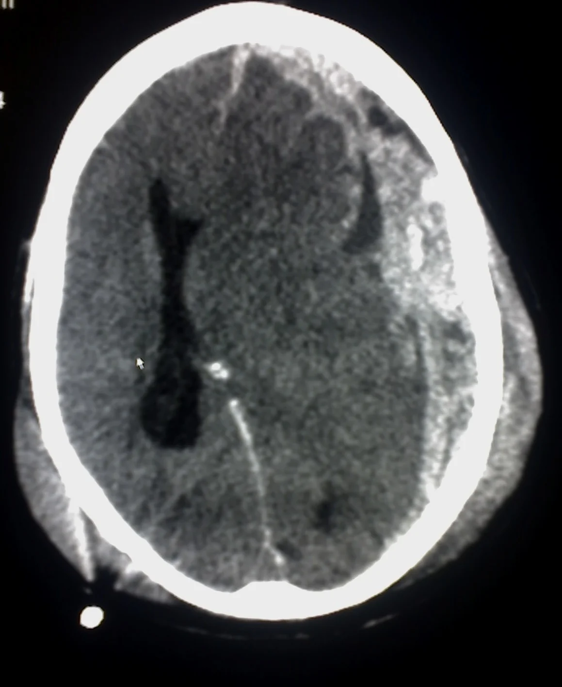
- CT appearance: a crescent-shaped (concave) collection that can cross suture lines, unlike an extradural haemorrhage.
- Classification by age: acute (hyperdense, days), subacute (isodense, 1-3 weeks - easy to miss) and chronic (hypodense, weeks to months).
- At-risk groups: older adults, chronic alcohol excess, and anticoagulated patients - all have fragile or stretched bridging veins and a higher bleeding risk from minimal or unwitnessed trauma.
- Management: conservative for small, asymptomatic collections; burr-hole evacuation or craniotomy for larger or symptomatic ones.
- Anticoagulation reversal: immediate, regardless of the agent, once any intracranial haemorrhage is confirmed.
- Presentation can be delayed: chronic subdurals may present weeks after a forgotten fall, with a fluctuating conscious level or progressive cognitive decline mistaken for dementia.
Introduction
Subdural haemorrhage (SDH) is bleeding into the potential space between the dura mater and the arachnoid mater, almost always caused by tearing of the cortical bridging veins that cross this space to drain into the dural venous sinuses.1
It is a classic presentation in older adults, in whom cerebral atrophy stretches the bridging veins and makes them vulnerable to rupture with only minor or even unrecalled trauma. This produces two very different clinical pictures that are both commonly examined: the acute subdural after a clear head injury, and the chronic subdural presenting weeks later with subtle, easily missed symptoms in a patient who may not remember falling at all. The reason a chronic subdural can grow so large before causing symptoms is the same reason it forms in the first place: cerebral atrophy leaves spare intracranial volume, so a slowly accumulating collection is accommodated for a long time before intracranial pressure rises. In a younger brain with no such reserve, the same volume of blood would cause symptoms far earlier.
SDH is a favourite topic because getting the CT appearance and the age-related shape (crescent, and its density over time) right is high-yield, and because it sits at the intersection of trauma, anticoagulation and an ageing population.
The subdural space itself is worth defining precisely, because the terminology causes confusion. It is a potential space rather than an anatomical one: the arachnoid is normally apposed to the dura, and a space only exists once blood or fluid separates them. This is why a subdural collection can spread widely over the convexity, tracking along the falx and tentorium and crossing suture lines freely - it is limited only by the dural reflections, not by the periosteal attachments that confine an extradural haematoma.
Aetiology and classification
The underlying mechanism is an acceleration-deceleration force that shears the bridging veins as the brain moves relative to the fixed dura. SDH is classified by the age of the blood, which changes both its clinical presentation and its CT density.
Chronic subdural haematomas are not simply old blood sitting passively in the subdural space. The collection becomes enclosed by vascularised neomembranes that form over weeks, and these fragile new vessels are prone to repeated small bleeds. This sets up a self-perpetuating cycle in which the haematoma slowly enlarges, which is why a chronic subdural can expand long after the original injury and why some appear on CT as mixed-density collections containing blood of several different ages.
Recognising this cycle explains two practical points. It is why a chronic subdural may continue to grow despite no further trauma, and it is the rationale behind newer treatments such as middle meningeal artery embolisation, which aims to cut off the blood supply to those neomembranes and reduce the high recurrence rate seen after simple drainage alone.
| Type | Timescale | CT density | Typical context |
|---|---|---|---|
| Acute | 0-3 days | Hyperdense (bright) | Significant trauma, often with other injuries |
| Subacute | 3 days-3 weeks | Isodense to brain - easy to miss | Falls in older adults; look for mass effect without an obvious collection |
| Chronic | >3 weeks | Hypodense (dark) | Minor or forgotten trauma in older adults, alcohol excess, anticoagulation |
Risk factors
- Increasing age - cerebral atrophy stretches the bridging veins, and even minor trauma (or none recalled) can cause a bleed
- Chronic alcohol excess - causes cerebral atrophy and coexisting coagulopathy from liver disease
- Anticoagulant or antiplatelet therapy
- Recurrent falls, including those from epilepsy, syncope or gait disturbance
- Infancy - shaken baby syndrome (abusive head trauma) is an important cause and should always be considered when the history is inconsistent with the injury
- Ventriculoperitoneal shunts - overdrainage of CSF can precipitate a subdural collection
- Coagulopathy from any cause
Clinical features
Presentation depends heavily on the age of the bleed and the patient's baseline reserve.
A practical trap is the bilateral chronic subdural. When collections are present on both sides, the mass effect is symmetrical and there may be no midline shift at all, so the scan can look deceptively unremarkable on a quick review. The clue is effacement of the sulci and small, compressed ventricles on both sides in a patient whose cerebral atrophy should have produced the opposite appearance.
- Acute SDH: reduced consciousness, headache and focal deficit following significant head trauma, often with a period of lucidity before deterioration, or continuously reduced GCS from the point of injury if there is associated brain injury
- Chronic SDH: an insidious, fluctuating picture over days to weeks - headache, personality change, cognitive slowing, gait disturbance, or a progressive focal deficit. The trauma may have been trivial and is frequently not recalled
- Fluctuating conscious level is a particularly characteristic and important feature of chronic SDH, and should always prompt consideration of the diagnosis in an older patient
- Seizures
- Signs of raised intracranial pressure in larger or rapidly expanding collections - headache, vomiting, papilloedema, reduced GCS
Clinical examination
- Conscious level (GCS), and whether it has fluctuated
- Pupils - a fixed, dilated pupil suggests uncal herniation from an expanding haematoma and is a neurosurgical emergency
- Focal neurology: hemiparesis, which may lateralise incorrectly if there is contralateral compression against the tentorium (a false localising sign)
- Signs of chronic alcohol use, liver disease or coagulopathy
- Scalp and skull for evidence of trauma, which may be absent or minimal even in a significant bleed
- Full trauma survey in the acute setting, since other injuries frequently coexist
Differential diagnosis
- Extradural haemorrhage - lens-shaped rather than crescent-shaped on CT, and does not cross suture lines
- Ischaemic or haemorrhagic stroke
- Dementia or delirium - especially for chronic SDH presenting with cognitive decline
- Normal pressure hydrocephalus - can mimic the gait and cognitive features of chronic SDH
- Alcohol intoxication or withdrawal
- Post-ictal state
- Cerebral contusion without an extra-axial collection
Investigations
- Non-contrast CT head - the key investigation, showing a crescent-shaped (concave) collection2 that, unlike an extradural haemorrhage, can cross suture lines because it is not constrained by dural attachments. Look for midline shift and effacement of the ventricles as markers of mass effect
- Clotting profile and platelet count - essential in anyone on anticoagulation or with suspected coagulopathy
- Group and save/crossmatch if surgery is anticipated
- MRI - more sensitive for small or isodense subacute collections, and for detecting membranes within a chronic subdural, though CT remains first-line in the acute setting
Management
Immediate
- ABCDE assessment and management of any life-threatening injury in the trauma setting
- Immediate reversal of anticoagulation - regardless of agent, as soon as any intracranial haemorrhage is confirmed3 (vitamin K and prothrombin complex concentrate for warfarin, and specific or non-specific reversal for DOACs per local protocol)
- Correct any coagulopathy or thrombocytopenia
- Neurosurgical discussion for any symptomatic collection, significant mass effect, or midline shift
Conservative management
Small, asymptomatic subdural collections with minimal mass effect can be managed conservatively with serial neurological observation and repeat imaging, particularly in frail patients where surgery carries a high risk. Many small chronic subdurals resorb spontaneously over weeks.
Surgical management
Surgery is indicated for a symptomatic collection, significant midline shift, or a deteriorating conscious level.
Post-operative care matters as much as the operation itself in determining whether the collection recurs. The brain in an older patient with cerebral atrophy may re-expand only slowly after drainage, leaving a residual subdural space that readily refills. Lying the patient flat for a period afterwards, ensuring adequate hydration, and leaving a subdural drain in situ for 24-48 hours are all directed at encouraging the brain to re-expand and obliterate that space. Residual fluid on an early post-operative scan is expected and is not, on its own, an indication to return to theatre.
- Burr-hole washout/drainage - the standard approach for most chronic subdurals, where liquefied blood can be drained through one or two small holes in the skull
- Craniotomy - required for acute subdurals with solid clot that cannot be evacuated through a burr hole, or where there is an associated contusion needing access
- Decompressive craniectomy - reserved for refractory raised intracranial pressure
- A subdural drain left in situ for 24-48 hours after burr-hole evacuation reduces the risk of recurrence
Complications
- Raised intracranial pressure and herniation (uncal or subfalcine)
- Recurrence after evacuation, particularly of chronic subdurals - a common reason for re-presentation
- Seizures, both acute and as a longer-term risk
- Persisting cognitive impairment, especially with delayed diagnosis
- Complications of surgery: infection, further haemorrhage, tension pneumocephalus
Red flags
The difficulty with subdural haemorrhage is that the patients most likely to have one are also those in whom the presentation is least typical. An older, anticoagulated patient with cognitive impairment may not report a fall, may not localise a headache, and may be assumed to have delirium from another cause. A low threshold for imaging in this group is more valuable than any individual clinical sign.
Prognosis
Outcome depends heavily on the age of the patient, the presence of associated brain injury, and how promptly the haemorrhage is diagnosed and treated. Acute SDH with underlying brain injury carries a significant mortality, particularly in older or anticoagulated patients. Chronic SDH treated with burr-hole evacuation generally has a good outcome, though recurrence occurs in a meaningful minority and cognitive recovery may be incomplete if diagnosis was delayed.
Outcome data can be misleading if read without context, since mortality figures for chronic subdural haematoma largely reflect the frailty and comorbidity of the population affected rather than the collection itself. A fit older adult with an isolated chronic subdural and no other pathology generally does very well after drainage.
An important part of managing chronic subdural haematoma is addressing why the patient fell in the first place, since a substantial proportion re-present with a further bleed. A falls assessment, medication review targeting sedatives and antihypertensives contributing to postural hypotension, visual and cognitive assessment, and correction of vitamin D deficiency all reduce that risk. Treating the haematoma without addressing the mechanism that produced it leaves the underlying problem untouched.
Decisions about anticoagulation afterwards are frequently the most difficult part of care, and are best framed explicitly as a trade-off between the risk of recurrent haemorrhage and the thromboembolic risk of withholding treatment - for example, stroke risk in atrial fibrillation or valve thrombosis in a metallic prosthesis. These decisions should be made jointly with the relevant specialties and, wherever possible, with the patient and family, rather than by a single team in isolation.
References
- NICE NG232. Head injury: assessment and early management. 2023. Available here
- NICE CKS. Head injury. Available here
- Karibe H, Kameyama M, Kawase M et al. Epidemiology of chronic subdural hematomas. Neurologia Medico-Chirurgica. 2014. Available here
This article is written for revision and education. It is not clinical guidance and must not be used to make decisions about the care of a patient. Always check current NICE guidance and local protocols.