
Bradyarrhythmias and Heart Block
Key points
- Bradycardia: a heart rate below 60 beats per minute. It is only significant if it is symptomatic, if there are adverse features, or if there is a risk of asystole.
- First degree AV block: a fixed PR interval above 200 ms with no dropped beats. Usually benign and needs no treatment.
- Mobitz I (Wenckebach): progressive PR lengthening then a dropped QRS. Block is within the AV node, often vagally mediated, and usually benign.
- Mobitz II: a constant PR interval with intermittently dropped QRS complexes. Block is below the AV node, it is unpredictable, and it frequently progresses to complete heart block. Pacing is indicated.
- Complete heart block: complete AV dissociation, with regular P waves and a slower independent escape rhythm. Cannon a waves and a variable first heart sound are the bedside signs.
- Escape rhythm width: a narrow escape from the AV junction at 40-60 per minute is relatively reliable; a broad ventricular escape at 20-40 per minute is not, and demands urgent pacing.
- Acute treatment: atropine 500 micrograms IV repeated to a maximum of 3 mg, then transcutaneous pacing, isoprenaline or adrenaline, and transvenous pacing if this fails.
- Risk of asystole: recent asystole, Mobitz II, complete heart block with a broad QRS, or a pause over 3 seconds. Any of these warrants escalation even without adverse features.
Introduction
A bradyarrhythmia is a heart rate below 60 beats per minute arising from failure of impulse generation at the sinus node, or failure of impulse conduction through the AV node and His-Purkinje system.
The most important principle is that a slow heart rate is not automatically abnormal. A resting rate of 45/min in a trained athlete, or 50/min during sleep, is physiological. What matters is whether the rate is adequate for the patient's needs, whether it causes symptoms, and whether the conducting system is showing signs that it may fail altogether.
This last point - the risk of progression - is what separates the benign bradyarrhythmias from the dangerous ones, and it turns almost entirely on where in the conducting system the problem lies. Disease at the level of the AV node is usually reversible, responsive to atropine, and backed up by a reliable junctional escape. Disease below the AV node, in the His-Purkinje system, is not reversible, does not respond to atropine, and is backed up only by an unreliable ventricular escape that may stop without warning.
Aetiology
Physiological
- Athletic training - high vagal tone with sinus bradycardia, and first degree or Mobitz I block during sleep
- Sleep - rates in the 40s are common and normal
- Vasovagal responses - during vomiting, straining, pain, or a needle procedure
Intrinsic conducting system disease
- Idiopathic fibrosis of the conducting system - the commonest cause of complete heart block in older people, sometimes named Lev disease (fibrosis of the cardiac skeleton) or Lenegre disease (fibrosis of the conducting tissue itself)
- Ischaemic heart disease - both acute and chronic. Inferior myocardial infarction commonly causes AV block, because the right coronary artery supplies the AV node in about 90% of people. This block is usually transient, atropine-responsive and does not require permanent pacing. Anterior infarction causing AV block indicates extensive septal necrosis affecting the bundle branches themselves - a much more ominous finding requiring pacing and carrying a poor prognosis.
- Infiltrative disease - amyloidosis, sarcoidosis and haemochromatosis. Cardiac sarcoidosis is an important and treatable cause of heart block in a younger patient.
- Infection - Lyme disease (a classic and reversible cause of AV block in a young patient with a rash or arthritis), infective endocarditis with an aortic root abscess, myocarditis, rheumatic fever, and Chagas disease
- Connective tissue disease - systemic lupus erythematosus, systemic sclerosis, rheumatoid arthritis, and maternal anti-Ro or anti-La antibodies causing congenital complete heart block
- Congenital complete heart block
- Cardiac surgery and interventions - aortic and mitral valve surgery, and TAVI, all risk damage to the conducting system, which lies close to the aortic valve
- Neuromuscular disease - myotonic dystrophy and Kearns-Sayre syndrome
Extrinsic and reversible causes
- Drugs - beta-blockers (including eye drops for glaucoma, which are systemically absorbed), rate-limiting calcium channel blockers, digoxin, amiodarone, ivabradine, adenosine, lithium, and cholinesterase inhibitors such as donepezil
- Hyperkalaemia - and other electrolyte disturbance
- Hypothyroidism
- Hypothermia - with J waves on the ECG
- Raised intracranial pressure - the Cushing reflex of bradycardia with hypertension
- Obstructive sleep apnoea - nocturnal bradycardia and pauses
- Hypoxia
- Cholestatic jaundice and anorexia nervosa
Sinus node disease
Sick sinus syndrome describes a spectrum of sinoatrial node dysfunction, most often from age-related fibrosis. It encompasses several ECG patterns, and one patient may show all of them at different times.
| Finding | Description |
|---|---|
| Sinus bradycardia | Persistent inappropriate slow sinus rate, without a physiological explanation |
| Sinus pause or arrest | The sinus node fails to fire; the ECG shows a flat line with no P wave until an escape beat or a resumed sinus beat. Pauses over 3 seconds while awake are significant. |
| Sinoatrial exit block | The impulse is generated but not conducted out of the node, giving a dropped P-QRS complex at a multiple of the underlying P-P interval |
| Chronotropic incompetence | Failure of the heart rate to rise appropriately with exertion, causing exercise intolerance |
| Tachy-brady syndrome | Alternating episodes of atrial tachyarrhythmia (usually AF) and bradycardia or long pauses, particularly on termination of the tachycardia |
Atrioventricular block
First degree AV block
A PR interval above 200 ms that is constant, with every P wave followed by a QRS. No beats are dropped - the term "block" is really a misnomer, since this is delay rather than block.
It is usually benign and asymptomatic, requiring no treatment. Consider reviewing rate-limiting drugs, and note that a markedly prolonged PR (over 300 ms) can occasionally cause symptoms resembling pacemaker syndrome, because atrial contraction occurs too early relative to ventricular filling.
Second degree AV block
Some P waves are not conducted. Distinguishing the two types is one of the highest-yield discriminations in cardiology, because the implications are entirely different.
| Feature | Mobitz I (Wenckebach) | Mobitz II |
|---|---|---|
| PR interval | Progressively lengthens until a beat is dropped, then resets | Constant, both before and after the dropped beat |
| Site of block | Within the AV node | Below the AV node, in the His-Purkinje system |
| QRS width | Usually narrow | Often broad |
| Response to atropine | Improves | May worsen - the sinus rate rises but conduction does not |
| Response to exercise | Improves | Worsens |
| Risk of progression | Low | High - frequently progresses to complete heart block |
| Typical cause | High vagal tone, athletes, inferior MI, rate-limiting drugs | Structural conducting system disease, anterior MI |
| Management | Usually observation; review drugs | Permanent pacemaker, even if asymptomatic |
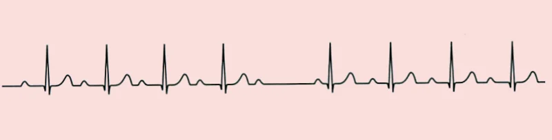

2:1 AV block - where every alternate P wave is conducted - cannot be classified as Mobitz I or II from the ECG alone, because there are never two consecutive conducted beats to compare PR intervals. Clues favouring a Mobitz II mechanism are a broad QRS and worsening with exercise or atropine. If in doubt, treat it as the more dangerous possibility and seek specialist assessment.
High-grade AV block describes two or more consecutive non-conducted P waves with some conduction preserved. It is managed as Mobitz II.
Third degree (complete) heart block
No atrial impulses reach the ventricles at all. The atria and ventricles beat entirely independently, each at its own intrinsic rate, and ventricular activity depends on an escape rhythm arising below the level of block.
- Regular P waves at the sinus rate, marching through the trace at a constant P-P interval
- Regular QRS complexes at a slower rate, with a constant R-R interval
- Complete dissociation - no fixed relationship between P waves and QRS complexes, and a variable PR interval. P waves may be buried in QRS complexes and T waves.
- The QRS width indicates the origin and reliability of the escape rhythm

| Escape origin | Rate | QRS | Reliability |
|---|---|---|---|
| Junctional (high in the AV junction) | 40-60/min | Narrow | Relatively stable; responds somewhat to atropine and exercise |
| Ventricular (below the bifurcation) | 20-40/min | Broad | Unreliable - may fail abruptly, causing Stokes-Adams attacks or asystole |
Clinical features
Symptoms
- Fatigue, lethargy and reduced exercise tolerance - often insidious, attributed to ageing, and only recognised in retrospect after pacing
- Dizziness and presyncope
- Syncope, classically as a Stokes-Adams attack
- Breathlessness - from reduced cardiac output, or frank heart failure
- Confusion - a common presentation of bradycardia in older patients
- Falls - bradycardia is an important and frequently missed cause of unexplained falls in older people
- Chest pain - if there is coexisting coronary disease
- Asymptomatic - many are found incidentally
Examination
- Slow, regular pulse - typically 30-40/min in complete heart block, and characteristically it does not increase with exertion
- Wide pulse pressure - a large stroke volume compensates for the slow rate
- Cannon a waves in the JVP - intermittent, irregular, large waves occurring when the atrium contracts against a closed tricuspid valve. A cardinal sign of AV dissociation.
- Variable intensity of the first heart sound - varying PR interval means varying valve position at the onset of systole
- Signs of heart failure if the rate is inadequate
- Signs of a cause - hypothyroidism, an erythema migrans rash, a new aortic regurgitation murmur suggesting an aortic root abscess, or a sternotomy scar
Investigations
- 12-lead ECG - the essential investigation. Measure the PR interval, look for dropped beats and their pattern, assess QRS width, and look for evidence of infarction and bifascicular block.
- Continuous cardiac monitoring - if there are symptoms, adverse features or any risk of asystole
- U&Es - particularly potassium; hyperkalaemia is a rapidly reversible cause
- Thyroid function tests
- Troponin - if ischaemia is suspected
- Digoxin level where relevant, and a full review of every rate-limiting drug
- Lyme serology - in a young patient, or with a suggestive exposure history or rash
- Blood cultures - if endocarditis with an aortic root abscess is a possibility
- Echocardiogram - to assess ventricular function, valve disease and structural causes
- Ambulatory monitoring or an implantable loop recorder - for intermittent symptoms, particularly unexplained syncope
- Cardiac MRI - where infiltrative disease such as sarcoidosis or amyloidosis is suspected
- Electrophysiological study - occasionally, to localise the level of block where it is uncertain
Acute management
Follow the Resuscitation Council UK bradycardia algorithm. Begin with an ABCDE assessment, give oxygen if hypoxic, obtain intravenous access and monitor the rhythm.1
Drug treatment
- Atropine 500 micrograms IV, repeated every 3-5 minutes to a maximum total of 3 mg
- If the response is inadequate, move to second-line measures without delay
Second-line measures
- Transcutaneous pacing - the fastest route to a guaranteed rate. It is painful, so give analgesia and sedation, and confirm mechanical capture by feeling a pulse, not just by seeing pacing spikes on the monitor.
- Isoprenaline infusion (5 micrograms/min, titrated) or adrenaline infusion (2-10 micrograms/min)
- Alternative drugs - aminophylline, dopamine, glucagon in beta-blocker or calcium channel blocker overdose, and digoxin-specific antibody fragments in digoxin toxicity
- Percussion pacing - a fist struck rhythmically over the lower left sternal edge, as a holding measure while awaiting equipment
- Transvenous pacing - the definitive temporary measure, arranged urgently through cardiology or critical care
Treat the cause
Stop rate-limiting drugs, correct hyperkalaemia, treat hypothyroidism, rewarm the hypothermic patient, treat Lyme disease with intravenous ceftriaxone, and reperfuse an acute infarct. Complete heart block complicating inferior myocardial infarction is often transient and resolves with reperfusion, which is why permanent pacing decisions are generally deferred for several days in that setting.
Permanent pacing
A permanent pacemaker is indicated where bradycardia is symptomatic and irreversible, or where there is a high risk of progression to asystole regardless of symptoms.2
| Condition | Indication |
|---|---|
| Complete heart block | Almost always paced, whether symptomatic or not, unless there is a reversible cause |
| Mobitz II second degree block | Paced even if asymptomatic, because progression is unpredictable |
| Mobitz I second degree block | Only if symptomatic, or if the block is demonstrated to be infranodal |
| Sick sinus syndrome | Paced if symptomatic. Asymptomatic bradycardia alone is not an indication. |
| Tachy-brady syndrome | Paced to permit rate-controlling drugs for the tachycardia |
| Bifascicular or trifascicular block | Paced if there is syncope, or documented intermittent complete heart block |
| Carotid sinus hypersensitivity | Paced if there is recurrent syncope with a documented cardioinhibitory response |
| Persistent AV block after anterior MI | Paced - indicates extensive septal infarction |
| AV block after inferior MI | Usually observed for several days, as it commonly resolves |
Device choice depends on the underlying rhythm: a single-chamber ventricular (VVI) device for permanent atrial fibrillation with slow ventricular response, a dual-chamber (DDD) device for AV block in sinus rhythm to preserve atrioventricular synchrony, and cardiac resynchronisation therapy where there is heart failure with a broad QRS. Pacemakers are covered in more detail in the article on pacemakers and implantable defibrillators.
Complications
- Asystole and sudden cardiac death - the outcome that pacing exists to prevent
- Syncope and consequent injury - fractures, head injury, and road traffic collisions
- Heart failure - cardiac output is the product of rate and stroke volume, and a persistently inadequate rate causes decompensation
- Bradycardia-related ventricular arrhythmia - a slow rate prolongs the QT and permits pause-dependent torsades de pointes
- Cognitive impairment and falls in older people from chronic cerebral hypoperfusion
- Complications of pacing - lead displacement, pneumothorax, infection, tamponade, and pacemaker syndrome from loss of AV synchrony in single-chamber ventricular pacing
Red flags
Prognosis
Prognosis depends on the level of block and whether the cause is reversible.
First degree block and Mobitz I in an asymptomatic patient carry an excellent prognosis and generally require nothing more than periodic review and attention to rate-limiting drugs.
Untreated complete heart block has a poor outlook, with a substantial annual mortality from asystole and heart failure, and syncope carries a high risk of injury in the interim. With permanent pacing, life expectancy approaches that of the general population, and the improvement in symptoms is often dramatic - patients frequently describe a return of energy they had attributed to ageing. Pacing for complete heart block is one of the clearest examples in medicine of a treatment that reliably restores both length and quality of life.
Reversible causes carry the prognosis of the underlying condition. AV block from drugs, hyperkalaemia, hypothyroidism or Lyme disease usually resolves completely with treatment, and identifying these avoids an unnecessary lifelong device. Conversely, complete heart block complicating anterior myocardial infarction carries a poor prognosis, not because of the block itself but because it indicates a large septal infarct with extensive myocardial loss.
References
- Resuscitation Council UK. Adult bradycardia algorithm. Available here
- Glikson M, Nielsen JC, Kronborg MB et al. 2021 ESC Guidelines on cardiac pacing and cardiac resynchronization therapy. European Heart Journal. 2021. Available here
- NICE CG109. Transient loss of consciousness (blackouts) in over 16s. 2010, updated 2023. Available here
- BNF. Atropine sulfate - indications and dosing. Available here
- NICE NG95. Lyme disease. 2018. Available here
- NICE NG185. Acute coronary syndromes. 2020, updated 2025. Available here
- DVLA. Assessing fitness to drive: a guide for medical professionals. Available here
- NICE Clinical Knowledge Summaries. Hyperkalaemia. Available here
This article is written for revision and education. It is not clinical guidance and must not be used to make decisions about the care of a patient. Always check current NICE guidance and local protocols.