
Spirometry and Lung Function Tests: Obstruction, Restriction and Transfer Factor
Key points
- The three numbers: FEV1, FVC and the FEV1/FVC ratio. The ratio decides whether the pattern is obstructive; the FVC and lung volumes decide whether it is restrictive.
- Obstructive pattern: FEV1/FVC below 0.7 with a disproportionately reduced FEV1. Asthma, COPD, bronchiectasis and cystic fibrosis.
- Restrictive pattern: FEV1/FVC normal or raised with a reduced FVC and, definitively, a reduced total lung capacity. Interstitial, chest wall, neuromuscular and pleural disease.
- Bronchodilator reversibility: an increase in FEV1 of 12% or more and 200 ml or more supports asthma. A rise of 400 ml or more is a NICE asthmatic feature in COPD.
- Transfer factor (TLCO): measures gas exchange across the alveolar-capillary membrane. Reduced in emphysema, interstitial lung disease, pulmonary vascular disease and anaemia.
- KCO: transfer factor corrected for the alveolar volume actually reached. It separates lung disease, where KCO falls, from extrapulmonary restriction, where KCO is normal or raised.
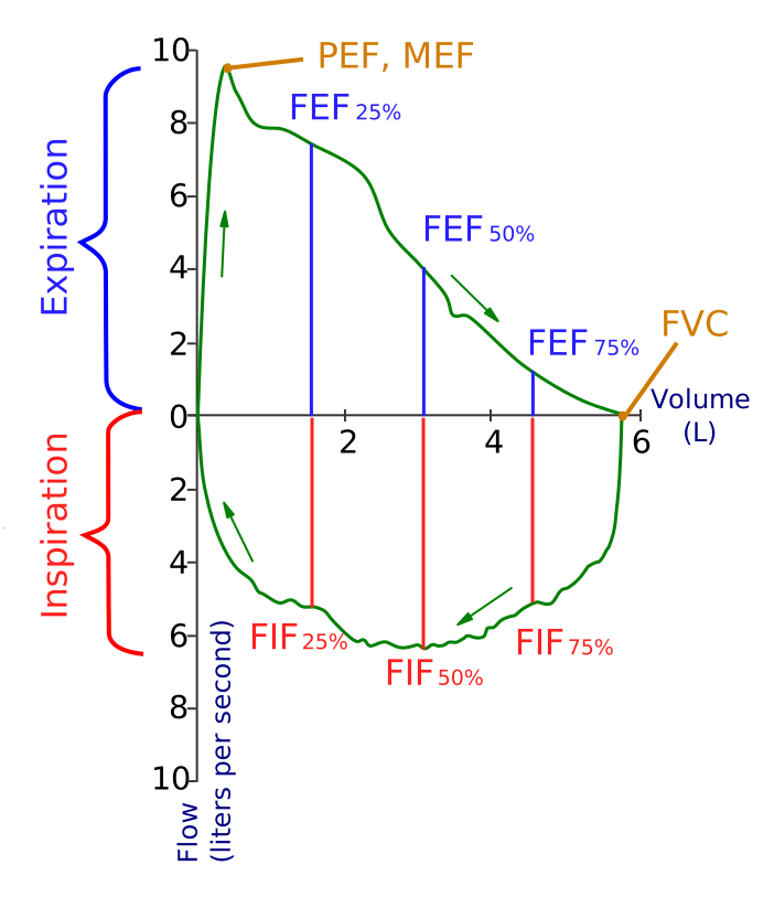
- Flow-volume loops: a scooped expiratory limb means airflow obstruction; flattening of both limbs means fixed large airway obstruction; a flattened inspiratory limb alone suggests a variable extrathoracic lesion.
- A normal test does not exclude disease: spirometry may be entirely normal between asthma attacks, and transfer factor may fall well before spirometry changes in interstitial disease.
Introduction
Lung function testing converts a clinical impression into a measurement. It answers three separate questions - is there airflow obstruction, is there restriction, and is gas exchange impaired - and it is the objective evidence on which the diagnosis of asthma, COPD and interstitial lung disease all rest.
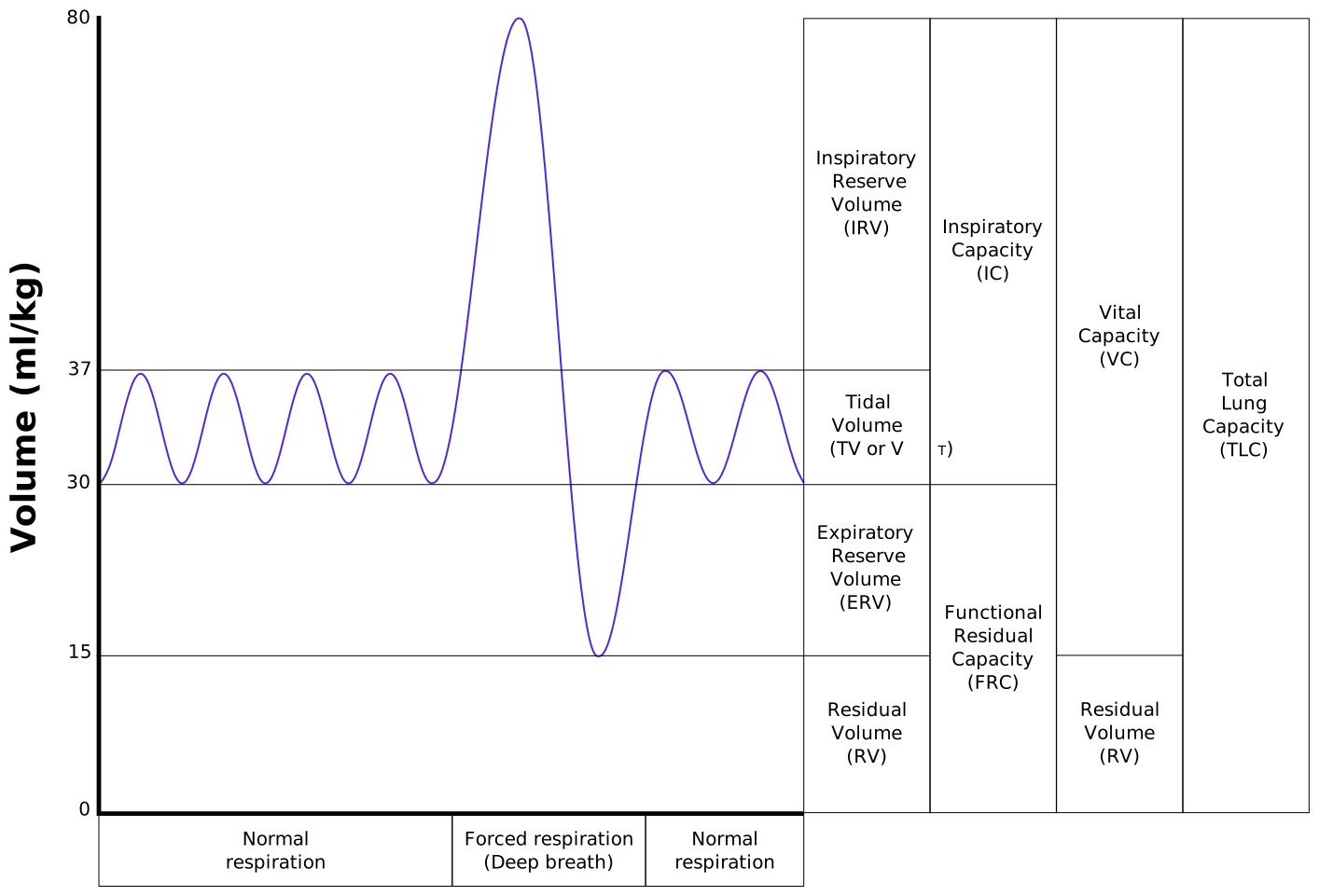
The tests form a hierarchy. Spirometry measures the volume and speed of air the patient can blow out and is available in every GP surgery. Lung volume measurement adds the air that cannot be exhaled and is what genuinely confirms restriction. Transfer factor assesses the alveolar-capillary membrane itself.
| Measurement | Definition |
|---|---|
| FVC - forced vital capacity | The total volume forcibly exhaled after a maximal inspiration |
| FEV1 | The volume exhaled in the first second of that manoeuvre |
| FEV1/FVC ratio | The proportion of the vital capacity exhaled in one second - normally above 0.7, and the marker of obstruction |
| PEF - peak expiratory flow | The maximum flow rate achieved, effort-dependent and used mainly for monitoring rather than diagnosis |
| TLC - total lung capacity | All the gas in the lungs at full inspiration. Reduced TLC defines restriction. |
| RV - residual volume | Gas remaining after maximal expiration. Raised in air trapping. |
| FRC - functional residual capacity | Volume at the end of a normal tidal expiration |
| TLCO (DLCO) - transfer factor | The rate at which carbon monoxide crosses from alveolus to haemoglobin - a measure of gas exchange |
| KCO - transfer coefficient | TLCO divided by the alveolar volume - transfer factor per unit of lung actually ventilated |
Performing and validating the test
Technique
- The patient sits upright with a nose clip, takes a maximal inspiration, seals their lips around the mouthpiece, and blows out as hard and as fast and for as long as possible
- At least three acceptable manoeuvres are required, with the two best FEV1 and FVC values agreeing within 150 ml
- Expiration should continue for at least 6 seconds, or until a plateau is reached - patients with severe obstruction may need considerably longer
- Results are compared with predicted values for age, sex, height and ethnicity, and reported as a percentage of predicted and as a z-score
Contraindications and cautions
- Recent myocardial infarction (within about a month), unstable angina or recent large pulmonary embolism
- Pneumothorax - current or recent
- Recent thoracic, abdominal, eye or neurosurgical surgery
- Thoracic, abdominal or cerebral aneurysm
- Active haemoptysis
- Active respiratory infection - both for accuracy and for infection control. Suspected or confirmed tuberculosis or COVID-19 requires appropriate precautions.
- The forced manoeuvre raises intrathoracic, intracranial, intraocular and intra-abdominal pressure, which is the common thread through that list
Obstruction and restriction
| Obstructive | Restrictive | |
|---|---|---|
| FEV1 | Reduced | Reduced |
| FVC | Normal or reduced | Reduced |
| FEV1/FVC ratio | Reduced (below 0.7) | Normal or increased (0.7 or above) |
| TLC | Normal or increased (air trapping) | Reduced |
| RV | Increased | Reduced |
| Flow-volume loop | Concave, scooped expiratory limb | Small loop of normal shape - tall and narrow |
| Typical causes | Asthma, COPD, bronchiectasis, cystic fibrosis, bronchiolitis obliterans | Interstitial lung disease, chest wall and neuromuscular disease, pleural disease, obesity |
Causes of an obstructive pattern
- Asthma - obstruction is variable and reversible, and spirometry may be entirely normal between episodes
- COPD - obstruction is persistent and only partly reversible, with a reduced TLCO in emphysema
- Bronchiectasis and cystic fibrosis
- Bronchiolitis obliterans - after transplantation, connective tissue disease or inhalation injury
- Upper airway obstruction - produces a distinctive flow-volume loop rather than a simple obstructive pattern
Causes of a restrictive pattern
It is worth dividing these by whether the lung itself is diseased, because the transfer factor separates them cleanly.
- Intrapulmonary (lung parenchyma diseased) - interstitial lung disease, sarcoidosis, pneumoconiosis, extensive consolidation, pulmonary oedema, lobectomy or pneumonectomy
- Extrapulmonary - chest wall - kyphoscoliosis, ankylosing spondylitis, obesity, circumferential burns
- Extrapulmonary - neuromuscular - motor neurone disease, myasthenia gravis, Guillain-Barre syndrome, muscular dystrophy, diaphragmatic paralysis
- Pleural - large effusion, extensive pleural thickening, empyema
Grading severity
| Stage | FEV1 % predicted | Severity |
|---|---|---|
| 1 | 80% or above with symptoms | Mild |
| 2 | 50-79% | Moderate |
| 3 | 30-49% | Severe |
| 4 | Below 30% | Very severe |
Reversibility and variability testing
Bronchodilator reversibility
- Spirometry is repeated 15 to 20 minutes after a short-acting bronchodilator (typically salbutamol 400 micrograms via a spacer)
- A significant response is an increase in FEV1 of 12% or more and at least 200 ml, which supports a diagnosis of asthma
- In a patient with COPD, NICE identifies an FEV1 variation of 400 ml or more as a feature suggesting steroid responsiveness and therefore a reason to include an inhaled corticosteroid
- A negative test does not exclude asthma - patients may be well controlled, already on treatment, or simply not reversible on the day. Continue down the diagnostic pathway rather than stopping.
Peak flow variability
- Twice-daily readings for 2 weeks, taking the best of three each time
- Variability of 20% or more is diagnostic of asthma in the NICE pathway
- Calculated as (highest minus lowest) divided by the highest, averaged over the period
- Also used to detect occupational asthma, with four-times-daily readings compared at and away from work
Other diagnostic tests
- FeNO (fractional exhaled nitric oxide) - a marker of eosinophilic airway inflammation. 50 ppb or more in adults, or 35 ppb or more in children, supports asthma. Suppressed by corticosteroids and by smoking.
- Bronchial challenge testing - histamine or methacholine, with a PC20 of 8 mg/ml or less indicating bronchial hyperresponsiveness. Used where the diagnosis remains uncertain.
- Corticosteroid trial - a course of oral or inhaled steroid with spirometry before and after, now used less often than objective testing
Transfer factor
The transfer factor is measured by having the patient inhale a trace of carbon monoxide, hold their breath for 10 seconds, and exhale. Because carbon monoxide binds haemoglobin avidly, the amount taken up reflects the surface area available for gas exchange, the thickness of the membrane, and the volume of blood in the pulmonary capillaries.
| Causes | |
|---|---|
| Reduced TLCO | Emphysema (loss of alveolar surface), interstitial lung disease (thickened membrane), pulmonary embolism and pulmonary hypertension (reduced capillary blood volume), anaemia, pneumonectomy, and low cardiac output |
| Raised TLCO | Pulmonary haemorrhage (blood in the alveoli avidly binds CO - the characteristic finding in Goodpasture disease), polycythaemia, asthma, left-to-right cardiac shunt, exercise, and obesity |
Always correct for haemoglobin. Anaemia lowers and polycythaemia raises the measured TLCO independently of any lung disease, and an uncorrected value is a common source of misinterpretation.
| Interstitial lung disease | Neuromuscular or chest wall disease | |
|---|---|---|
| FVC | Reduced | Reduced |
| FEV1/FVC | Normal or raised | Normal or raised |
| TLC | Reduced | Reduced |
| TLCO | Reduced | Reduced |
| KCO | Reduced | Normal or raised |
| Additional clue | Crackles, clubbing, HRCT changes | Weak cough, a fall in FVC of over 25% when lying flat, generalised weakness |
Measuring lung volumes
- Body plethysmography - the patient sits in a sealed booth and pants against a shutter. It measures all the gas in the thorax, including gas trapped behind closed airways, and is the reference method.
- Helium dilution or nitrogen washout - measure only gas that communicates with the airways, so they underestimate lung volumes in bullous disease and severe emphysema. A large discrepancy between plethysmographic and dilutional volumes is itself a measure of trapped gas.
Flow-volume loops
The flow-volume loop plots airflow against volume through a full forced expiration and inspiration. Its shape is often more informative than any single number, and it is the only reliable way to detect large airway obstruction on lung function testing.
| Pattern | Appearance | Causes |
|---|---|---|
| Obstructive | Concave, scooped-out expiratory limb with a reduced peak, shifted towards higher lung volumes | Asthma, COPD, bronchiectasis |
| Restrictive | A small loop of normal shape - narrow and tall, shifted towards lower lung volumes | Interstitial lung disease, chest wall and neuromuscular disease |
| Fixed large airway obstruction | Flattening of both the inspiratory and expiratory limbs, giving a boxed appearance | Tracheal stenosis, a large goitre compressing the trachea, tracheal tumour |
| Variable extrathoracic obstruction | Flattened inspiratory limb only - inspiration draws the lesion inwards while expiration splints it open | Vocal cord dysfunction (inducible laryngeal obstruction), vocal cord palsy, laryngeal tumour |
| Variable intrathoracic obstruction | Flattened expiratory limb only - expiration compresses the airway around the lesion | Tracheomalacia, a lower tracheal or main bronchial tumour |
Other tests of respiratory function
- Serial FVC, sitting and lying - the key monitoring test in neuromuscular disease. A fall of more than 25% on lying flat indicates diaphragmatic weakness, and an absolute FVC below 20 ml/kg should prompt critical care review in Guillain-Barre syndrome and myasthenic crisis.
- Maximal inspiratory and expiratory pressures (MIP and MEP) and sniff nasal inspiratory pressure (SNIP) - more sensitive measures of respiratory muscle strength than FVC alone
- Six-minute walk test - measures the distance walked in 6 minutes with continuous oximetry. Used in interstitial lung disease, pulmonary hypertension and pre-transplant assessment. Desaturation on exertion is often the earliest sign of significant disease in a patient whose resting saturations look reassuring.
- Incremental and endurance shuttle walk tests - standardised, externally paced alternatives
- Cardiopulmonary exercise testing (CPET) - measures peak oxygen uptake and the anaerobic threshold, distinguishes cardiac from respiratory limitation, and is used before major surgery and for transplant assessment
- Overnight oximetry and capnography - for sleep-disordered breathing and nocturnal hypoventilation
- Arterial or capillary blood gases - the definitive assessment of gas exchange and ventilation
Preoperative assessment
- Before lung resection, both FEV1 and TLCO are measured and the predicted postoperative values calculated from the number of segments to be removed
- A predicted postoperative FEV1 or TLCO below about 40% of predicted indicates high risk and prompts formal exercise testing
- CPET with a peak oxygen uptake below 10 ml/kg/min indicates a very high risk of perioperative death
- Spirometry alone is a poor predictor of risk for non-thoracic surgery, where functional capacity and comorbidity matter more
Pitfalls in interpretation
Putting it together
A structured way to read any lung function report:
- Check the test is valid - acceptable and repeatable manoeuvres, and a sensible-looking flow-volume loop
- Look at the FEV1/FVC ratio. Below 0.7 or the LLN means obstruction - grade it by FEV1 percentage predicted and check reversibility.
- If the ratio is normal, look at the FVC and TLC. A reduced TLC means restriction; a reduced FVC with a normal or high TLC means air trapping, not restriction.
- Look at the shape of the flow-volume loop for large airway obstruction, which the numbers alone will miss
- Look at TLCO and KCO. Together they tell you whether the lung tissue is diseased or whether the lung is simply not being expanded.
- Compare with previous tests. A single result describes a state; serial results describe a trajectory, and it is the trajectory that usually determines management.
- Interpret alongside the patient - the history, examination and imaging. Lung function that does not fit the clinical picture usually means the test, not the patient, needs repeating.
References
- NICE NG115. Chronic obstructive pulmonary disease in over 16s: diagnosis and management. 2018, updated 2019. Available here
- NICE NG245. Asthma: diagnosis, monitoring and chronic asthma management (BTS, NICE, SIGN). 2024. Available here
- Graham BL, Steenbruggen I, Miller MR et al. Standardization of spirometry 2019 update. An official ATS and ERS technical statement. Available here
- Stanojevic S, Kaminsky DA, Miller MR et al. ERS/ATS technical standard on interpretive strategies for routine lung function tests. European Respiratory Journal. 2022. Available here
- Association for Respiratory Technology and Physiology (ARTP). Spirometry standards and certification. Available here
- British Thoracic Society. Guidelines on the radical management of patients with lung cancer - preoperative assessment. Available here
- NICE CG163. Idiopathic pulmonary fibrosis in adults: diagnosis and management. 2013, updated 2017. Available here
- Vihsadas, derivative work by rscottweekly, CC BY-SA 3.0, via Wikimedia Commons. Available here
- SPhotographer and Jmarchn, CC BY-SA 3.0, via Wikimedia Commons. Available here
This article is written for revision and education. It is not clinical guidance and must not be used to make decisions about the care of a patient. Always check current NICE guidance and local protocols.